. 2016 Sep 13;113(37):10304-9.
doi: 10.1073/pnas.1608069113. Epub 2016 Aug 22.

Precision glycocalyx editing as a strategy for cancer immunotherapy

Affiliations

Precision glycocalyx editing as a strategy for cancer immunotherapy

Han Xiao et al. Proc Natl Acad Sci U S A. .

Abstract

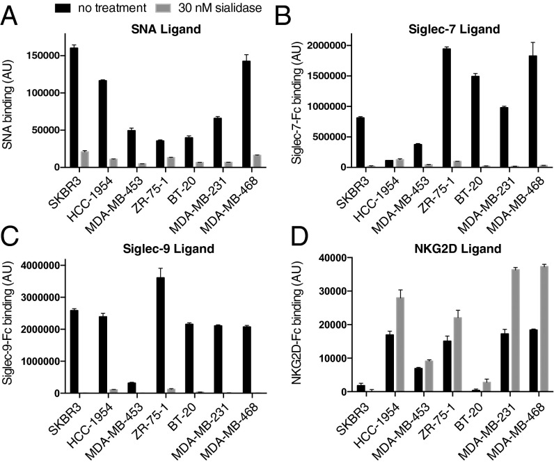
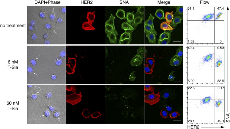
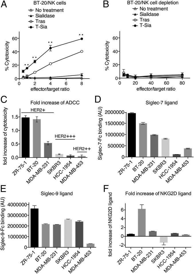
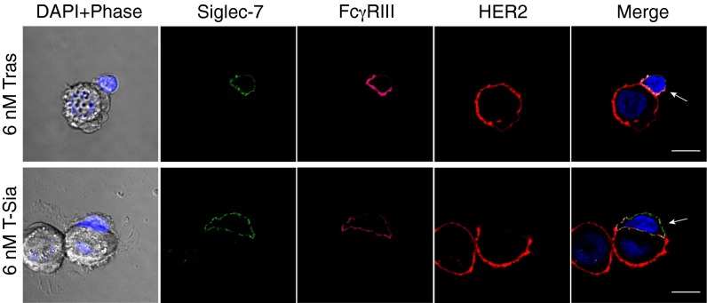
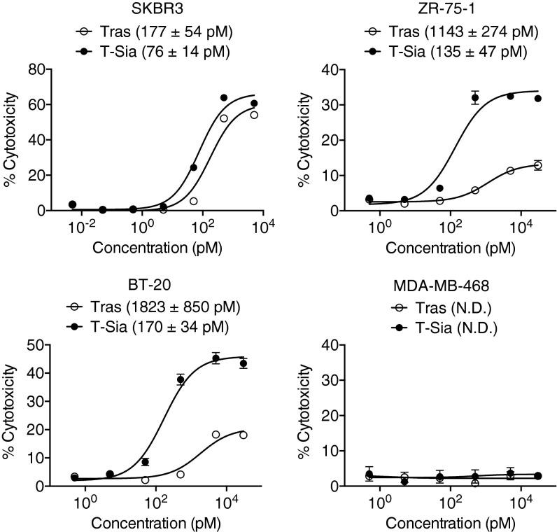
Cell surface sialosides constitute a central axis of immune modulation that is exploited by tumors to evade both innate and adaptive immune destruction. Therapeutic strategies that target tumor-associated sialosides may therefore potentiate antitumor immunity. Here, we report the development of antibody-sialidase conjugates that enhance tumor cell susceptibility to antibody-dependent cell-mediated cytotoxicity (ADCC) by selective desialylation of the tumor cell glycocalyx. We chemically fused a recombinant sialidase to the human epidermal growth factor receptor 2 (HER2)-specific antibody trastuzumab through a C-terminal aldehyde tag. The antibody-sialidase conjugate desialylated tumor cells in a HER2-dependent manner, reduced binding by natural killer (NK) cell inhibitory sialic acid-binding Ig-like lectin (Siglec) receptors, and enhanced binding to the NK-activating receptor natural killer group 2D (NKG2D). Sialidase conjugation to trastuzumab enhanced ADCC against tumor cells expressing moderate levels of HER2, suggesting a therapeutic strategy for cancer patients with lower HER2 levels or inherent trastuzumab resistance. Precision glycocalyx editing with antibody-enzyme conjugates is therefore a promising avenue for cancer immune therapy.

Keywords: ADCC; Siglec; cancer immune therapy; sialic acid; trastuzumab.


Conflict of interest statement

The authors declare no conflict of interest.

Figures

Fig. 1.
Fig. 1.
A glycocalyx approach to cancer immunotherapy targeting the sialic acid axis of immune modulation. (A) In sialic acid-overexpressing cancer cells, hypersialylated glycans interact with NK inhibitory receptors, leading to inhibition of NK cell activation. (B) Removal of cell-surface sialic acids by an antibody–sialidase conjugate (T-Sia) abolishes the interaction of sialylated glycans and NK-inhibitory Siglec receptors and increases the binding between NK-activating NKG2D receptor and its ligands, thereby enhancing the tumor cell susceptibility to NK cell-mediated ADCC.
Fig. 2.
Fig. 2.
Levels of SNA, Siglec-7, Siglec-9, and NKG2D ligands on breast cancer cell lines. (A) Analysis of cell-surface sialylation levels of different breast cancer cell lines with or without sialidase treatment. (B) Ligand levels of Siglec-7 on different breast cancer cell lines with or without sialidase treatment. (C) Ligand levels of Siglec-9 on different breast cancer cell lines with or without sialidase treatment. (D) Ligand levels of NKG2D on different breast cancer cell lines with or without sialidase treatment.
Fig. 3.
Fig. 3.
Preparation and characterization of antibody–sialidase conjugate. (A) Humanized trastuzumab (Tras) bearing an aldehyde tag was produced by genetically introducing the consensus sequence SLCTPSRGS at the C terminus of the heavy chain. The cysteine residue within the sequence was converted to formylglycine in situ by action of ER-resident formylglycine generating enzyme (FGE) during expression. The aldehyde-tagged Tras was coupled to the heterobifunctional linker aminooxy-TEG-N3 by oxime ligation. Separately, recombinant V. cholerae sialidase was reacted at lysine side chains with bicyclononyne-N-hydroxysuccinimide ester (BCN-NHS). Finally, the two functionalized proteins were conjugated via copper-free click chemistry. (B) SDS/PAGE analysis of sialidase, Tras, and trastuzumab–sialidase conjugate (T-Sia) under nonreducing (lanes 3, 4, and 5) and reducing (lanes 6, 7, and 8) conditions visualized by Coomassie staining. Multiple bands observed for Tras and T-sia reflect heterogeneity in antibody glycosylation. Prestained protein ladder: lanes 1 and 2.
Fig. 4.
Fig. 4.
Visualization and quantitation of T-Sia’s tissue-specific selective activity. Cell-surface sialic acid on the HER2-high expressing cell line, SKBR3 (white arrows), can be selectively removed using 6 nM T-Sia in the presence of HER2-negative cell lines MDA-MB-468. SKBR3 and MDA-MB-468 cells were cocultured and then treated or not with T-Sia for 1 h and costained with Alexa Fluor 647-labeled anti-HER2 antibody, FITC-SNA, and DAPI before imaging by confocal microscopy (Left). Cell-surface sialylation and HER2 levels were also quantitatively determined by flow cytometry (Right). (Scale bars: 25 μm.)
Fig. 5.
Fig. 5.
In vitro activity of trastuzumab and T-Sia against different HER2-expressing cancer cells. (A) Cytotoxicity assays performed with BT-20 cells in the absence or presence of sialidase (30 nM), Tras (30 nM), and T-Sia (30 nM) using NK cells at various E/T ratios. (B) Cytotoxicity assays performed with BT-20 cells in the absence or presence of sialidase (30 nM), Tras (30 nM), and T-Sia (30 nM) using NK cell-depleted PBMCs. (C) The trend seen in the enhancement of ADCC correlated with Siglec-7-Fc (D) and Siglec-9-Fc (E), but not NKG2D-Fc (F) binding levels for various breast cancer cell lines.**P < 0.005.
Fig. 6.
Fig. 6.
Fluorescence microscopy analysis of Siglec-7 distribution on NK cells with trastuzumab or T-Sia treatments. Siglec-7 displayed recruitment to the NK cell (white arrows) synapse with Tras treatment. After removing sialic acids on SKBR3 cells using T-Sia, Siglec-7 recruitment to the NK–tumor synapse was lost. (Scale bars: 10 μm.)
Fig. 7.
Fig. 7.
Cytotoxic activity of NK cells against different HER2-expressing cancer cells with trastuzumab or T-Sia as a function of concentration. Cytotoxicity assays were performed with NK cells and target cells at a 4:1 ratio in the presence of different concentrations of Tras or T-Sia. Cytolytic activity was determined by measuring the amount of lactate dehydrogenase (LDH) released into cultured media. Error bars represent SD of triplicate samples. N.D., not determined.

Comment in

  • Cutting back on the carbs.
    Trang VH, Senter PD. Trang VH, et al. Proc Natl Acad Sci U S A. 2016 Sep 13;113(37):10228-30. doi: 10.1073/pnas.1612432113. Epub 2016 Sep 1. Proc Natl Acad Sci U S A. 2016. PMID: 27588908 Free PMC article. No abstract available.

References

    1. Vanneman M, Dranoff G. Combining immunotherapy and targeted therapies in cancer treatment. Nat Rev Cancer. 2012;12(4):237–251. - PMC - PubMed
    1. Pardoll DM. The blockade of immune checkpoints in cancer immunotherapy. Nat Rev Cancer. 2012;12(4):252–264. - PMC - PubMed
    1. Restifo NP, Dudley ME, Rosenberg SA. Adoptive immunotherapy for cancer: Harnessing the T cell response. Nat Rev Immunol. 2012;12(4):269–281. - PMC - PubMed
    1. Bhatia A, Kumar Y. Cellular and molecular mechanisms in cancer immune escape: A comprehensive review. Expert Rev Clin Immunol. 2014;10(1):41–62. - PubMed
    1. Raulet DH, Vance RE. Self-tolerance of natural killer cells. Nat Rev Immunol. 2006;6(7):520–531. - PubMed

Publication types

MeSH terms