. 2016 Sep 6;113(36):E5261-70.
doi: 10.1073/pnas.1605844113. Epub 2016 Aug 22.

Autophagy and ubiquitin-proteasome system contribute to sperm mitophagy after mammalian fertilization

Affiliations

Autophagy and ubiquitin-proteasome system contribute to sperm mitophagy after mammalian fertilization

Won-Hee Song et al. Proc Natl Acad Sci U S A. .

Abstract

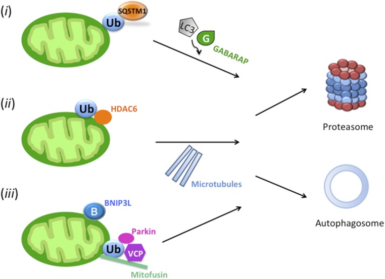
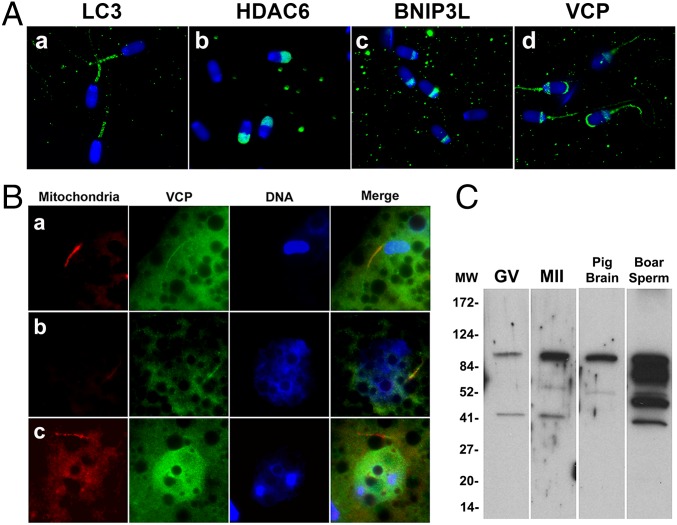
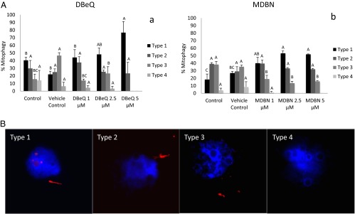
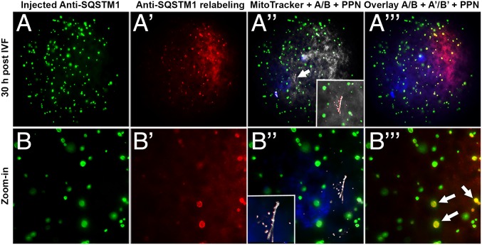
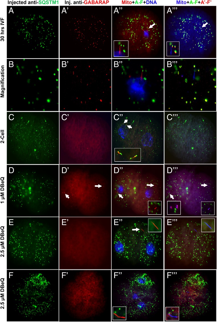
Maternal inheritance of mitochondria and mtDNA is a universal principle in human and animal development, guided by selective ubiquitin-dependent degradation of the sperm-borne mitochondria after fertilization. However, it is not clear how the 26S proteasome, the ubiquitin-dependent protease that is only capable of degrading one protein molecule at a time, can dispose of a whole sperm mitochondrial sheath. We hypothesized that the canonical ubiquitin-like autophagy receptors [sequestosome 1 (SQSTM1), microtubule-associated protein 1 light chain 3 (LC3), gamma-aminobutyric acid receptor-associated protein (GABARAP)] and the nontraditional mitophagy pathways involving ubiquitin-proteasome system and the ubiquitin-binding protein dislocase, valosin-containing protein (VCP), may act in concert during mammalian sperm mitophagy. We found that the SQSTM1, but not GABARAP or LC3, associated with sperm mitochondria after fertilization in pig and rhesus monkey zygotes. Three sperm mitochondrial proteins copurified with the recombinant, ubiquitin-associated domain of SQSTM1. The accumulation of GABARAP-containing protein aggregates was observed in the vicinity of sperm mitochondrial sheaths in the zygotes and increased in the embryos treated with proteasomal inhibitor MG132, in which intact sperm mitochondrial sheaths were observed. Pharmacological inhibition of VCP significantly delayed the process of sperm mitophagy and completely prevented it when combined with microinjection of autophagy-targeting antibodies specific to SQSTM1 and/or GABARAP. Sperm mitophagy in higher mammals thus relies on a combined action of SQSTM1-dependent autophagy and VCP-mediated dislocation and presentation of ubiquitinated sperm mitochondrial proteins to the 26S proteasome, explaining how the whole sperm mitochondria are degraded inside the fertilized mammalian oocytes by a protein recycling system involved in degradation of single protein molecules.

Keywords: autophagy; mitochondria; mitophagy; mtDNA; ubiquitin.


Conflict of interest statement

The authors declare no conflict of interest.

Figures

Fig. 1.
Fig. 1.
Putative pathways participating in the elimination of sperm mitochondria by coordinated activities of autophagy and the ubiquitin–proteasome system. (i) The ubiquitin-binding autophagy receptor SQSTM1 could recognize ubiquitinated mitochondrial protein cargo and interact with autophagosome-binding ubiquitin-like modifiers, such as LC3 and/or GABARAP to transport them toward autophagosome. (ii) Ubiquitinated proteins could be extracted from mitochondria and form aggresomes, the protein aggregates induced by ubiquitin-binding adaptor protein HDAC6 that transports them along the microtubules to the autophagophore. (iii) Protein dislocase VCP could extract and present the ubiquitinated mitochondrial membrane proteins to the 26S proteasome.
Fig. 2.
Fig. 2.
The ubiquitin-binding autophagy receptor SQSTM1 associated with sperm mitochondria after porcine (A and B) and rhesus monkey (C) fertilization. (A) The SQSTM1 protein (green) was not detectable in the zona-bound spermatozoa before sperm incorporation into oocyte cytoplasm (a); it became associated with sperm mitochondria by 9 h postfertilization (b; very early stage of paternal pronucleus formation was shown) and remained detectable at 12 (c) and 30 h postfertilization (d). (Original magnification, 1,000×.) (B) Western blotting detected the predicted 75-kDa SQSTM1 protein band in porcine GV and MII oocytes, cumulus cells and zygotes (IVF, 30 h), but not in boar spermatozoa (boar sperm). (C) The SQSTM1 protein was detected in the sperm mitochondria at the pronuclear stages of zygotic development in rhesus monkey ICSI embryos (a), but not yet associated with the sperm mitochondrial sheath early after ICSI (b).
Fig. 3.
Fig. 3.
Affinity purification and identification of the ubiquitinated mitochondrial proteins from boar sperm tails. (A) Boar sperm heads and tails were solubilized with RIPA buffer and the resultant extracts were incubated with a synthetic SQSTM1-derived UBA domain, immobilized on agarose beads. The eluted sperm proteins were separated on the gel, then stained with Coomassie blue. Arrows mark protein bands that are conspicuously different between sperm head and tail fractions and were excised for proteomic identification. (B) Immunofluorescence localization of HADHA, ACO2, and ATP5B (all in green), three proteins prevalent in the ubiquitinated sperm tail protein fraction, with high ion scores. (C) Western blotting confirmed the presence of HADHA, ACO2, and ATP5B in the ubiquitinated sperm tail protein fraction. Bands of anticipated size were observed for each of the identified proteins.
Fig. 4.
Fig. 4.
Detection of ubiquitin-like protein modifier GABARAP, a mitophagy receptor, in control and MG132-treated porcine zygotes. (A) The accumulation of GABARAP-containing aggregates/autophagophores (green) was detected around the sperm tail (red; MitoTracker) and paternal pronuclei (blue; DAPI) at 30 h postinsemination in untreated control (a and a′), at 50 μM MG132 (b and b′), 100 μM MG132 (c and c′), and in vehicle control (d and d′; EtOH). Note the increased GABARAP particle content in the MG132-treated zygotes. Note that very few of the GABARAP+ green particles were colocalized with the red fluorescent sperm mitochondria. (Original magnification, 1,000×.) (B) Western blotting of GABARAP in porcine zygotes cultured for 30 h after fertilization (i.e., 36 h after sperm addition), with or without MG132 (10/100 μM). The GABARAP protein migrated as a triplet of bands. The lowest mass band was amplified in the presence of MG132. Last lane is a vehicle control (EtOH). (C) Ooplasmic, autophagophore-associated GABARAP (red) recruited along sperm aster microtubules (green) in a fertilized oocyte cultured for 30 h after IVF. Well-developed sperm aster microtubules were formed in the vicinity of paternal pronucleus, showing that GABARAP recruitment to the sperm aster was concomitant with the pronuclear development and degradation of the sperm mitochondria. Image was processed by 2D deconvolution. PPN, paternal pronucleus; TUBB, β-tubulin.
Fig. 5.
Fig. 5.
Immunolocalization and inhibition of autophagy determinants implicated in sperm mitophagy. (A) Immunolocalization of candidate mitophagy determinants in the boar spermatozoa before fertilization: LC3 (a) was present in the midpiece/mitochondrial sheath of the sperm tail. HDAC6 (b) was present in the acrosomal region of the sperm head. BNIP3L (c) was also detectable in the sperm head, particularly in the postacrosomal sheath. VCP (d) was detected in the apical ridge of the acrosome, in the sperm head postacrosomal sheath and in the sperm tail midpiece. Nuclear DNA was counterstained with DAPI (blue). (B) Colocalization of VCP (green) with the sperm mitochondria (red) in porcine zygotes at the early stage of sperm-nuclear decondensation (a), at 30 h after IVF (b), and at 30 h after IVF in the presence of MDBN, a specific inhibitor of VCP (c). Intact sperm mitochondria were present in embryos cultured for 30 h in the presence of MDBN. (C) Western blotting of VCP in the porcine oocytes and sperm extracts. The anticipated band of 97 kDa, corresponding to VCP protein, was detected in GV oocytes, MII-oocytes, pig brain (third lane), and boar spermatozoa (fourth lane).
Fig. 6.
Fig. 6.
(A) The effect of specific VCP inhibitors: DBeQ (reversible inhibitor) (a) and MDBN (irreversible) (b) on sperm mitophagy in porcine zygotes examined at 30 h after IVF. (A) Porcine oocytes were fertilized for 6 h with or without VCP inhibitors and then washed and cultured in an inhibitor-free embryo culture medium for an additional 24 h. The incidence of type 1, intact sperm mitochondrial sheaths gradually increased with increasing dose of both VCP inhibitors. Experiments were repeated three times for each treatment group (10–20 oocytes per group). Values are shown as the mean percentages of sperm mitophagy ± SEM. Different superscripts A, B, and C in each diagram present a significant difference at P < 0.05. (B) Four different phenotypes of the sperm mitochondrial sheath, reflective of the progression of sperm mitochondrion degradation after fertilization. Type 1 displays a straight, intact mitochondrial sheath. Type 2 represents a distorted, corkscrew-shaped mitochondrial sheath. Type 3 shows scattered or clumped, nearly completely degraded sperm mitochondria. Type 4 represents the absence of sperm mitochondria; only the remains of outer dense fibers that take up the red MitoTracker stain nonspecifically are present.
Fig. 7.
Fig. 7.
Preinjection of MII oocytes with anti-SQSTM1antibody prevented the postfertilization binding of ooplasmic SQSTM1 to sperm mitochondria. Oocytes preinjected with mouse anti-SQSTM1 antibody (green) were cultured for 30 h after fertilization, fixed, labeled with green fluorescent anti-mouse secondary antibody, then washed, and relabeled with anti-SQSTM1 antibody followed by red fluorescent anti-mouse IgG. In oocytes preinjected with anti-SQSTM1 antibody, SQSTM1+ aggregates (A and B, green) were formed throughout ooplasm. Neither the injected anti-SQSTM1 antibody (A′′ and B′′) nor the ooplasmic SQSTM1 (A′′′ and B′′′) associated with sperm mitochondria (MitoTracker; arrow in A′′) in the oocytes preinjected with anti-SQSTM1 antibody. Some of the aggregates triggered by antibody injection appeared to attract ooplasmic SQSTM1, as shown by red–green overlap (arrows in B′′′). PPN, paternal pronucleus.
Fig. 8.
Fig. 8.
Concomitant inhibition of autophagy and substrate–proteasome presentation prevented postfertilization sperm mitophagy without affecting embryo cleavage. (AB′′′) The formation of large ooplasmic aggregates in which SQSTM1 and GABARAP colocalized throughout zygotic cytoplasm without obvious association of either protein with the mitochondrial sheath was observed in one-cell zygotes obtained by IVF of the oocytes preinjected with a combination of mouse anti-SQSTM1 and rabbit anti-GABARAP antibodies. (CC′′′) The aforementioned co-injection delayed sperm mitophagy until the two-cell stage, at which the association of SQSTM1 with sperm mitochondria reappeared. (DF′′′) Sperm mitophagy was blocked in the early embryos grown from the in vitro fertilized oocytes preinjected with anti-SQSTM1 and anti-GABARAP antibodies and cultured in the presence of the specific VCP inhibitor DBeQ (DD′′′, 1 µM DBeQ; EF′′′, 2.5 µM DBeQ) until the blastocyst stage. In some cases, intact sperm mitochondria were clearly observed in the resultant two-cell embryos (arrows in D′′, D′′′, and E′′ and E′′′) and occasionally displayed the binding of injected anti-GABARAP antibody (Insets in D′′′). Intact mitochondrial sheaths were still present in embryos transitioning from two- to four-cell stages (FF′′′).

References

    1. Giles RE, Blanc H, Cann HM, Wallace DC. Maternal inheritance of human mitochondrial DNA. Proc Natl Acad Sci USA. 1980;77(11):6715–6719. - PMC - PubMed
    1. Basse CW. Mitochondrial inheritance in fungi. Curr Opin Microbiol. 2010;13(6):712–719. - PubMed
    1. Sutovsky P, et al. Ubiquitinated sperm mitochondria, selective proteolysis, and the regulation of mitochondrial inheritance in mammalian embryos. Biol Reprod. 2000;63(2):582–590. - PubMed
    1. Sutovsky P, McCauley TC, Sutovsky M, Day BN. Early degradation of paternal mitochondria in domestic pig (Sus scrofa) is prevented by selective proteasomal inhibitors lactacystin and MG132. Biol Reprod. 2003;68(5):1793–1800. - PubMed
    1. Seglen PO, Gordon PB, Holen I. Non-selective autophagy. Semin Cell Biol. 1990;1(6):441–448. - PubMed

Publication types

MeSH terms