. 2016 Sep 6;113(36):10079-84.
doi: 10.1073/pnas.1611956113. Epub 2016 Aug 22.

Clues to the mechanism of cholesterol transfer from the structure of NPC1 middle lumenal domain bound to NPC2

Affiliations

Clues to the mechanism of cholesterol transfer from the structure of NPC1 middle lumenal domain bound to NPC2

Xiaochun Li et al. Proc Natl Acad Sci U S A. .

Abstract

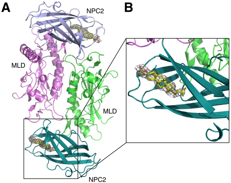
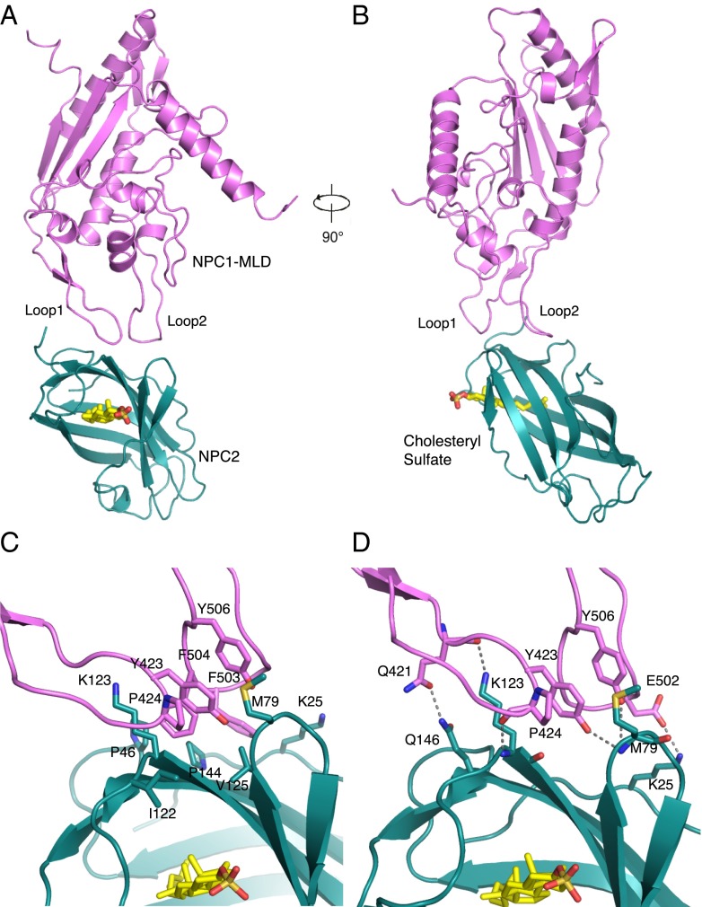
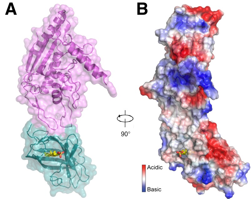
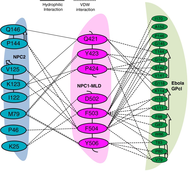
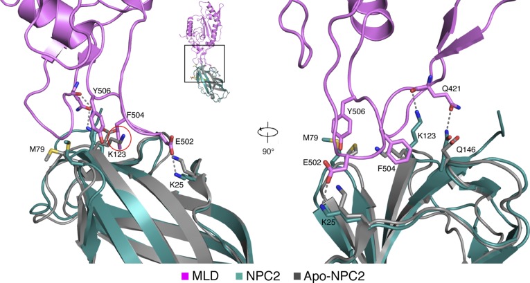
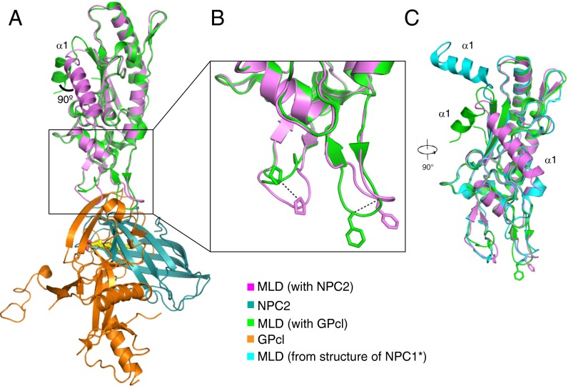
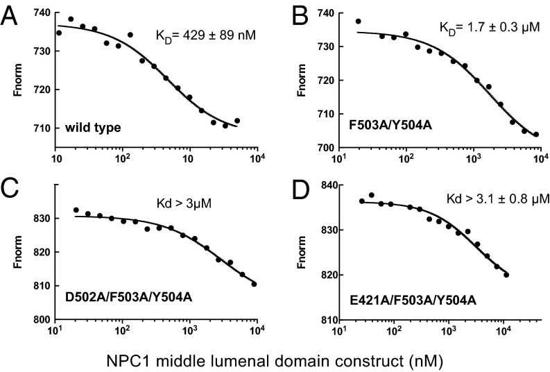
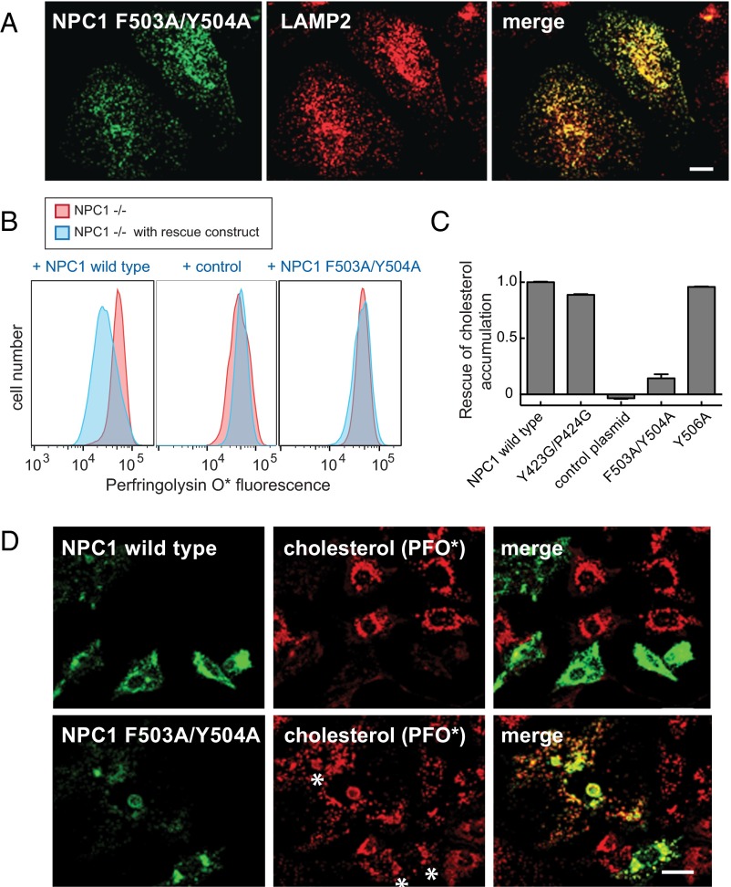
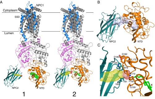
Export of LDL-derived cholesterol from lysosomes requires the cooperation of the integral membrane protein Niemann-Pick C1 (NPC1) and a soluble protein, Niemann-Pick C2 (NPC2). Mutations in the genes encoding these proteins lead to Niemann-Pick disease type C (NPC). NPC2 binds to NPC1's second (middle), lumenally oriented domain (MLD) and transfers cholesterol to NPC1's N-terminal domain (NTD). Here, we report the 2.4-Å resolution crystal structure of a complex of human NPC1-MLD and NPC2 bearing bound cholesterol-3-O-sulfate. NPC1-MLD uses two protruding loops to bind NPC2, analogous to its interaction with the primed Ebola virus glycoprotein. Docking of the NPC1-NPC2 complex onto the full-length NPC1 structure reveals a direct cholesterol transfer tunnel between NPC2 and NTD cholesterol binding pockets, supporting the "hydrophobic hand-off" cholesterol transfer model.

Keywords: Ebola virus glycoprotein; Niemann–Pick type C disease; cholesterol trafficking; crystal structure.


Conflict of interest statement

The authors declare no conflict of interest.

Figures

Fig. S1.
Fig. S1.
The Omit map for cholesteryl sulfate in NPC2. (A) Overall structure of MLD–NPC2 complex in one asymmetry unit. (B) Close-up view of SA-Omit map (Fo-Fc densities) for cholesteryl sulfate (gray mesh) contoured at σ level 2.0.
Fig. 1.
Fig. 1.
Structural overview of the NPC1 MLD–NPC2 complex. (A) NPC1–MLD (magenta), NPC2 (cyan), and cholesteryl sulfate (yellow) are shown. (B) Complex rotated 90° relative to A. (C) Close-up view of the interface. Residues involved in hydrophobic interactions are indicated. (D) Residues involved in hydrophilic interactions are indicated; dotted lines represent hydrogen bonds.
Fig. S2.
Fig. S2.
NPC1 MLD–NPC2 complex in a space-filling representation. (A) Surface is oriented as in Fig. 1A. (B) Electrostatic surface representation in the same orientation as Fig. 1B. Cholesteryl sulfate (yellow) is shown.
Fig. S3.
Fig. S3.
The interaction detail of NPC1–MLD and NPC2. Residues involved at the interface between NPC1–MLD (cyan), NPC2 (magenta), and Ebola GPcl (green) are shown. Black dotted lines indicate hydrophilic interactions; dotted lines indicate van der Waals interactions.
Fig. S4.
Fig. S4.
Structural comparison of NPC2–cholesteryl sulfate in the NPC1–MLD–NPC2 complex versus apo-NPC2. NPC1–MLD (magenta), NPC2 (cyan), and cholesteryl sulfate (yellow) and apo-NPC2 (gray, without cholesteryl sulfate, PDB ID code 1NEP) are shown. K123 of NPC2 clashes with F504 of NPC1 (red circle) before binding cholesteryl sulfate. Upon binding cholesteryl sulfate, K25, M79, K123, and Q146 of NPC2 change their conformations significantly to bind Q421 and E502 of NPC1 (12).
Fig. 2.
Fig. 2.
Comparison of NPC1 MLD structures. (A) Overlay of MLD–NPC2 and MLD–GPcl (PDB ID code 5F1B). (B) Close-up view of the conformational changes of the two protruding loops; the shift of Cα is indicated by dotted lines. (C) Overlay of NPC1 MLDs from MLD-NPC2, MLD–GPcl, and the NPC1* crystal structure (PDB ID code 5I31), rotated 90° relative to A.
Fig. 3.
Fig. 3.
Microscale thermophoresis analysis of murine NPC1–MLD interaction with bovine NPC2–cholesteryl sulfate. (A) Wild type. (B) F503A/Y504A. (C) D502A/F503A/Y504A. (D) E421A/F503A/Y504A.
Fig. 4.
Fig. 4.
Functional analysis of NPC1 MLD mutant proteins. (A) Confocal immunofluorescence microscopy analysis of the localization of mouse NPC1 F503A/Y504A and LAMP2 proteins in HeLa cells; a single stack is shown. (Scale bar, 20 µm.) (B and C) Flow cytometry of NPC1−/− CHO cells transfected for 48 h with GFP-tagged versions of either mouse wild-type NPC1, control plasmid encoding human GCC185 residues 1–889 (30), or NPC1 F503A/Y504A protein. Cells were labeled with AF-647 labeled perfringolysin O* (20) after fixation. More than 5,000 GFP-positive cells were analyzed; shown in C are mean values for the data presented in B, and error bars represent SEM. (D) Confocal immunofluorescence microscopy of the rescue experiment analyzed in B and C. NPC1−/− CHO cells were transfected with the indicated plasmids for 48 h before fixation; endogenous GFP fluorescence and AF647-PFO* labeling are shown. Asterisks denote regions that show mutant protein expression and residual cholesterol accumulation. Images represent maximum projections. (Scale bar, 20 µm.)
Fig. 5.
Fig. 5.
Hypothetical model for cholesterol transfer between NPC2 and NPC1. (A) Model of NPC2 and full-length NPC1 complex. State 1 was generated by alignment of major MLD structures from the NPC1 MLD–NPC2 complex and cryo-EM NPC1 structure (PDB ID code 3JD8); state 2 was generated by alignment of two protruding loops of both structures, creating a cholesterol transfer state. The sterol ligand is shown in stick representation, and the second sterol-binding site is shown in mesh. (B) Putative cholesterol transfer tunnel in state 2 (light purple); note that this tunnel is composed of two independent, adjacent cholesterol binding sites. (C) Potential interacting residues between NPC2 and NPC1-NTD in State 2.

References

    1. Brown MS, Goldstein JL. A receptor-mediated pathway for cholesterol homeostasis. Science. 1986;232(4746):34–47. - PubMed
    1. Goldstein JL, Dana SE, Faust JR, Beaudet AL, Brown MS. Role of lysosomal acid lipase in the metabolism of plasma low density lipoprotein. Observations in cultured fibroblasts from a patient with cholesteryl ester storage disease. J Biol Chem. 1975;250(21):8487–8495. - PubMed
    1. Rosenbaum AI, Maxfield FR. Niemann-Pick type C disease: Molecular mechanisms and potential therapeutic approaches. J Neurochem. 2011;116(5):789–795. - PMC - PubMed
    1. Carette JE, et al. Ebola virus entry requires the cholesterol transporter Niemann-Pick C1. Nature. 2011;477(7364):340–343. - PMC - PubMed
    1. Côté M, et al. Small molecule inhibitors reveal Niemann-Pick C1 is essential for Ebola virus infection. Nature. 2011;477(7364):344–348. - PMC - PubMed

Publication types

MeSH terms

Associated data