. 2016 Aug 8:7:298.
doi: 10.3389/fimmu.2016.00298. eCollection 2016.

Structure and Function of HLA-A*02-Restricted Hantaan Virus Cytotoxic T-Cell Epitope That Mediates Effective Protective Responses in HLA-A2.1/K(b) Transgenic Mice

Affiliations

Structure and Function of HLA-A*02-Restricted Hantaan Virus Cytotoxic T-Cell Epitope That Mediates Effective Protective Responses in HLA-A2.1/K(b) Transgenic Mice

Ying Ma et al. Front Immunol. .

Abstract

Hantavirus infections cause severe emerging diseases in humans and are associated with high mortality rates; therefore, they have become a global public health concern. Our previous study showed that the CD8(+) T-cell epitope aa129-aa137 (FVVPILLKA, FA9) of the Hantaan virus (HTNV) nucleoprotein (NP), restricted by human leukocyte antigen (HLA)-A*02, induced specific CD8(+) T-cell responses that controlled HTNV infection in humans. However, the in vivo immunogenicity of peptide FA9 and the effect of FA9-specific CD8(+) T-cell immunity remain unclear. Here, based on a detailed structural analysis of the peptide FA9/HLA-A*0201 complex and functional investigations using HLA-A2.1/K(b) transgenic (Tg) mice, we found that the overall structure of the peptide FA9/HLA-A*0201 complex displayed a typical MHC class I fold with Val2 and Ala9 as primary anchor residues and Val3 and Leu7 as secondary anchor residues that allow peptide FA9 to bind tightly with an HLA-A*0201 molecule. Residues in the middle portion of peptide FA9 extruding out of the binding groove may be the sites that allow for recognition by T-cell receptors. Immunization with peptide FA9 in HLA-A2.1/K(b) Tg mice induced FA9-specific cytotoxic T-cell responses characterized by the induction of high expression levels of interferon-γ, tumor necrosis factor-α, granzyme B, and CD107a. In an HTNV challenge trial, significant reductions in the levels of both the antigens and the HTNV RNA loads were observed in the liver, spleen, and kidneys of Tg mice pre-vaccinated with peptide FA9. Thus, our findings highlight the ability of HTNV epitope-specific CD8(+) T-cell immunity to control HTNV and support the possibility that the HTNV-NP FA9 peptide, naturally processed in vivo in an HLA-A*02-restriction manner, may be a good candidate for the development HTNV peptide vaccines.

Keywords: CD8+ T-cell response; HLA-A*02; HLA-A2.1/Kb transgenic mice; Hantaan virus; crystal structure; cytotoxic T-cell epitope.


Figures

Figure 1
Figure 1
Binding affinity of the HTNV nucleoprotein FA9 peptide to HLA-A*0201 molecule. (A) MHC stabilization assay with T2 cells was used to quantify the peptide-binding affinity of HTNV nucleoprotein (NP) FA9 (aa129–aa137, FVVPILLKA) to HLA-A*0201 molecule via flow cytometry. HLA-B*35-restricted HTNV-NP VY9 (aa131–aa139, VPILLKALY) unrelated peptides served as negative controls. The red curve indicates T2 cells incubated without peptide. The blue curve indicates T2 cells incubated with peptide VY9. The orange curve indicates T2 cells incubated with peptide FA9. The overlay of the three conditions in histograms clearly demonstrate that the curve of T2 cells incubated with peptide FA9 is shifted more to the right than that incubated with peptide VY9 or without peptide. The results shown are representative of three independent experiments. (B) In vitro refolding of the HTNV-NP FA9 peptide with the HLA-A*0201 heavy chain and β2m. The refolded complex was analyzed by gel filtration chromatography using a Superdex 200 16/60 column. The peak of the HLA complex as indicated by the arrow with the expected molecular mass of 45 kDa eluted at the estimated volume of 15.9 ml. mAU, milli-absorbance units.
Figure 2
Figure 2
The structure of HLA-A*0201 complexed with the HTNV nucleoprotein FA9 peptide. The crystals were obtained from vapor-diffusion hanging drops and the data were processed and scaled using the HKL2000. (A) Overview of the three-dimensional structure of the FA9/HLA-A*0201complex showing a typical MHC class I fold. The antigenic peptide-binding groove was constituted by antiparallel α1 and α2 helices in the extracellular region of the HLA-A*0201 heavy chain, and supported by an eight-stranded β-sheet bedplate. The HTNV nucleoprotein (NP) FA9 (aa129–aa137, FVVPILLKA) peptide was presented in the peptide-binding cleft. The α3 domain and β2m occupied the standard positions below the bedplate. (B) Structural superposition of the FA9/HLA-A*0201 complex and the previously determined HLA-A*0201 structure complexed with peptide GV9 (GLMWLSYFV, PDB code: 3I6G). The structure of FA9/HLA-A*0201 is similar to the structure of GV9/HLA-A*0201 with a low root mean square difference (RMSD) of 0.415 Å. (C) The alignment of peptide FA9 with peptide GV9. The mainchain conformation of FA9 is similar to GV9. The residues Val2 and Ala9 (red arrows) act as the primary anchor residues of FA9, which exhibit the same conformations as Leu2 and Val9 of GV9. The residues Val3 and Leu7 (green arrows) act as secondary anchor residues for peptide FA9, whereas the residues Met3 and Ser6 might be the secondary anchors for the peptide GV9. The residues Phe1, Pro4, Ile5, Leu6, and Lys8 (blue arrows) of peptide FA9 pointing upwards from the HLA-A*0201 surface are the most probable recognition sites for TCRs. (D) The electron density for peptide FA9 at σ = 1.0 contour level.
Figure 3
Figure 3
The analysis of interferon (IFN)-γ secretion by splenocytes of HLA-A2.1/Kb transgenic mice against HTNV nucleoprotein peptide FA9. Comparison of the magnitudes (y-axis) of ex vivo ELISPOT IFN-γ responses to the HTNV nucleoprotein (NP) FA9 peptide (aa129–aa137, FVVPILLKA) among the different groups (x-axis). The HLA-A2.1/Kb transgenic mice were divided into four groups (n = 6 each), including the mice immunized with the HLA-A*02-restricted HTNV-NP FA9 peptide, the HLA-B*35-restricted HTNV-NP VY9 (aa131–aa139, VPILLKALY) as an unrelated peptide control, the HTNV vaccine as a positive control and PBS as a negative control, respectively. Three immunization injections were administered to each mouse at intervals of 2 weeks. Ten days after the last immunization, the splenocytes were isolated for IFN-γ detection by ELISPOT assay. The magnitudes of the responses are represented as the spot-forming cells (SFC)/106 splenocytes. A02-FA9 indicates the HLA-A2.1/Kb transgenic mice immunized with the HLA-A*02-restricted HTNV-NP FA9 peptide. B35-VY9 indicates the transgenic mice immunized with the HLA-B*35-restricted HTNV-NP VY9 peptide. The Wilcoxon rank-sum test was used for statistical evaluation. ns, not significant.
Figure 4
Figure 4
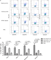
The cytokine and cytolytic mediator production of splenocytes in HLA-A2.1/Kb transgenic mice after peptide immunization. The HLA-A2.1/Kb transgenic mice were divided into four groups (n = 6 each) according to the different immunization, including the HLA-A*02-restricted HTNV-NP FA9 peptide (aa129–aa137, FVVPILLKA), the HLA-B*35-restricted HTNV-NP VY9 peptide (aa131–aa139, VPILLKALY), the HTNV vaccine and PBS immunized mice groups, respectively. (A) Representative flow cytometric plots of the cytokines interferon (IFN)-γ and tumor necrosis factor (TNF)-α or cytotoxic mediator granzyme B-producing and CD107a-expressing CD8+ T cells in splenocytes (vertical) after three immunizations with the HLA-A*02-restricted HTNV-NP FA9 peptide, the HLA-B*35-restricted HTNV-NP VY9 peptide, the HTNV vaccine or PBS (horizontal). The numbers indicate the percentage of cells within the boxed regions. (B) Comparison of the frequencies (y-axis) of cytokines IFN-γ and TNF-α and cytotoxic mediator granzyme B and the expression percentage of CD107a (x-axis) in the peptide FA9-specific CD8+ T cells between the peptide FA9-immunized mice and control mice, including peptide VY9-immunized mice as an unrelated peptide control, HTNV-vaccinated mice as a positive control and PBS-administered mice as a negative control, respectively. The results shown are representative of three independent experiments. The Wilcoxon rank-sum test was used for statistical evaluation. ns, not significant.
Figure 5
Figure 5
Detection of HTNV antigen with ELISA in the organs of HLA-A2.1/Kb transgenic mice after HTNV challenge. The HLA-A2.1/Kb transgenic mice were divided into four groups (n = 6 each) according to the different immunization, including the mice immunized with the HLA-A*02-restricted HTNV-NP peptide FA9 (aa129–aa137, FVVPILLKA), the HLA-B*35-restricted HTNV-NP peptide VY9 (aa131–aa139, VPILLKALY), the HTNV vaccine or PBS, respectively. Ten days after the final immunization booster, the mice were challenged with the HTNV 76-118 strain and sacrificed 4 days following HTNV challenge. (A) The HTNV antigen (y-axis) was detected in the lungs, liver, cerebrum, spleen, kidneys, and heart (x-axis) of HLA-A2.1/Kb transgenic mice after HTNV infection in the four groups, respectively. Few or no detectable virus was found in lungs, cerebrum, and heart in all four groups of immunized transgenic mice after HTNV challenge detected via ELISA. (B) Comparison of the HTNV antigen levels (y-axis) in the tissue supernatant of the liver, spleen, and kidneys in each immunized group (x-axis). (C) Comparison of the HTNV antigen levels (y-axis) between the HLA-A*02-restricted HTNV-NP peptide FA9immunized mice group and HLA-B*35-restricted HTNV-NP peptide VY9-immunized mice group or PBS-injected mice group (x-axis) in the organs liver, spleen and kidneys, respectively. The results shown are representative of three independent experiments. A P/N ratio >2.1 is considered positive. The Wilcoxon rank-sum test was used for statistical evaluation. ns, not significant.
Figure 6
Figure 6
Detection of HTNV RNA loads via real-time PCR in the organs of HTNV-challenged HLA-A2.1/Kb transgenic mice. The HLA-A2.1/Kb transgenic mice were divided into four groups (n = 6 each), including the mice immunized with HLA-A*02-restricted HTNV-NP peptide FA9 (aa129–aa137, FVVPILLKA), HLA-B*35-restricted HTNV-NP peptide VY9 (aa131–aa139, VPILLKALY), HTNV vaccine or PBS, respectively. Ten days after the final immunization booster, the mice were challenged with the HTNV 76-118 strain and sacrificed 4 days following HTNV challenge. (A) The detection of HTNV RNA loads (y-axis) in the lungs, liver, cerebrum, spleen, kidneys, and heart (x-axis) of HLA-A2.1/Kb transgenic mice after HTNV challenge in the four groups. Comparison of the HTNV RNA loads that could be detected in liver, spleen, and kidneys of the immunized transgenic mice after HTNV challenge among four immunization mice groups. (B) The HTNV RNA loads (y-axis) in the liver, spleen, and kidneys of each immunized group (x-axis) and comparison of the HTNV RNA loads between the liver and spleen or between the liver and kidneys in the HLA-A*02-restricted HTNV-NP peptide FA9-immunized mice group. The results shown were recorded as cycle time (Ct) and quantified by 2−ΔΔCt and were representative of three independent experiments. The Wilcoxon rank-sum test was used for statistical evaluation. ns, not significant.

Similar articles

Cited by

References

    1. Vaheri A, Strandin T, Hepojoki J, Sironen T, Henttonen H, Makela S, et al. Uncovering the mysteries of hantavirus infections. Nat Rev Microbiol (2013) 11(8):539–50.10.1038/nrmicro3066 - DOI - PubMed
    1. Watson DC, Sargianou M, Papa A, Chra P, Starakis I, Panos G. Epidemiology of Hantavirus infections in humans: a comprehensive, global overview. Crit Rev Microbiol (2014) 40(3):261–72.10.3109/1040841X.2013.783555 - DOI - PubMed
    1. Krautkramer E, Zeier M, Plyusnin A. Hantavirus infection: an emerging infectious disease causing acute renal failure. Kidney Int (2013) 83(1):23–7.10.1038/ki.2012.360 - DOI - PubMed
    1. Vaheri A, Henttonen H, Voutilainen L, Mustonen J, Sironen T, Vapalahti O. Hantavirus infections in Europe and their impact on public health. Rev Med Virol (2013) 23(1):35–49.10.1002/rmv.1722 - DOI - PubMed
    1. Macneil A, Nichol ST, Spiropoulou CF. Hantavirus pulmonary syndrome. Virus Res (2011) 162(1–2):138–47.10.1016/j.virusres.2011.09.017 - DOI - PubMed