

Forkhead containing transcription factor Albino controls tetrapyrrole-based body pigmentation in planarian
- PMID: 27551436
- PMCID: PMC4969599
- DOI: 10.1038/celldisc.2016.29
Forkhead containing transcription factor Albino controls tetrapyrrole-based body pigmentation in planarian
Abstract
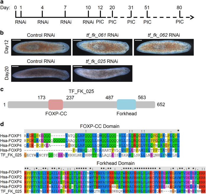
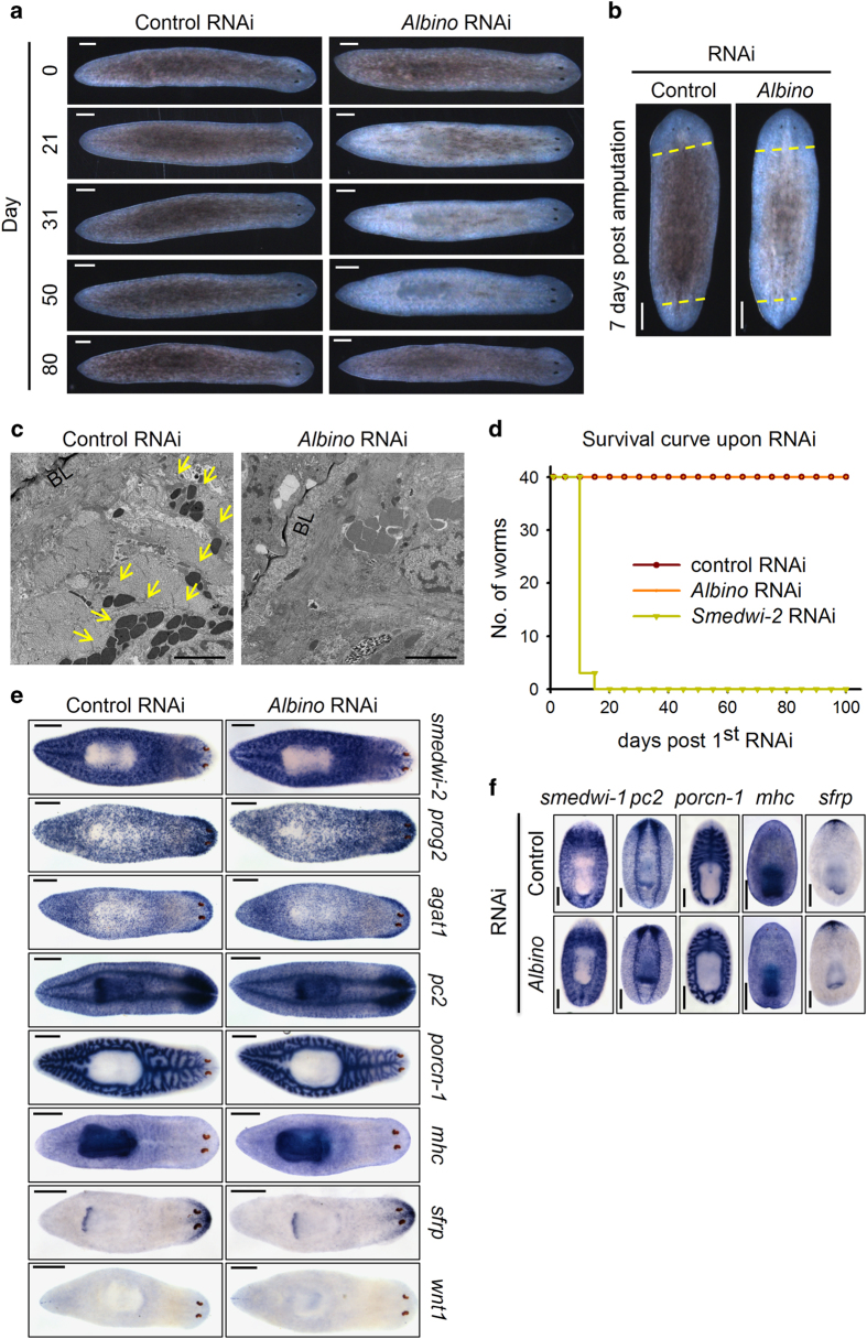
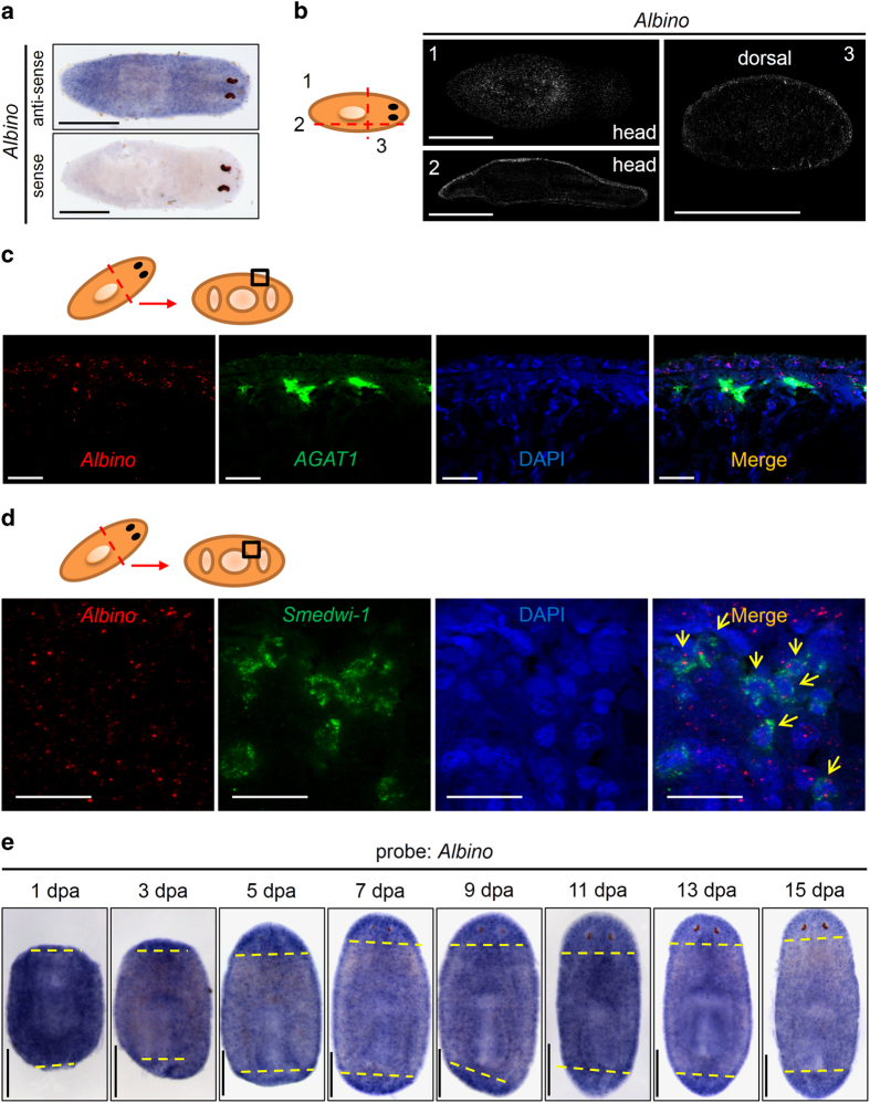
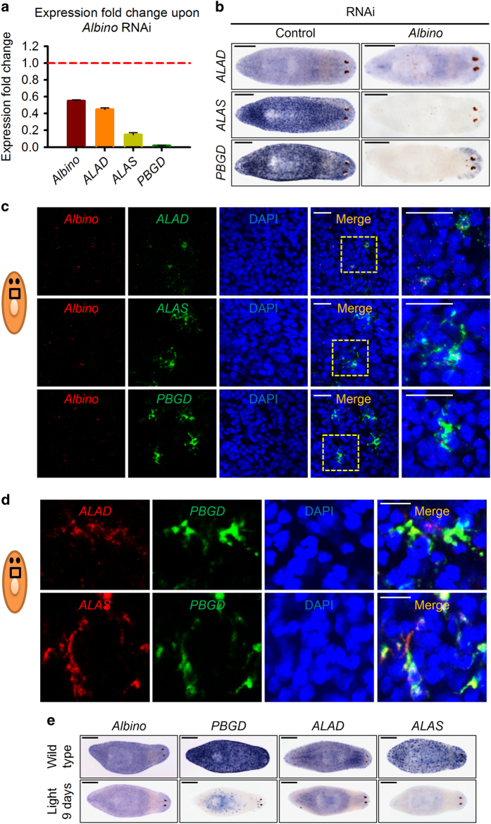
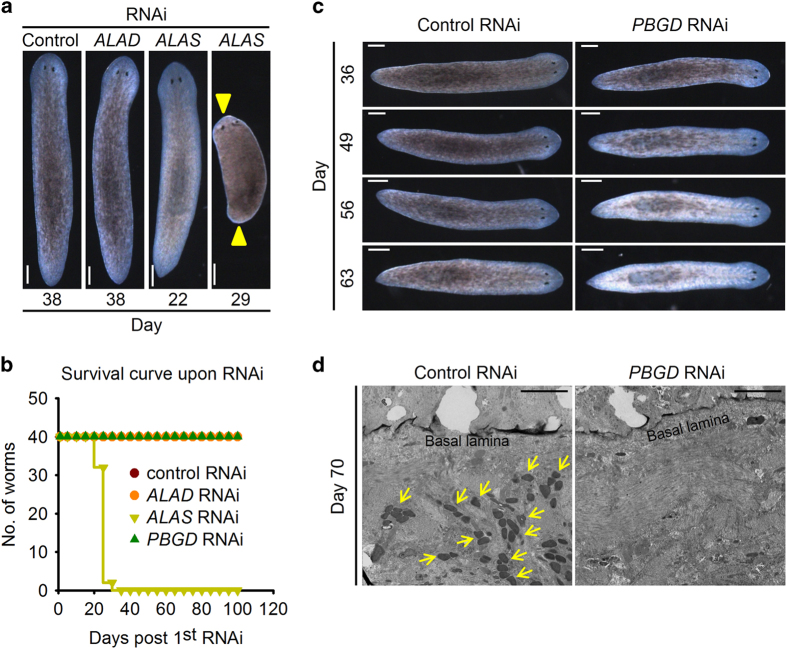
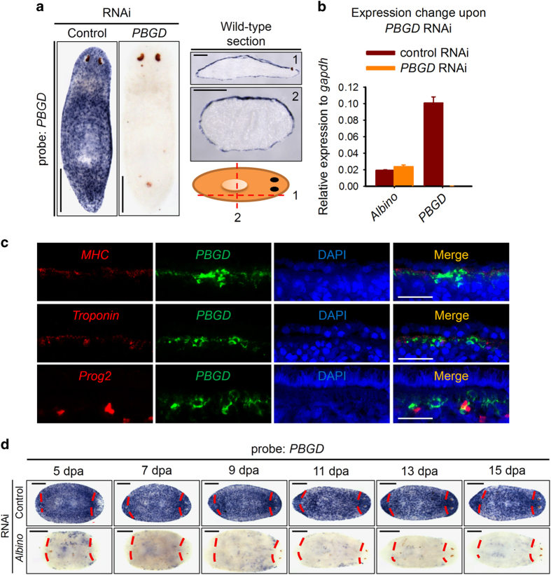
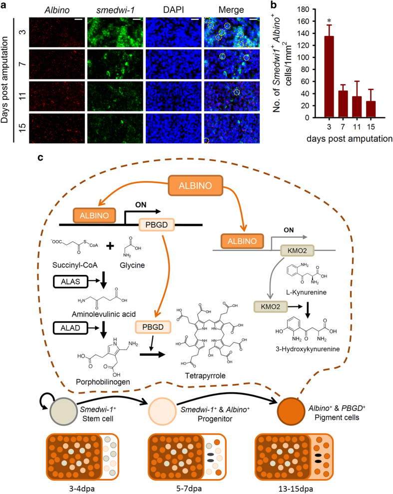
Pigmentation processes occur from invertebrates to mammals. Owing to the complexity of the pigmentary system, in vivo animal models for pigmentation study are limited. Planarians are capable of regenerating any missing part including the dark-brown pigments, providing a promising model for pigmentation study. However, the molecular mechanism of planarian body pigmentation is poorly understood. We found in an RNA interference screen that a forkhead containing transcription factor, Albino, was required for pigmentation without affecting survival or other regeneration processes. In addition, the body color recovered after termination of Albino double stranded RNA feeding owing to the robust stem cell system. Further expression analysis revealed a spatial and temporal correlation between Albino and pigmentation process. Gene expression arrays revealed that the expression of three tetrapyrrole biosynthesis enzymes, ALAD, ALAS and PBGD, was impaired upon Albino RNA interference. RNA interference of PBGD led to a similar albinism phenotype caused by Albino RNA interference. Moreover, PBGD was specifically expressed in pigment cells and can serve as a pigment cell molecular marker. Our results revealed that Albino controls planarian body color pigmentation dominantly via regulating tetrapyrrole biogenesis. These results identified Albino as the key regulator of the tetrapyrrole-based planarian body pigmentation, suggesting a role of Albino during stem cell-pigment cell fate decision and provided new insights into porphyria pathogenesis.
Keywords: FoxP; PBGD; body color; pigmentation; planarian; tetrapyrrole.
Figures
Similar articles
-
The true colours of the flatworm: Mechanisms of pigment biosynthesis and pigment cell lineage development in planarians.Semin Cell Dev Biol. 2019 Mar;87:37-44. doi: 10.1016/j.semcdb.2018.05.010. Epub 2018 May 21. Semin Cell Dev Biol. 2019. PMID: 29758350 Review.
-
RNA Interference by Ingested Dsrna-Expressing Bacteria to Study Porphyrin Pigmentation in Crassostrea gigas.Int J Mol Sci. 2021 Jun 6;22(11):6120. doi: 10.3390/ijms22116120. Int J Mol Sci. 2021. PMID: 34204154 Free PMC article.
-
FOX and ETS family transcription factors regulate the pigment cell lineage in planarians.Development. 2017 Dec 15;144(24):4540-4551. doi: 10.1242/dev.156349. Epub 2017 Nov 20. Development. 2017. PMID: 29158443 Free PMC article.
-
Ingestion of bacterially expressed double-stranded RNA inhibits gene expression in planarians.Proc Natl Acad Sci U S A. 2003 Sep 30;100 Suppl 1(Suppl 1):11861-5. doi: 10.1073/pnas.1834205100. Epub 2003 Aug 13. Proc Natl Acad Sci U S A. 2003. PMID: 12917490 Free PMC article.
-
[Molecular mechanism of brain regeneration and reconstruction of dopaminergic neural network in planarians].Brain Nerve. 2008 Apr;60(4):307-17. Brain Nerve. 2008. PMID: 18421972 Review. Japanese.
Cited by
-
LIM-HD transcription factors control axial patterning and specify distinct neuronal and intestinal cell identities in planarians.Open Biol. 2023 Dec;13(12):230327. doi: 10.1098/rsob.230327. Epub 2023 Dec 13. Open Biol. 2023. PMID: 38086422 Free PMC article.
-
Discovery of a body-wide photosensory array that matures in an adult-like animal and mediates eye-brain-independent movement and arousal.Proc Natl Acad Sci U S A. 2021 May 18;118(20):e2021426118. doi: 10.1073/pnas.2021426118. Proc Natl Acad Sci U S A. 2021. PMID: 33941643 Free PMC article.
-
Solar and terrestrial radiations explain continental-scale variation in bird pigmentation.Oecologia. 2018 Nov;188(3):683-693. doi: 10.1007/s00442-018-4238-8. Epub 2018 Aug 9. Oecologia. 2018. PMID: 30094635
-
Neoblast-enriched zinc finger protein FIR1 triggers local proliferation during planarian regeneration.Protein Cell. 2019 Jan;10(1):43-59. doi: 10.1007/s13238-018-0512-0. Epub 2018 Mar 20. Protein Cell. 2019. PMID: 29557542 Free PMC article.
-
Positional Information and Stem Cells Combine to Result in Planarian Regeneration.Cold Spring Harb Perspect Biol. 2022 May 17;14(4):a040717. doi: 10.1101/cshperspect.a040717. Cold Spring Harb Perspect Biol. 2022. PMID: 34518341 Free PMC article. Review.
References
-
- Reddien PW , Sanchez Alvarado A . Fundamentals of planarian regeneration. Annu Rev Cell Dev Biol 2004; 20: 725–757. - PubMed
-
- Salo E , Baguna J . Regeneration and pattern formation in planarians. I. The pattern of mitosis in anterior and posterior regeneration in Dugesia (G) tigrina, and a new proposal for blastema formation. J Embryol Exp Morphol 1984; 83: 63–80. - PubMed
LinkOut - more resources
Full Text Sources
Other Literature Sources
