. 2015 Aug 3:1:15003.
doi: 10.1038/cddiscovery.2015.3. eCollection 2015.

WWOX dysfunction induces sequential aggregation of TRAPPC6AΔ, TIAF1, tau and amyloid β, and causes apoptosis

Affiliations

WWOX dysfunction induces sequential aggregation of TRAPPC6AΔ, TIAF1, tau and amyloid β, and causes apoptosis

J-Y Chang et al. Cell Death Discov. .

Abstract

Aggregated vesicle-trafficking protein isoform TRAPPC6AΔ (TPC6AΔ) has a critical role in causing caspase activation, tau aggregation and Aβ generation in the brains of nondemented middle-aged humans, patients with Alzheimer's disease (AD) and 3-week-old Wwox gene knockout mice. WWOX blocks neurodegeneration via interactions with tau and tau-phosphorylating enzymes. WWOX deficiency leads to epilepsy, mental retardation and early death. Here, we demonstrated that TGF-β1 induces shuttling of endogenous wild-type TPC6A and TPC6AΔ in between nucleoli and mitochondria (~40-60 min per round trip), and WWOX reduces the shuttling time by 50%. TGF-β1 initially maximizes the binding of TPC6AΔ to the C-terminal tail of WWOX, followed by dissociation. TPC6AΔ then undergoes aggregation, together with TIAF1 (TGF-β1-induced antiapoptotic factor), in the mitochondria to induce apoptosis. An additional rescue scenario is that TGF-β1 induces Tyr33 phosphorylation and unfolding of WWOX and its the N-terminal WW domain slowly binds TPC6AΔ to block aggregation and apoptosis. Similarly, loss of WWOX induces TPC6AΔ polymerization first, then aggregation of TIAF1, amyloid β and tau, and subsequent cell death, suggesting that a cascade of protein aggregation leads to neurodegeneration.


Figures

Figure 1
Figure 1
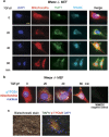
TPC6A protein aggregation in subcellular compartment. (a) Ectopic expression of wild-type TPC6A and an isoform TPC6AΔ in COS7 and SCC-9 cells results in localization of the proteins in the nucleus and cytoplasm. Perinuclear protein aggregation is shown in most cases (see white arrows). Control cells expressing EGFP do not exhibit protein aggregation. (b) Basal cell carcinoma (BCC) cells were exposed to UV light, followed by incubation and then harvesting at indicated times. UV irradiation quickly induced formation of endogenous trimeric TPC6A in both cytosol and nucleus in 10–20 min. A representative data from two experiments is shown. (c) B16F10 melanoma cells were treated with TGF-β1 (5 ng/ml) for various durations. Presence of aggregates of TIAF1 and TPC6A and its Ser35 phosphorylation is shown (non-reducing SDS-PAGE). No ubiquitin attachment to these proteins was observed (data not shown). Protein size markers are on the left column. Monomeric TIAF1 is 12 kDa and TPC6A is 17–20 kDa. A representative data from two experiments is shown. (d and e) COS7 cells were treated with TGF-β1 (5 ng/ml) for indicated durations and stained with a specific antibody against TPC6A and MitoTracker Red for mitochondria, respectively. Confocal microscopy analysis revealed that TGF-β1 induced TPC6A relocation to the mitochondria, as revealed by increased yellow fluorescence in co-localization analysis. (f) PMA at 10 μM stimulated relocation of TPC6A and WWOX, along with their phosphorylated forms, to the mitochondria in Jurkat T cells during the treatment for 90 min.
Figure 2
Figure 2
TGF-β1 induces a two-way shuttling of endogenous TPC6A in between nucleoli and mitochondria. (a) Wwox−/− MEF cells were exposed to TGF-β1 (5 ng/ml), and then stained with specific antibodies for TPC6A and TIAF1. Mitochondria were stained with MitoTracker Red. Wild-type TPC6A shuttles in between nucleoli to mitochondria. Each round trip takes ~60 min. Endogenous TIAF1 is mainly retained in the mitochondria. Approximately 100–120 cells were examined. (b) Similar results were observed by staining Ser35-phosphorylated TPC6A with a specific antibody. (c) TIAF1 co-localizes with TPC6A with Ser35 phosphorylation in the extracellular matrix as aggregates in the human AD hippocampus (also see Supplementary Figure S1).
Figure 3
Figure 3
TPC6A physically binds the C-terminal D3 tail of WWOX. (a and b) Transiently overexpresssed EGFP-WWOX localized mainly in the perinuclear area in COS7 cells. Co-expression of WWOX and TPC6AΔ results in increased nuclear accumulation of WWOX compared with WWOX expression alone (~100 cells counted, mean±S.D., n=3; **P<0.05, Student’s t-test). (c and d) HEK293 cells were cultured in 10% FBS/medium and grown in a 10-cm dish to 100% confluence. Cell lysates were prepared and processed for co-immunoprecipitation using specific antibodies, respectively, against TPC6A and WWOX. Western blotting analysis showed TPC6A physically interacted with WWOX in resting cells. PreIP=~40 μg of protein in the input loading. (e) COS7 cells were co-transfected with EYFP-TPC6AΔ and ECFP-WWOX or indicated domains of WWOX. FRET microscopy revealed that TPC6AΔ binds WWOX to its C-terminal D3 tail (see increased FRETc). The relative binding strength is shown in a color scale, where the highest binding energy is indicated in red. (f) Designated WWOX domains for interacting with TPC6AΔ are determined by FRET microscopy. Ten cells from each experimental set were analyzed by Image Pro Plus 6.1 (mean±S.D.; **P<0.05, Student’s t-test).
Figure 4
Figure 4
TPC6A fails to induce apoptosis synergistically with WWOX. (a) Transiently overexpressed TPC6AΔ and TPC6A were equally potent in inducing apoptosis of SK-N-SH and HEK293 cells in a dose-dependent manner. No ep indicates no electroporation. (b and c) SK-N-SH cells were transiently overexpressed with EYFP-TPC6AΔ and EYFP-WWOX, and both proteins failed to induce apoptosis in an additive manner (mean±S.D.; n=3; *P>0.1, ***P<0.005; Student’s t-test). (d) An assay for NF-κB-governed promoter activation was carried out (Li et al.). WWOX and its WW domain-induced promoter activation were significantly blocked by TPC6AΔ (n=10).
Figure 5
Figure 5
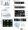
WWOX binds and blocks TPC6AΔ aggregation, and TGF-β1 dissociates TPC6AΔ from WWOX for leading to aggregation. (a) COS7 cells were co-transfected with expression constructs for EYFP, EYFP-TPC6AΔ, EYFP-TPC6AΔ(S35G) and/or EYFP-TPC6AΔ(Y112F), followed by culturing for 24 h and then treating with or without TGF-β1 (5 ng/ml) for another 24 h. The extent of protein aggregation was measured. TGF-β1-induced protein aggregation was statistically analyzed (control versus treated samples; ~100 cells analyzed; n=3). (b) COS7 cells were transfected with ECFP-WWOX and/or EGFP-TPC6AΔ by electroporation. Forty-eight hours later, ectopic TPC6AΔ underwent aggregation, and WWOX suppressed the aggregation by ~50%. Dominant negative WWOX (dn-WWOX) failed to block the aggregation of TPC6AΔ. Approximately 300 green fluorescent cells in total were counted, and the average is shown. (c and d) COS7 cells were transiently expressed with EYFP-WWOX and ECFP-TPC6AΔ. After 24 h in culture, cells were treated with TGF-β1 (5 ng/ml) and time-lapse FRET microscopy was performed. TGF-β1 increased the binding of WWOX with TPC6AΔ initially in the nucleus for 6 h, followed by reduction in the binding and increased TPC6AΔ aggregation in the cytoplasm. Also, see Supplementary Videos 1,2,3. (e and f) COS7 cells were transiently expressed with ECFP-WWOX and EYFP-TPC6AΔ or EYFP-TPC6AΔ(S35G). Upon exposure to TGF-β1 (5 ng/ml), ectopic WWOX and TPC6AΔ became aggregated in the nucleolus, whereas no aggregation was shown for WWOX and TPC6AΔ(S35G). The cell started undergoing apoptosis at hour 15 (see Supplementary Video 6). (g) Similarly, aggregation of EYFP-TPC6AΔ in the nucleolus (top panel) or cytoplasm (middle panel) was observed upon exposure of COS7 cells to TGF-β1 (5 ng/ml). Alteration of Tyr112 to Phe112 resulted in the failure of relocation of the mutant protein to the cytoplasm. (h) Under similar conditions, TGF-β2 (5 ng/ml) induced aggregation of EYFP-TPC6AΔ in the nucleus in 4 h. (i) In summary, two-way endogenous TPC6A shuttling is illustrated. In response to TGF-β1, nuclear TPC6A undergoes Ser35 phosphorylation for entering the nucleoli and then relocates out to the mitochondria as a dimer, which probably requires phosphorylation at Tyr112. Again, TPC6A migrates back to the nucleoli. Ectopic TPC6AΔ, tagged with EGFP or EYFP, undergoes one-way trafficking from the nuclei to the mitochondria only. This is due to TPC6AΔ protein aggregation in the mitochondria.
Figure 6
Figure 6
TPC6AΔ and D3 act synergistically in TGF-β1-mediated apoptosis, whereas the WW domain counteracts the event. (a) To induce mitochondrial apoptosis, ECFP-TPC6AΔ expressing COS7 cells were treated with CCCP. Time-lapse microscopy was carried out to chase the loss of mitochondrial membrane permeability (stained with MitoTracker Red) and relocation of ECFP-TPC6AΔ to the cytoplasm, along with aggregation (see white arrows), with time (also see Supplementary Videos 10 and 11). (b and c) COS7 cells, transiently overexpressing with TPC6AΔ and SDR/D3, were treated with TGF-β1 for time-lapse FRET microscopy (also see Supplementary Videos 12,13,14,15). TGF-β1 increased the binding of TPC6AΔ with SDR/D3 with time followed by reduction and occurrence of cell death. During cell death, increased secretion of exosome-like particles to the extracellular space is shown (Supplementary Video 14). (d) TGF-β1 failed to induce the binding of TPC6AΔ with SDR domain and no cell death occurred (data not shown). (e and f) Interestingly, TGF-β1 increased the binding of TPC6AΔ with WW domain during exposure for 8–12 h, whereas no cell death occurred.
Figure 7
Figure 7
Binding of TPC6AΔ with TIAF1 in vivo. (a) By antibody-FRET, we determined the binding of TPC6A plaques (red) with TIAF1 aggregates (green) in the brain cortex of Wwox−/− mice. The binding affinity is expressed as FRETc (n=6; mean±S.D.). (b) In negative controls, no binding was observed. No primary antibodies were used. (c) Ser35-phosphorylated TPC6A also binds TIAF1. (d) By immunohistochemistry, we showed the pS35-TPC6A plaque in the brain cortex of Wwox−/− mice. (e and f) Transient overexpression of EGFP-TIAF1 in SH-SY5Y cells resulted in upregulation of TPC6A expression and their binding in the cytoplasm, but not in the nucleus (n=15, mean±S.D.). (g) COS7 cells were transiently expressed with ECFP-TPC6AΔ and EYFP-WWOX, EYFP-TPC6AΔ or EYFP-TIAF1. By FRET microscopy, we showed TIAF1 bound strongly with TPC6AΔ, but the binding was weak in other combinations (n=15, mean±S.D.). No binding was shown for ECFP and EYFP only.

Similar articles

Cited by

References

    1. Chang JY, Lee MH, Lin SR, Yang LY, Sun HS, Sze CI et al. Trafficking protein particle complex 6A delta (TRAPPC6∆) is an extracellular plaque-forming protein in the brain. Oncotarget 2015; 6: 3578–3589. - PMC - PubMed
    1. Kümmel D, Müller JJ, Roske Y, Misselwitz R, Büssow K, Heinemann U. The structure of the TRAPP subunit TPC6 suggests a model for a TRAPP subcomplex. EMBO Rep 2005; 6: 787–793. - PMC - PubMed
    1. Gwynn B, Smith RS, Rowe LB, Taylor BA, Peters LL. A mouse TRAPP-related protein is involved in pigmentation. Genomics 2006; 88: 196–203. - PubMed
    1. Sacher M, Kim YG, Lavie A, Oh BH, Segev N. The TRAPP complex: insights into its architecture and function. Traffic 2008; 9: 2032–2042. - PMC - PubMed
    1. Hamilton G, Harris SE, Davies G, Liewald DC, Tenesa A, Starr JM et al. Alzheimer's disease genes are associated with measures of cognitive ageing in the lothian birth cohorts of 1921 and 1936. Int J Alzheimers Dis 2011; 2011: 505984. - PMC - PubMed

LinkOut - more resources