. 2015 Jul 27:1:15011.
doi: 10.1038/cddiscovery.2015.11. eCollection 2015.

Modulation of dendritic cell function by the radiation-mediated secretory protein γ-synuclein

Affiliations

Modulation of dendritic cell function by the radiation-mediated secretory protein γ-synuclein

S-M Kang et al. Cell Death Discov. .

Abstract

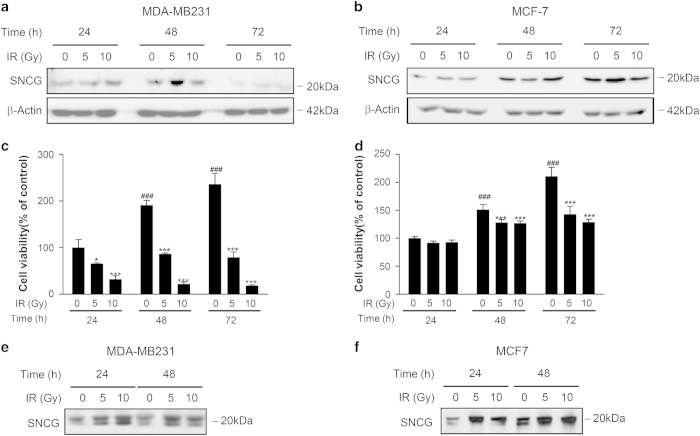
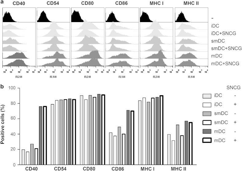
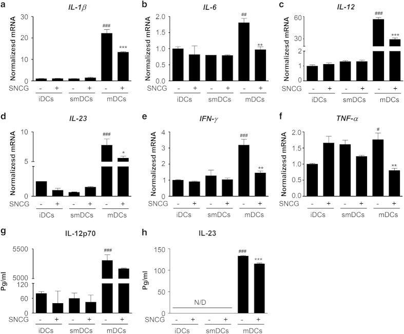
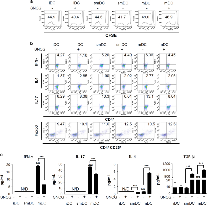
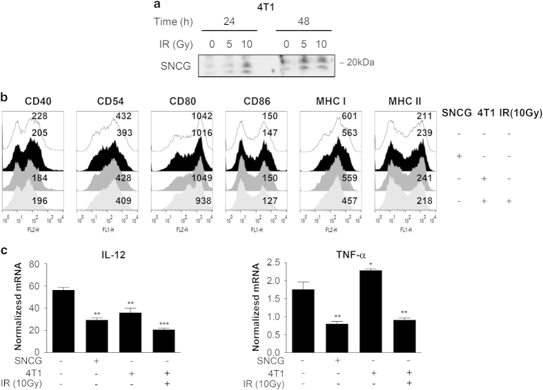
Recently, γ-synuclein (SNCG), which is also known as breast cancer-specific gene-1, has been demonstrated to be an adverse and aggressive marker in breast cancer. In our previous study, SNCG was significantly upregulated in irradiated human breast cancer cells. The aim of this study was to investigate whether radiation-induced, tumor-derived SNCG can influence dendritic cell (DC) function in immune systems. The phenotypical and functional changes of DCs in the presence or absence of SNCG were investigated by FACS analysis, ELISA, and real-time PCR. The ability of SNCG-treated DCs to influence T cells was also examined by coculturing with T cells. The treatment of DCs with SNCG protein inhibited the surface expression of the co-stimulatory molecules CD40 and CD86, and decreased the mRNA levels of pro-inflammatory cytokines. The SNCG-treated DCs inhibited T-cell proliferation slightly, but distinctively increased the population of regulatory T cells. In addition, the production of TGF-β from T cells was significantly increased when they were cocultured with SNCG-treated DCs. Taken together, these results demonstrate that tumor-derived SNCG contributes to immunosuppressive effects via the inhibition of DC differentiation and activation, thus making it a potential target for cancer treatment.


Figures

Figure 1
Figure 1
SNCG was increased by irradiated breast cancer cell lines. (a and b) Human breast cancer cell lines (MDA-MB231 and MCF-7) were exposed to 5 or 10 Gy doses of γ-irradiation, and the expression of SNCG was detected by western blotting at 24, 48 and 72 h after radiation. (c and d) The viability of MDA-MB231 and MCF-7 cells was determined by an MTT assay. (e and f) The secreted SNCG was detected in the conditioned media from MDA-MB231 and MCF-7 cells using immunoblotting. The data represents the mean ± S.D. of three independent experiments. ### P<0.001 was compared with 24h 0 Gy group; while ***P<0.001 and *P<0.05 were compared with the 0 Gy group in their seperate time frames. Significance was determined by one-way ANOVA followed by Tukey's multiple comparison tests.
Figure 2
Figure 2
SNCG altered the phenotypic changes of DCs. BMDCs were generated by culturing with 10 ng/ml GM-CSF and 10 ng/ml IL-4 for 6 days. Then, the cells were treated with TNF-α (10 ng/ml) or LPS (1 μg/ml) in the absence or presence of SNCG (1 μg/ml) for 24 h. (a) The antigen-presenting (MHC-I and –II) and representative co-stimulatory molecules of DCs, including CD40, CD80, and CD86, and the CD54 adhesion molecule, were evaluated by flow cytometry. (b) The above histogram data were analyzed and depicted as a bar graph of positively stained cells.
Figure 3
Figure 3
SNCG decreased immunostimulatory cytokines from mDCs. The iDCs, smDCs, or mDCs were treated with SNCG (1 μg/ml) for 24 h. (af) The mRNA levels of IL-1β, IL-6, IL-12, IL-23, IFN-γ, and TNF-α were normalized to the 18S mRNA level. (g and h) The culture supernatants were evaluated for the production of IL-12p70 and IL-23 by ELISA. The data represent the mean±S.D. of three independent experiments. ###P<0.001, ##P<0.01 and #P<0.05 were compared with the imDC group; ***P<0.001, **P<0.01 and *P<0.05 compared with the mDC group. Significance was determined by a one-way ANOVA followed by Tukey’s multiple comparison tests.
Figure 4
Figure 4
SNCG exhibits an immunosuppressive effect on DCs cocultured with CD4+ T cells. Spleens were isolated from 6- to 8-week-old BALB/c mice, and CD4+ T cells were isolated using a pluriBead KIT (pluriSelect, Deutscher Pl). (a) TNF-α (10 ng/ml) or LPS (1 μg/ml) stimulated DCs were treated with SNCG (1 μg/ml) and cocultured with CFSE-labeled CD4+ T cells (1 : 10) in 96-well round-bottom plates for 5 days. The proliferation of the T cells was determined by flow cytometry. Representative data are shown as histograms. (b) The differentiated DCs in the presence or absence of SNCG were cultured with T cells (1 : 10) in six-well plates for 5 days, and then the populations of IFN-γ-, IL-4-, and IL-17-secreting CD4+ T cells, as well as CD4+CD25+Foxp3+ Treg cells, were determined by flow cytometry. The number in each panel indicates the percentage of double-positive cells. One of three similar results is shown. (c) The supernatants from coculturing DCs and T cells were evaluated for IFN-γ, IL-4, IL-17, and TGF-β production by ELISA. The data represents the mean ± S.D. of three independent experiments. ###P<0.001 compared with the imDC group; ***P<0.001 compared with SNCG negative DC group. Significance was determined by a one-way ANOVA followed by Tukey's multiple comparison tests.
Figure 5
Figure 5
Irradiated tumor cells decreased DC maturation and activation. (a) The secreted SNCG was detected in the conditioned media from murine mammary 4T1 tumor cells using immunoblotting. (b) The mDCs (1×10⁶ cells) were stimulated with SNCG (1 μg/ml) or irradiated 4T1 murine breast carcinoma cells (10 Gy, 2×10⁵ cells) for 48 h in a Transwell system. The surface maturation markers of the DCs were determined by FACS analysis. (c) The expression of IL-12 and TNF-α mRNAs isolated from DCs cocultured with SNCG or irradiated 4T1 tumor cells was investigated by real-time reverse transcription PCR and was normalized to 18S mRNA. The data represents the mean ± S.D. of three independent experiments. ***P<0.001, **P<0.01 and *P<0.05 compared with the mDC group. Significance was determined by a one-way ANOVA followed by Tukey's multiple comparison tests.

References

    1. Barnett GC , West CM , Dunning AM , Elliott RM , Coles CE , Pharoah PD et al. Normal tissue reactions to radiotherapy: towards tailoring treatment dose by genotype. Nat Rev Cancer 2009; 9: 134–142. - PMC - PubMed
    1. Begg AC , Stewart FA , Vens C . Strategies to improve radiotherapy with targeted drugs. Nat Rev Cancer 2011; 11: 239–253. - PubMed
    1. Eriksson D , Stigbrand T . Radiation-induced cell death mechanisms. Tumour Biol 2010; 31: 363–372. - PubMed
    1. Buller S . Lasting attachments. Nursing Times 1989; 85: 36–37. - PubMed
    1. Ding Y , Yan Q , Ruan JW , Zhang YQ , Li WJ , Zeng X et al. Bone marrow mesenchymal stem cells and electroacupuncture downregulate the inhibitor molecules and promote the axonal regeneration in the transected spinal cord of rats. Cell Transplant 2011; 20: 475–491. - PubMed