. 2015 Sep 14:1:15019.
doi: 10.1038/cddiscovery.2015.19. eCollection 2015.

Pro-survival function of MEF2 in cardiomyocytes is enhanced by β-blockers

Affiliations

Pro-survival function of MEF2 in cardiomyocytes is enhanced by β-blockers

S Hashemi et al. Cell Death Discov. .

Abstract

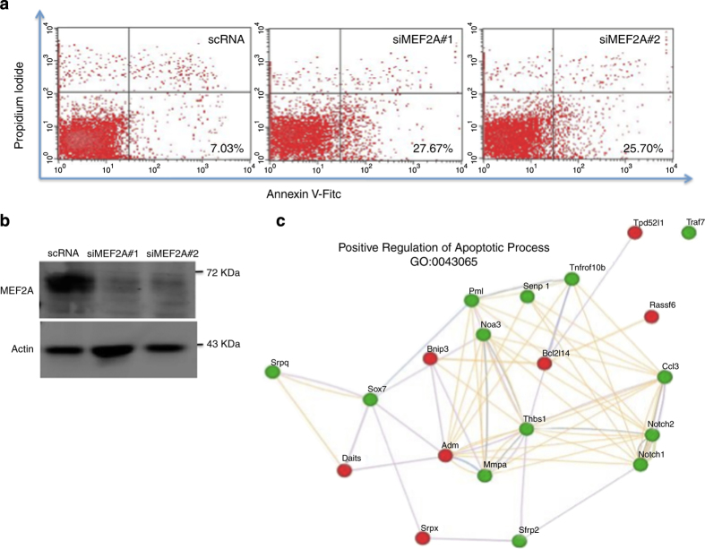
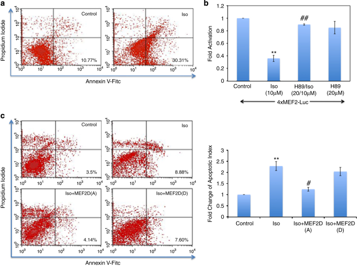
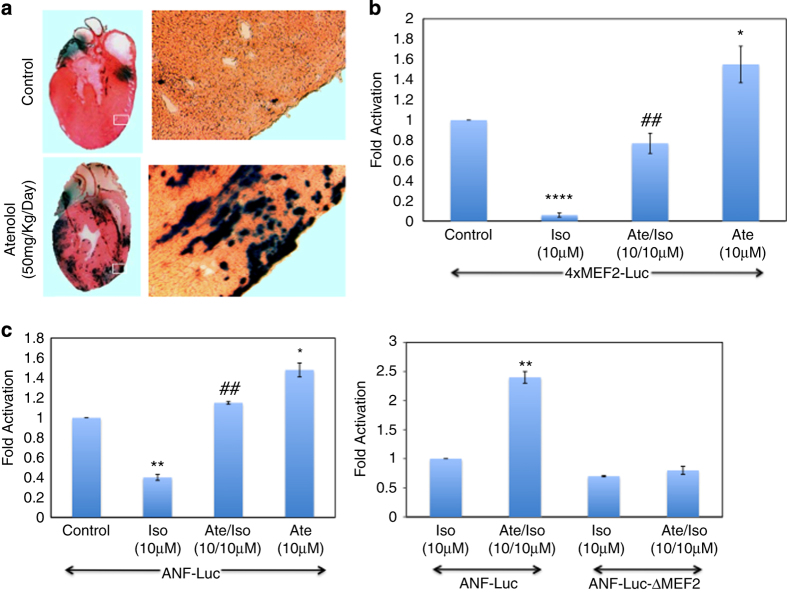
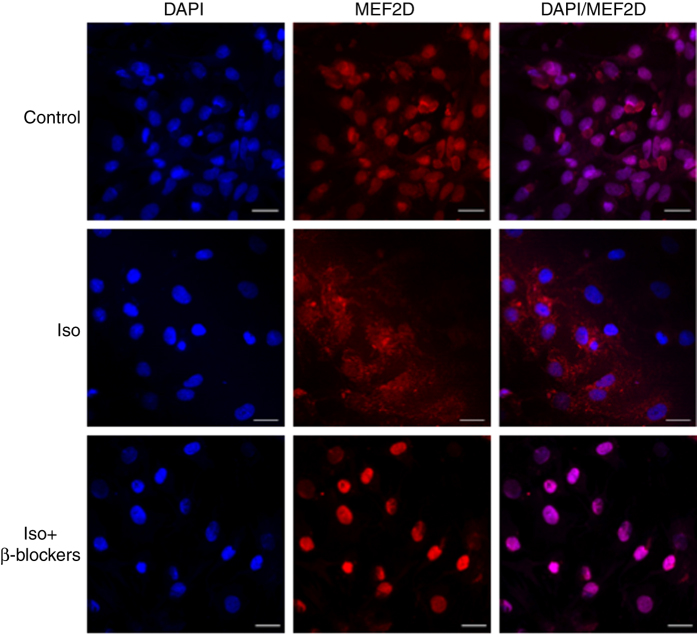
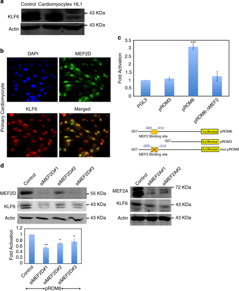
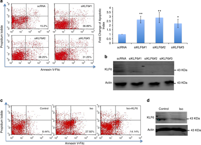
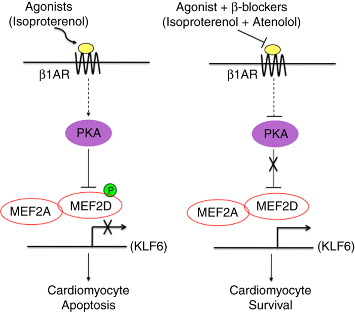
β1-Adrenergic receptor (β1-AR) stimulation increases apoptosis in cardiomyocytes through activation of cAMP/protein kinase A (PKA) signaling. The myocyte enhancer factor 2 (MEF2) proteins function as important regulators of myocardial gene expression. Previously, we reported that PKA signaling directly represses MEF2 activity. We determined whether (a) MEF2 has a pro-survival function in cardiomyocytes, and (b) whether β-adrenergic/PKA signaling modulates MEF2 function in cardiomyocytes. Initially, we observed that siRNA-mediated gene silencing of MEF2 induces cardiomyocyte apoptosis as indicated by flow cytometry. β1-AR activation by isoproterenol represses MEF2 activity and promotes apoptosis in cultured neonatal cardiomyocytes. Importantly, β1-AR mediated apoptosis was abrogated in cardiomyocytes expressing a PKA-resistant form of MEF2D (S121/190A). We also observed that a β1-blocker, Atenolol, antagonizes isoproterenol-induced apoptosis while concomitantly enhancing MEF2 transcriptional activity. β-AR stimulation modulated MEF2 cellular localization in cardiomyocytes and this effect was reversed by β-blocker treatment. Furthermore, Kruppel-like factor 6, a MEF2 target gene in the heart, functions as a downstream pro-survival factor in cardiomyocytes. Collectively, these data indicate that (a) MEF2 has an important pro-survival role in cardiomyocytes, and (b) β-adrenergic signaling antagonizes the pro-survival function of MEF2 in cardiomyocytes and β-blockers promote it. These observations have important clinical implications that may contribute to novel strategies for preventing cardiomyocyte apoptosis associated with heart pathology.


Figures

Figure 1
Figure 1
MEF2A knockdown induces apoptosis in cardiomyocytes. (a) Annexin V detection is upregulated in MEF2A-depleted cardiomyocytes. Primary cardiomyocytes were transfected with two independent MEF2A siRNAs or a control scRNA. Cells were stained with annexin V-FITC and PI using annexin V-FITC apoptosis detection kit 48 h after transfection. Apoptosis was measured using flow cytometry analysis (FACS analyzer). (b) siRNA-mediated gene silencing reduces MEF2A protein. Equal amounts of total protein were used for western blot analysis. The levels of the indicated proteins were assessed by a standard immunoblotting technique using specific primary antibodies for each as indicated. (c) RNA-seq analysis of MEF2A knockdown in cardiomyocytes. siMEF2A#1 or a control scRNA were transfected in cardiomyocytes in duplicate and prepared for RNA-seq analysis. The gene ontology (GO) term, positive regulation of apoptotic process, was observed. The 20 genes within this category with the lowest P-value are shown in a network created by the program GeneMania. Green and red nodes indicate down- and upregulated genes, respectively, whereas connections between nodes are as follows: purple, co-expression; orange, predicted; blue, co-localization; green, shared protein domains; gray, other.
Figure 2
Figure 2
Activation of β1-AR induces cardiomyocytes apoptosis through the PKA pathway. (a) Primary cardiomyocytes were treated with Iso (10 μM) for 48 h and then stained with annexin V-FITC and PI using annexin V-FITC apoptosis detection kit. Apoptosis was measured using flow cytometry analysis. (b) Cardiomyocytes were transfected with 4xMEF2-Luc reporter gene and treated with Iso (10 μM) alone or in combination with PKA inhibitor (H89, 20 μM). Luciferase activity was assessed using the respective reporter gene and normalized to β-galactosidase (β-gal) activity. Data are the mean±S.E. The quantification data is between the same batch with n=3, **P≤0.01 comparing Iso with control, ##P≤0.01 comparing H89/Iso to Iso. (c) PKA-resistant MEF2D rescues cardiomyocytes. Primary cardiomyocytes were transfected with empty vector or mutated forms of MEF2D S121/190A (MEF2D(A), neutralizing) and S121/190D (MEF2D(D), phospho-mimetic) and then treated with with Iso (10 μM). Cells were prepared for FACS analysis as in a. n=3, **P≤0.01 Iso versus control, #P≤0.05 Iso+MEF2D(A) versus Iso.
Figure 3
Figure 3
Ate enhances MEF2 transcriptional activity in cardiomyoytes. (a) Animal treatment with Ate in vivo. MEF2-LacZ transgenic mice were fed daily with Ate (50 mg/Kg per day) or water for 48 h. After treatment, mice were killed and hearts were fixed with 2% paraformaldehyde in PBS for 30 min. The samples were then incubated with X-Gal solution overnight and visualized for MEF2 activity. The dark blue stain indicates MEF2 activity. (b) Cardiomyocytes were transfected with 4xMEF2-Luc reporter gene and treated with Iso (10 μM), Ate (10 μM) alone or in combination. Luciferase activity was assessed using the respective reporter gene and normalized to β-galactosidase. Data are the mean±S.E. n=3, ****P≤0.0001 comparing Iso to control, **P≤0.01 comparing Ate/Iso to Iso, *P≤0.05 comparing Ate to control. (c) Ate enhances transactivation of the ANF promoter through MEF2. The effect of Iso (10 μM), Ate (10 μM) alone and in combination was assessed on ANF-Luc or an analog with the MEF2 site mutated (ANF-Luc ΔMEF2) in cardiomyocytes. Data are the mean±S.E. n=3, ****P≤0.0001 Iso versus control, ## P≤0.01 Ate/Iso versus Iso, *P≤0.05 Ate versus control.
Figure 4
Figure 4
β-AR activation modulates cellular localization of MEF2D in cardiomyocytes. Primary cardiomyocytes were treated with solvent or Iso (10 μM) alone and in combination with β-blockers Ate (10 μM) and ICI118551 (1 μM). After treatment, cells were fixed with 4% paraformaldehyde and immunofluorescence analysis was performed using a primary antibody to MEF2D (red). DAPI (4,6-diamidino-2-phenylindole) was used to identify nuclei (blue). The merged pictures demonstrate localization of MEF2D (red) in respect to Iso (10 μM), β-blockers Ate (10 μM) and ICI118551 (1 μM) treatment, counterstained with DAPI. Scale bars, 20 μm.
Figure 5
Figure 5
MEF2 regulates KLF6 expression in cardiomyocytes (a) KLF6 protein is expressed in cardiomyocytes. Cell lysates of primary cardiomyocytes and HL1 cells and C2C12 (as control) were prepared and equal amounts of total protein were used for western blot analysis. The levels of the indicated proteins were assessed by a standard immunoblotting technique using specific primary antibodies for each as indicated. (b) Cellular localization of MEF2D and KLF6 in cardiomyocytes. Primary cardiomyocytes were fixed with 4% paraformaldehyde. Double immunofluorescence labeling demonstrating KLF6 (red) and MEF2D (green) and DAPI (4,6-diamidino-2-phenylindole) was used to identify nuclei (blue). (c) MEF2-dependant induction of the KLF6 promoter in cardiomyocytes. Schematic illustrations of KLF6 reporter gene constructs used in reporter assays are indicated in the lower panel. All KLF6 promoter constructs were cloned into the pGL3-basic reporter vector (pGL3-KLF6-Luc). Primary cardiomyocytes were transfected with various constructs of the KLF6 promoter, pROM6, pROM3 and pROM6 with the MEF2 site mutated. Cell extracts were prepared and MEF2-mediated transcriptional activity was determined by luciferase and β-gal assays. n=3, ***P≤0.001 pROM6 versus pGL3. (d) KLF6 expression is reduced in MEF2 depleted cardiomyocytes. Primary cardiomyocytes were transfected with three independent MEF2D siRNAs (left panel) or two independent MEF2A siRNAs (right panel). Forty-eight hours after transfection equal amounts of total protein were used for western blot analysis. The levels of the indicated proteins were assessed by a standard immunoblotting technique using specific primary antibodies for each as indicated. KLF6 reporter gene expression is reduced in MEF2D depleted cells. Luciferase activity was assessed using KLF6 promoter driving luciferase (pROM6-Luc) and normalized to β-galactosidase. Data are the mean±S.E. (n=3), **P≤0.01, *P≤0.05 comparing to control.
Figure 6
Figure 6
Role of KLF6 in cardiomyocyte survival. (a) siRNA-mediated depletion of KLF6 expression enhances apoptosis in primary cardiomyoytes. Primary cardiomyocytes were transfected with three independent KLF6 siRNAs and a control scRNA. Seventy-two hours after transfection, cells were stained with annexin V-FITC and PI using annexin V-FITC apoptosis detection kit. Cardiomyocytes apoptosis was measured using flow cytometry analysis (FACS analyzer). Changes in the number of apoptotic cells is indicated in a bar graph (left panel). n=3, **P≤0.01,*P≤0.05 comparing to control. (b) Equal amounts of total protein were used for western blot analysis to validate KLF6 knockdown. The levels of the indicated proteins were assessed by a standard immunoblotting technique using specific primary antibodies for each as indicated. (c) Exogenous KLF6 expression rescues cardiomyocytes apoptosis. Primary cardiomyocytes were transfected with KLF6 expression vector or a control vector and then treated with Iso (10 μM). Cells were then prepared for FACS analysis. (d) Activation of β1-AR suppresses KLF6 protein expression. Primary cardiomyocytes were treated with Iso (10 μM) or solvent. Cells were prepared for western blot analysis as in b.
Figure 7
Figure 7
Summary of MEF2 regulation by β-adrenergic signaling in cardiomyocyte survival. On the left side, acute activation of β-adrenergic receptors invokes cAMP-mediated PKA activation in cardiomyocytes, resulting in suppression of MEF2 transcriptional activity by direct phosphorylation. Expression of pro-survival genes such as KLF6 is prevented resulting in enhanced cardiomyocyte death. On the right side, β-blockers, such as Atenolol, competitively inhibit the activation of the β-adrenergic receptors by agonists resulting in enhanced MEF2 activity, thereby promoting cardiomyocyte survival.

Similar articles

Cited by

References

    1. Dorn GW , Molkentin JD . Manipulating cardiac contractility in heart failure—Data from mice and men. Circulation 2004; 109: 150–158. - PubMed
    1. Iwai-Kanai E , Hasegawa K , Araki M , Kakita T , Morimoto T , Sasayama S . alpha- and beta-Adrenergic pathways differentially regulate cell type-specific apoptosis in rat cardiac myocytes. Circulation 1999; 100: 305–311. - PubMed
    1. Zaugg M , Xu WM , Lucchinetti E , Shafiq SA , Jamali NZ , Siddiqui MAQ . Beta-adrenergic receptor subtypes differentially affect apoptosis in adult rat ventricular myocytes. Circulation 2000; 102: 344–350. - PubMed
    1. Krijnen PAJ , Nijmeijer R , Meijer CJLM , Visser CA , Hack CE , Niessen HWM . Apoptosis in myocardial ischaemia and infarction. J Clin Pathol 2002; 55: 801–811. - PMC - PubMed
    1. DeGeorge BR Jr. , Koch WJ . Beta blocker specificity: a building block toward personalized medicine. J Clin Invest 2007; 117: 86–89. - PMC - PubMed

LinkOut - more resources