. 2015 Dec 21:1:15059.
doi: 10.1038/cddiscovery.2015.59. eCollection 2015.

Selective cytoprotective effect of histamine on doxorubicin-induced hepatic and cardiac toxicity in animal models

Affiliations

Selective cytoprotective effect of histamine on doxorubicin-induced hepatic and cardiac toxicity in animal models

D J Martinel Lamas et al. Cell Death Discov. .

Abstract

The aim of the present work was to evaluate the potential protective effect of histamine on Doxorubicin (Dox)-induced hepatic and cardiac toxicity in different rodent species and in a triple-negative breast tumor-bearing mice model. Male Sprague Dawley rats and Balb/c mice were divided into four groups: control (received saline), histamine (5 mg/kg for rats and 1 mg/kg for mice, daily subcutaneous injection starting 24 h before treatment with Dox), Dox (2 mg/kg, intraperitoneally injected three times a week for 2 weeks) and Dox+histamine (received both treatments). Tissue toxicity was evaluated by histopathological studies and oxidative stress and biochemical parameters. The combined effect of histamine and Dox was also investigated in vitro and in vivo in human MDA-MB-231 triple-negative breast cancer model. Heart and liver of Dox-treated animals displayed severe histological damage, loss of tissue weight, increased TBARS levels and DNA damage along with an augment in serum creatine kinase-myocardial band. Pretreatment with histamine prevented Dox-induced tissue events producing a significant preservation of the integrity of both rat and mouse myocardium and liver, through the reduction of Dox-induced oxidative stress and apoptosis. Histamine treatment preserved anti-tumor activity of Dox, exhibiting differential cytotoxicity and increasing the Dox-induced inhibition of breast tumor growth. Findings provide preclinical evidence indicating that histamine could be a promising candidate as a selective cytoprotective agent for the treatment of Dox-induced cardiac and hepatic toxicity, and encourage the translation to clinical practice.


Figures
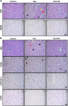
Figure 1
Figure 1
Histamine decreases doxorubicin-induced cardiotoxicity and hepatotoxicity in rats and mice. (A) (a, d) Normal histological appearance of untreated heart. (b, e) Heart of Dox-treated animals displaying focal necrotic cell death (black arrow), congestion-hemorrhage (red arrow), myocytolysis (yellow arrow) and myofribrillolysis (blue arrow) with fibrils de-arrangement. (c, f) Heart of Dox+HA-treated animals showing evident preservation of heart structure with reduced myofibrillolysis (blue arrow) and muscle bands with normal appearance. (a–c) Representative H&E stained specimens and (d–f) immunohistochemical images of γH2AX sections are shown. (B) (a, d, g, j) Normal histological appearance of liver from untreated animals. (b, e, h, k) Liver of Dox-treated rats displaying de-arrangement of hepatic trabecula (red arrow), cellular edema and focal necrosis (black arrow). (c, f, i, l) Liver of Dox+HA-treated animals showing preservation of tissue structure with mild cellular edema. (a–c) Representative H&E-stained sections are shown. (d–f) Representative immunohistochemical images of TUNEL, (g–i) caspase 3 and (j–l) γH2AX in paraffin-embedded liver specimens. Arrows indicate TUNEL-positive cells. (Six to eight rats per group). (C) (a) Normal histological appearance of WT mice untreated heart. (b) Heart of Dox-treated animals displaying vascular damage with perivascular edema, myofribrillolysis (blue arrow), cell recruitment (yellow arrows) and focal necrosis (black arrows). (c) Heart of Dox+HA-treated animals showing minimal focal damage with muscle bands with normal appearance. (d) Normal histological appearance of untreated heart of H4R−/− mice. (e) Heart of Dox-treated H4R−/− mice displaying myofibrillolysis (blue arrow), cell mobilization (yellow arrows) and focal necrosis (black arrow). (f) Heart of Dox+HA-treated H4R−/− mice showing minimal focal damage (non-diffused), including rippled and non-extended myofibrillolysis, with vasculature with normal appearance. (D) (a) Normal histological appearance of untreated liver of WT mice. (b) Liver of Dox-treated animals displaying two different areas, an extended necrotic region and a normal area, between them cellular mobilization as a band (arrow) (c) Liver of Dox+HA-treated WT mice showing almost normal characteristics. (d) Normal histological appearance of liver of untreated H4R−/− mice. (e) Liver of Dox-treated H4R−/− displaying reduced histological damage with focal and non-diffuse necrotic areas without cell mobilization. (f) Liver of Dox+HA-treated H4R−/− showing preservation of hepatic structure. Representative H&E stained sections are shown. x630 Original magnification. Scale bar, 20 μm. Inset: image at x100-fold magnification. (8–12 Mice per group).
Figure 1
Figure 1
Histamine decreases doxorubicin-induced cardiotoxicity and hepatotoxicity in rats and mice. (A) (a, d) Normal histological appearance of untreated heart. (b, e) Heart of Dox-treated animals displaying focal necrotic cell death (black arrow), congestion-hemorrhage (red arrow), myocytolysis (yellow arrow) and myofribrillolysis (blue arrow) with fibrils de-arrangement. (c, f) Heart of Dox+HA-treated animals showing evident preservation of heart structure with reduced myofibrillolysis (blue arrow) and muscle bands with normal appearance. (a–c) Representative H&E stained specimens and (d–f) immunohistochemical images of γH2AX sections are shown. (B) (a, d, g, j) Normal histological appearance of liver from untreated animals. (b, e, h, k) Liver of Dox-treated rats displaying de-arrangement of hepatic trabecula (red arrow), cellular edema and focal necrosis (black arrow). (c, f, i, l) Liver of Dox+HA-treated animals showing preservation of tissue structure with mild cellular edema. (a–c) Representative H&E-stained sections are shown. (d–f) Representative immunohistochemical images of TUNEL, (g–i) caspase 3 and (j–l) γH2AX in paraffin-embedded liver specimens. Arrows indicate TUNEL-positive cells. (Six to eight rats per group). (C) (a) Normal histological appearance of WT mice untreated heart. (b) Heart of Dox-treated animals displaying vascular damage with perivascular edema, myofribrillolysis (blue arrow), cell recruitment (yellow arrows) and focal necrosis (black arrows). (c) Heart of Dox+HA-treated animals showing minimal focal damage with muscle bands with normal appearance. (d) Normal histological appearance of untreated heart of H4R−/− mice. (e) Heart of Dox-treated H4R−/− mice displaying myofibrillolysis (blue arrow), cell mobilization (yellow arrows) and focal necrosis (black arrow). (f) Heart of Dox+HA-treated H4R−/− mice showing minimal focal damage (non-diffused), including rippled and non-extended myofibrillolysis, with vasculature with normal appearance. (D) (a) Normal histological appearance of untreated liver of WT mice. (b) Liver of Dox-treated animals displaying two different areas, an extended necrotic region and a normal area, between them cellular mobilization as a band (arrow) (c) Liver of Dox+HA-treated WT mice showing almost normal characteristics. (d) Normal histological appearance of liver of untreated H4R−/− mice. (e) Liver of Dox-treated H4R−/− displaying reduced histological damage with focal and non-diffuse necrotic areas without cell mobilization. (f) Liver of Dox+HA-treated H4R−/− showing preservation of hepatic structure. Representative H&E stained sections are shown. x630 Original magnification. Scale bar, 20 μm. Inset: image at x100-fold magnification. (8–12 Mice per group).
Figure 2
Figure 2
Histamine blocks doxorubicin-induced cytotoxic and oxidative damage in rat’s heart. (a) Heart weight determined as percentage of body weight. (b) Serum cholesterol levels. (c) Serum CK-MB levels. (d) TBARS levels expressed as nmol/mg of cardiac tissue. (e) Thiols content expressed as nmol/mg of tissue. (f) SOD activity expressed as U/mg of heart proteins. (Six to eight rats per group, *P<0.05, **P<0.01, ***P<0.001 versus control; # P<0.05, ## P<0.01 versus Dox).
Figure 3
Figure 3
Histamine alleviates doxorubicin-induced cytotoxic and oxidative damage in a rat's liver. (a) Liver weight determined as percentage of body weight. (b) The number of Kupffer cells and (c) the number of TUNEL-positive cells were determined by counting 10 random fields. (d) TBARS levels expressed as nmol/mg of cardiac tissue. (e) Thiols content expressed as nmol/mg of tissue. (f) SOD activity expressed as U/mg of liver proteins. (Six to eight rats per group, *P<0.05, **P<0.01, ***P<0.001 versus control; # P<0.05, ### P<0.001 versus Dox).
Figure 4
Figure 4
Doxorubicin and histamine effects on TBARS levels of H4R−/− mice compared with WT Balb/c mice. TBARS levels were determined in mice (a) heart and (b) liver of WT and KO mice. Data are expressed as nmol/mg of tissue. (8–12 mice per group, *P<0.05 versus WT Control; # P<0.05, ## P<0.01 versus WT Dox).
Figure 5
Figure 5
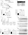
Histamine enhances anti-proliferative properties of doxorubicin in vitro. (a) Proliferation was evaluated by the clonogenic assay in human TNBC MDA-MB-231 cells treated with Dox (0.01–10 nM) in the absence (closed circles) or presence (open circles) of 10 μM histamine. Proliferation was expressed as a percentage relative to untreated cells (n=3, *P<0.01 versus Dox; two-way ANOVA and Bonferroni post test). (b) Incorporation of BrdU, (c) TUNEL and (d) Annexin-V staining assays were evaluated in MDA-MB-231 cells that were left untreated (control; C) or were treated with histamine (HA, 10 μM) and/or doxorubicin (Dox, 10 nM) for 48 h. (e) The mRNA expression levels of p21, p27, cyclin D1 and cyclin E2 were determined 24 h after treatments using qPCR and the expression levels were normalized to the expression of β-2-microglobulin. The ΔΔCt method was used to calculate the fold change. (g) Oxidative DNA damage was evaluated by measuring 8-OHdG formation and (h) intracellular ROS levels were determined 24 h after HA and/or Dox treatments using flow cytometry. (n=3-5, *P<0.05, **P<0.01, ***P<0.001 versus control; # P<0.05, ## P<0.01 versus Dox). Time course effects of Dox and HA on (i) γH2AX (15 kDa) and (f) phospho-MAPKs (p-ERK1/2, 42/44 kDa and p-p38) were assayed by western blot. Total ERK1/2, p38 and β-actin (42 kDa) were used as loading control. Semiquantitative analyses of band intensities are shown (n=2).
Figure 6
Figure 6
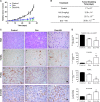
Combined effect of histamine and doxorubicin on triple-negative breast tumors induced in nude mice. (A) Relative tumor volume of the control group versus doxorubicin (Dox, 2 mg/kg) or the combination of Dox (2 mg/kg) and histamine (HA, 5 mg/kg). (6 mice per group, **P<0.01 versus Control; T-Test). (B) Median tumor doubling time of each group is depicted numerically (*P<0.05, ***P<0.001 versus Control; ## P<0.01 versus Dox. T-Test). (C) Histopathological and immunohistochemical analyses of tumor tissues. (a–c) Representative H&E-stained sections are shown. (a) Untreated tumors presented undifferentiated adenocarcinoma cells with marked anisokaryosis and anisocytosis. (b) Dox increased necrosis and the nuclear optical density. (c) The combined treatment Dox+HA reduced tumor cellularity, with evident nuclear hyperchromasia, that was replaced by extracellular matrix. Representative immunohistochemical images of (d–f) TUNEL, (g–i) PCNA and (j–l) 8-dOHG in paraffin-embedded tumor tissues. x630 Original magnification. Scale bar, 20 μm. (D) The number of tumor cells and the percentage of TUNEL, PCNA and 8-OHdG-positive stained cells were quantified by counting 10 random fields. (*P<0.05, ***P<0.001 versus Control; ## P<0.01, ### P<0.001 versus Dox). (E) Representative H&E stained sections of heart and liver are shown. (a) Normal histological appearance of untreated heart. (b) Heart of Dox-treated animals displaying severe myocytolysis (red arrow), areas of necrosis (black arrow) and reduced striated muscle bands. (c) Heart of Dox+HA-treated animals showing preservation of the structure with reduced myocytolysis, nuclei and muscle bands with normal appearance. (d) Normal histological appearance of untreated liver. (e) Liver of Dox-treated animals displaying focal necrosis (black arrow), sinusoidal atrophy (white arrow), inflammatory infiltrates, and fibrosis (red arrow). (f) Liver of Dox+HA-treated animals showing reduced sinusoidal disarrangement, displaying similar characteristics of the untreated liver. x1000 Original magnification. Scale bar, 20 μm.
Figure 6
Figure 6
Combined effect of histamine and doxorubicin on triple-negative breast tumors induced in nude mice. (A) Relative tumor volume of the control group versus doxorubicin (Dox, 2 mg/kg) or the combination of Dox (2 mg/kg) and histamine (HA, 5 mg/kg). (6 mice per group, **P<0.01 versus Control; T-Test). (B) Median tumor doubling time of each group is depicted numerically (*P<0.05, ***P<0.001 versus Control; ## P<0.01 versus Dox. T-Test). (C) Histopathological and immunohistochemical analyses of tumor tissues. (a–c) Representative H&E-stained sections are shown. (a) Untreated tumors presented undifferentiated adenocarcinoma cells with marked anisokaryosis and anisocytosis. (b) Dox increased necrosis and the nuclear optical density. (c) The combined treatment Dox+HA reduced tumor cellularity, with evident nuclear hyperchromasia, that was replaced by extracellular matrix. Representative immunohistochemical images of (d–f) TUNEL, (g–i) PCNA and (j–l) 8-dOHG in paraffin-embedded tumor tissues. x630 Original magnification. Scale bar, 20 μm. (D) The number of tumor cells and the percentage of TUNEL, PCNA and 8-OHdG-positive stained cells were quantified by counting 10 random fields. (*P<0.05, ***P<0.001 versus Control; ## P<0.01, ### P<0.001 versus Dox). (E) Representative H&E stained sections of heart and liver are shown. (a) Normal histological appearance of untreated heart. (b) Heart of Dox-treated animals displaying severe myocytolysis (red arrow), areas of necrosis (black arrow) and reduced striated muscle bands. (c) Heart of Dox+HA-treated animals showing preservation of the structure with reduced myocytolysis, nuclei and muscle bands with normal appearance. (d) Normal histological appearance of untreated liver. (e) Liver of Dox-treated animals displaying focal necrosis (black arrow), sinusoidal atrophy (white arrow), inflammatory infiltrates, and fibrosis (red arrow). (f) Liver of Dox+HA-treated animals showing reduced sinusoidal disarrangement, displaying similar characteristics of the untreated liver. x1000 Original magnification. Scale bar, 20 μm.

Similar articles

Cited by

References

    1. Ferlay J , Soerjomataram I , Dikshit R , Eser S , Mathers C , Rebelo M et al. Cancer incidence and mortality worldwide: sources, methods and major patterns in GLOBOCAN 2012. Int J Cancer 2015; 136: E359–E386. - PubMed
    1. Kim JH , Jenrow KA , Brown SL . Mechanisms of radiation-induced normal tissue toxicity and implications for future clinical trials. Radiat Oncol J 2014; 32: 103–115. - PMC - PubMed
    1. Octavia Y , Tocchetti CG , Gabrielson KL , Janssens S , Crijns HJ , Moens AL . Doxorubicin-induced cardiomyopathy: from molecular mechanisms to therapeutic strategies. J Mol Cell Cardiol 2012; 52: 1213–1225. - PubMed
    1. Stěrba M , Popelová O , Vávrová A , Jirkovský E , Kovaříková P , Geršl V et al. Oxidative stress, redox signaling, and metal chelation in anthracycline cardiotoxicity and pharmacological cardioprotection. Antioxid Redox Signal 2013; 18: 899–929. - PMC - PubMed
    1. Yang F , Teves SS , Kemp CJ , Henikoff S . Doxorubicin, DNA torsion, and chromatin dynamics. Biochim Biophys Acta 2014; 1845: 84–89. - PMC - PubMed

LinkOut - more resources