. 2016 Mar 21:2:16021.
doi: 10.1038/cddiscovery.2016.21. eCollection 2016.

Cytotoxic L-amino-acid oxidases from Amanita phalloides and Clitocybe geotropa induce caspase-dependent apoptosis

Affiliations

Cytotoxic L-amino-acid oxidases from Amanita phalloides and Clitocybe geotropa induce caspase-dependent apoptosis

A Pišlar et al. Cell Death Discov. .

Abstract

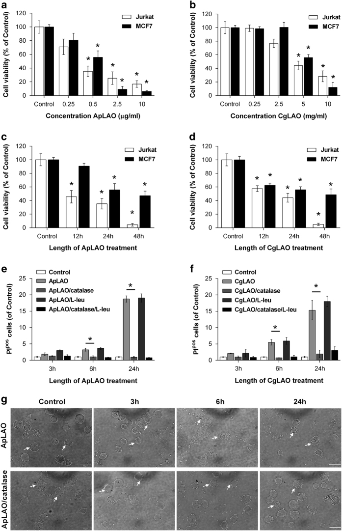
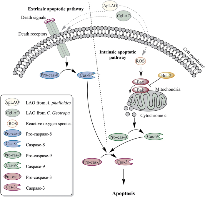
L-amino-acid oxidases (LAO) purified from fungi induce cell death in various mammalian cells including human tumor cell lines. The mechanism, however, remains poorly understood. In this study, we aimed to define a precise mechanism of cell death induced in Jurkat and MCF7 cancer cell lines by ApLAO and CgLAO, LAOs isolated from Amanita phalloides and Clitocybe geotropa, respectively. Cell death induced by both LAOs is shown to be concentration- and time-dependent, with higher toxic effects in Jurkat cells. LAO activity is required for the cytotoxicity. Detailed study on Jurkat cells further demonstrated that ApLAO and CgLAO both induce the intrinsic mitochondrial pathway of apoptosis, accompanied by a time-dependent depolarization of the mitochondrial membrane through the generation of reactive oxygen species. Treatment with the LAOs resulted in an increased ratio of the expression of proapoptotic Bax to that of antiapoptotic Bcl-2, subsequently leading to the activation of caspase-9 and -3. However, the pancaspase inhibitor, Z-VAD-FMK, did not completely abolish the cell death induced by either ApLAO or CgLAO, suggesting an alternative pathway for LAO-induced apoptosis. Indeed, caspase-8 activity in ApLAO- and CgLAO-treated cells was increased. Further, Fas/FasL (Fas ligand) antagonist caused a slight reduction in toxin-induced cell death, supporting the involvement of ApLAO and CgLAO in death-receptor-mediated apoptosis. These results thus provide new evidence that ApLAO and CgLAO induce apoptosis in Jurkat cells via both the intrinsic and extrinsic pathways, although the significantly higher increase of caspase-9 over caspase-8 activity suggests that it is the intrinsic pathway that is the predominant mode of ApLAO- and CgLAO-induced apoptosis.


Figures

Figure 1
Figure 1
Effects of toxins ApLAO and CgLAO on survival of Jurkat and MCF7 cells. (a and b) Jurkat cells and MCF7 cells were exposed to the increasing concentrations of ApLAO (0.25–10 μg/ml) and CgLAO (0.25–10 μg/ml). After 24 h of treatment, cell viability was assessed by MTS assay. Results are the means±S.D. of three independent assays. *P<0.05. (c and d) Jurkat cells and MCF7 cells were exposed to 0.5 μg/ml of ApLAO and 5 μg/ml of CgLAO for the time period indicated, followed by MTS assay. Cells were treated in quadruplicate. Results are the means±S.D. of three independent assays. *P<0.05. (e and f) Jurkat cells were treated with 0.5 μg/ml ApLAO (left) or 5 μg/ml CgLAO (right) in the absence or presence of catalase (1000 U/ml) and/or L-Leu (790 μg/ml) for the time indicated. They were then harvested and labeled with PI and the percentage of PIpos cells was determined by flow cytometry. Cells were treated in duplicate. Results are the means±S.D. of two independent assays. *P<0.05. (g) Representative images of morphological changes of cell membrane integrity (white arrows) following treatment of Jurkat cells with ApLAO (0.5 μg/ml) in the absence or presence of catalase (1000 U/ml) at indicated times. Cells were observed under an inverted phase-contrast microscope. Scale bars=20 μm.
Figure 2
Figure 2
ApLAO- and CgLAO-induced apoptosis in Jurkat cells. (a and b) Cells were treated with 0.5 μg/ml ApLAO (a) and 5 μg/ml CgLAO (b) in the absence or presence of catalase (1000 U/ml) for 16 h. Percentages of apoptotic cells were determined by flow cytometry using Annexin V and PI staining. The quadrant threshold was set according to control Jurkat cells and cells treated with toxin, and early apoptotic cells (Annexin Vpos, PIneg) and late apoptotic cells (Annexin Vpos, PIpos) were determined. Graphs (right panels) show quantified analysis and represent the percentage of the fraction of cells showing early apoptosis, late apoptosis and total apoptosis. Results are the means±S.D. of two independent assays. *P<0.05.
Figure 3
Figure 3
Effects catalase and L-Leu on ApLAO- and CgLAO-induced ROS generation. (a and b) Jurkat cells were exposed to ApLAO (0.5 μg/ml) (a) or CgLAO (5 μg/ml) (b) in the absence or presence of catalase (1000 U/ml) and/or L-Leu (790 μg/ml) for the time period indicated. The intracellular ROS levels were measured by flow cytometry using fluorescent DCFH-DA probe. Representative images (left panels) of flow cytometric analysis of ROS generation, where thin dashed line represents control Jurkat cells, thin solid line cells treated with ApLAO (a) or CgLAO (b) and thick solid line cells treated with toxin in the presence of catalase for 24 h. Graphs (right panels) present flow cytometric analysis of ROS levels as a relative percentage of DCF-positive cells normalized to appropriate control. Results are the means±S.D. of at least two independent assays. *P<0.05.
Figure 4
Figure 4
ApLAO- and CgLAO-induced caspase-dependent apoptosis. (a–d) Jurkat cells and MCF7 cells were pretreated with pan-specific caspase inhibitor Z-VAD-FMK (10 μM) for 30 min, followed by ApLAO (0.5 μg/ml) and CgLAO (5 μg/ml) treatment. (a and b) After 24 h, cell viability in Jurkat (a) and MCF7 cells (b) was assessed by MTS assay. Results are the means±S.D. of three independent assays. *P<0.05. (c and d) After 16 h of treatment, apoptotic cells were assessed by flow cytometry using Annexin V and PI staining. Graphs show quantified analysis of apoptosis in Jurkat cells (c) and MCF7 cells (d) and represent the percentage of the fraction of cells showing total apoptosis. Results are the means±S.D. of least two independent assays. *P<0.05. (e–g) Jurkat cells were treated with ApLAO (0.5 μg/ml) and CgLAO (5 μg/ml) for 12 h. Caspase activity in cell lysates was determined fluorometrically using the specific substrates for caspase-3/7 (AC-DEVD-AFC) (e), caspase-8 (z-IETD-AFC) (f) and caspase-9 (Ac-LEHD-AFC) (g). The results are presented as changes in fluorescence as a function of time (ΔFt). Cells were treated in duplicate. Results are the means±S.D. of at least two independent assays. *P<0.05.
Figure 5
Figure 5
ApLAO- and CgLAO-induced loss of mitochondrial function in Jurkat cells. (a and b) Cells were exposed to ApLAO (0.5 μg/ml) (a) or CgLAO (5 μg/ml) (b) in the absence or presence of catalase (1000 U/ml) and/or L-Leu (790 μg/ml) for the time period indicated. Mitochondrial transmembrane potential (Δψm) was then measured by flow cytometry using mitochondria-sensitive CMXRos dye. Cells were treated in duplicate. Results are the means±S.D. of two independent assays. *P<0.05. (c and d) Western blots showing the effect of ApLAO (0.5 μg/ml) (c) and CgLAO (5 μg/ml) (d) treatment at the time period indicated on the protein levels of Bax (upper panel) and Bcl-2 (lower panel) in Jurkat cells. The bar plot shows densitometric analysis of protein expression and represents the ratio of Bax to Bcl-2 expression (Bax/Bcl-2) relative to that in control cells. Results are the means±S.D. of two independent assays. *P<0.05.
Figure 6
Figure 6
Effects of ApLAO and CgLAO in death receptor-mediated apoptosis of Jurkat cells. (a) Representative images of the localization of ApLAO conjugated with FITC (ApLAO-FITC; green fluorescence) in control Jurkat cells and in cells treated with ApLAO-FITC for 1, 10 and 60 min (white arrows), as analyzed by fluorescence microscopy. Scale bars=20 μm (b and c) Jurkat cells were pretreated with Fas/FasL inhibitor Kp7-6 (0.1–0.5 mM) for 1 h, followed by ApLAO (0.5 μg/ml) or CgLAO (5 μg/ml) treatment. After 24 h, cell viability was assessed by MTS assay (b). Cells were treated in quadruplicate. Results are the means±S.D. of three independent assays. *P<0.05. Cytotoxicity was assessed by staining cells with PI and the percentage of PIpos cells was determined by flow cytometry (c). Cells were treated in duplicate. Results are the means±S.D. of two independent assays. *P<0.05.
Figure 7
Figure 7
Proposed molecular mechanism of the apoptotic action of LAOs in Jurkat cells. ApLAO and CgLAO can induce apoptosis by both the intrinsic and extrinsic pathways. The intrinsic mitochondrial pathway is triggered by mitochondrial dysfunctions, including generation of intracellular ROS, decreased Δψm, increased Bax/Bcl-2 ratio and caspase-9 activation, leading to increased caspase-3 and consequently to apoptosis. Extrinsic, death receptor-mediated apoptosis is triggered by caspase-8 activation.

References

    1. Fesik SW. Promoting apoptosis as a strategy for cancer drug discovery. Nat Rev Cancer 2005; 5: 876–885. - PubMed
    1. Elmore S. Apoptosis: a review of programmed cell death. Toxicol Pathol 2007; 35: 495–516. - PMC - PubMed
    1. Walczak H, Krammer PH. The CD95 (APO-1/Fas) and the TRAIL (APO-2L) apoptosis systems. Exp Cell Res 2000; 256: 58–66. - PubMed
    1. Eldering E, Mackus WJ, Derks IA, Evers LM, Beuling E, Teeling P et al. Apoptosis via the B cell antigen receptor requires Bax translocation and involves mitochondrial depolarization, cytochrome C release, and caspase-9 activation. Eur J Immunol 2004; 34: 1950–1960. - PubMed
    1. Fulda S, Debatin KM. Extrinsic versus intrinsic apoptosis pathways in anticancer chemotherapy. Oncogene 2006; 25: 4798–4811. - PubMed