

Ultrashort Echo-Time Magnetic Resonance Imaging Is a Sensitive Method for the Evaluation of Early Cystic Fibrosis Lung Disease
- PMID: 27551814
- PMCID: PMC5122478
- DOI: 10.1513/AnnalsATS.201603-203OC
Ultrashort Echo-Time Magnetic Resonance Imaging Is a Sensitive Method for the Evaluation of Early Cystic Fibrosis Lung Disease
Abstract
Rationale: Recent advancements that have been made in magnetic resonance imaging (MRI) improve our ability to assess pulmonary structure and function in patients with cystic fibrosis (CF). A nonionizing imaging modality that can be used as a serial monitoring tool throughout life can positively affect patient care and outcomes.
Objectives: To compare an ultrashort echo-time MRI method with computed tomography (CT) as a biomarker of lung structure abnormalities in young children with early CF lung disease.
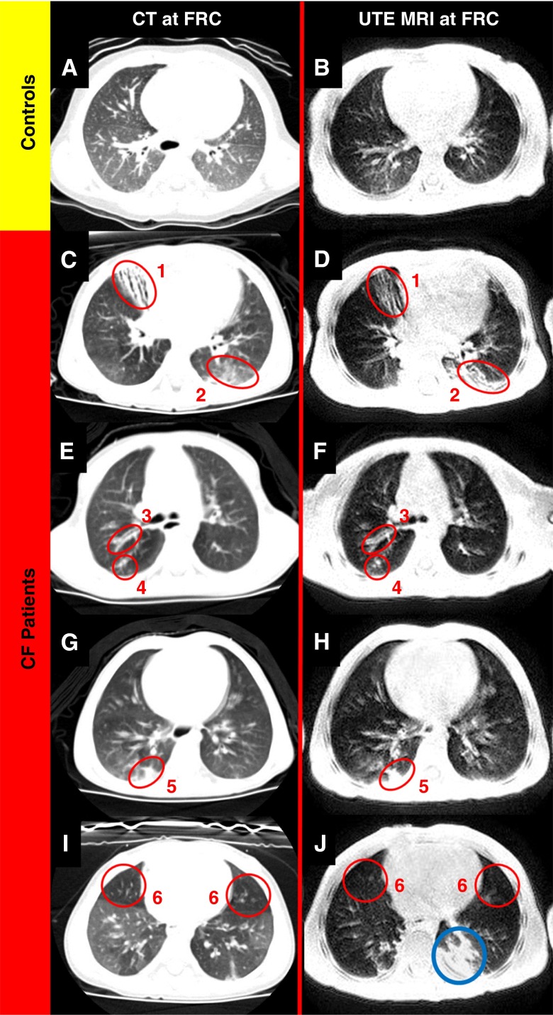
Methods: Eleven patients with CF (mean age, 31.8 ± 5.7 mo; median age, 33 mo; 7 male and 4 female) were imaged via CT and ultrashort echo-time MRI. Eleven healthy age-matched patients (mean age, 22.5 ± 10.2 mo; median age, 23 mo; 5 male and 6 female) were imaged via ultrashort echo-time MRI. CT scans of 13 additional patients obtained for clinical indications not affecting the heart or lungs and interpreted as normal provided a CT control group (mean age, 24.1 ± 11.7 mo; median age, 24 mo; 6 male and 7 female). Studies were scored by two experienced radiologists using a well-validated CF-specific scoring system for CF lung disease.
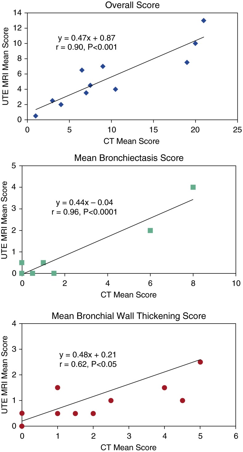
Measurements and main results: Correlations between CT and ultrashort echo-time MRI scores of patients with CF were very strong, with P values ≤0.001 for bronchiectasis (r = 0.96) and overall score (r = 0.90), and moderately strong for bronchial wall thickening (r = 0.62, P = 0.043). MRI easily differentiated CF and control groups via a reader CF-specific scoring system.
Conclusions: Ultrashort echo-time MRI detected structural lung disease in very young patients with CF and provided imaging data that correlated well with CT. By quantifying early CF lung disease without using ionizing radiation, ultrashort echo-time MRI appears well suited for pediatric patients requiring longitudinal imaging for clinical care or research studies. Clinical Trial registered with www.clinicaltrials.gov (NCT01832519).
Keywords: cystic fibrosis; lung; magnetic resonance imaging; ultrashort echo-time.
Figures
References
-
- Bradley WG., Jr Magnetic resonance imaging in the central nervous system: comparison with computed tomography. Magn Reson Annu. 1986:81–122. - PubMed
-
- Bergin CJ, Noll DC, Pauly JM, Glover GH, Macovski A. MR imaging of lung parenchyma: a solution to susceptibility. Radiology. 1992;183:673–676. - PubMed
-
- Kauczor HU, Kreitner KF. MRI of the pulmonary parenchyma. Eur Radiol. 1999;9:1755–1764. - PubMed
Publication types
MeSH terms
Supplementary concepts
Associated data
Grants and funding
LinkOut - more resources
Full Text Sources
Other Literature Sources
Medical
