Review
. 2016 Dec:41:194-202.
doi: 10.1016/j.sbi.2016.07.009. Epub 2016 Aug 20.

Resolution advances in cryo-EM enable application to drug discovery

Affiliations
Review

Resolution advances in cryo-EM enable application to drug discovery

Sriram Subramaniam et al. Curr Opin Struct Biol. 2016 Dec.

Abstract

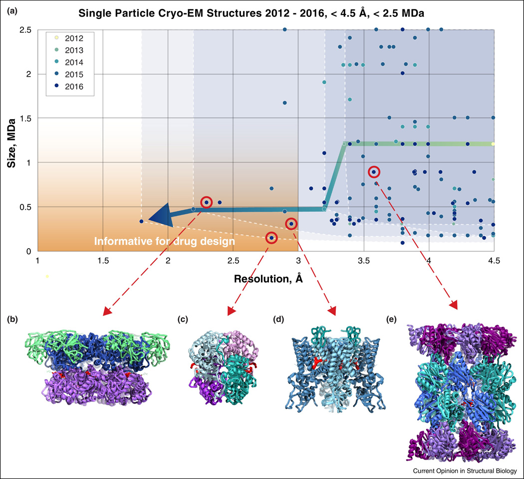
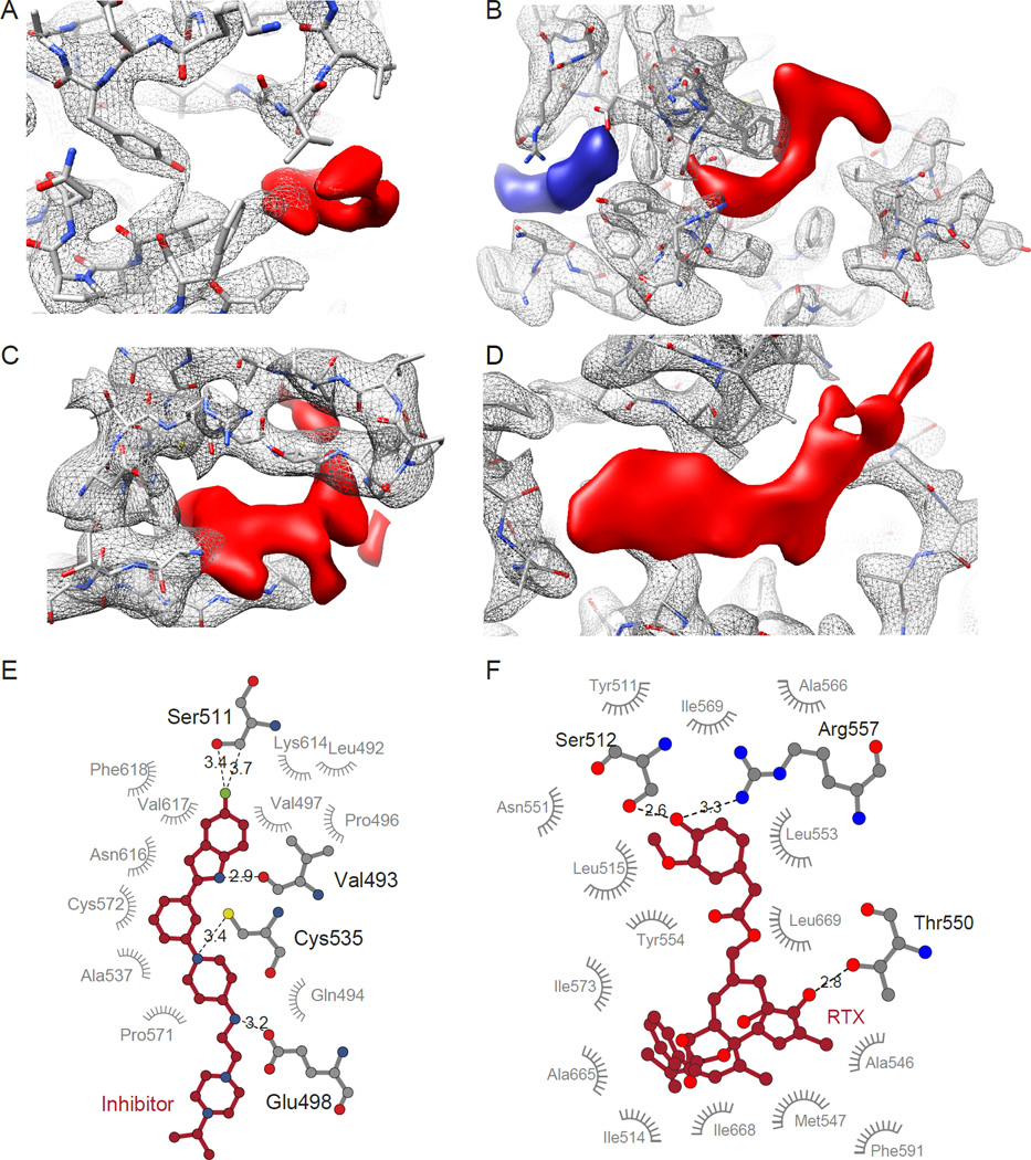
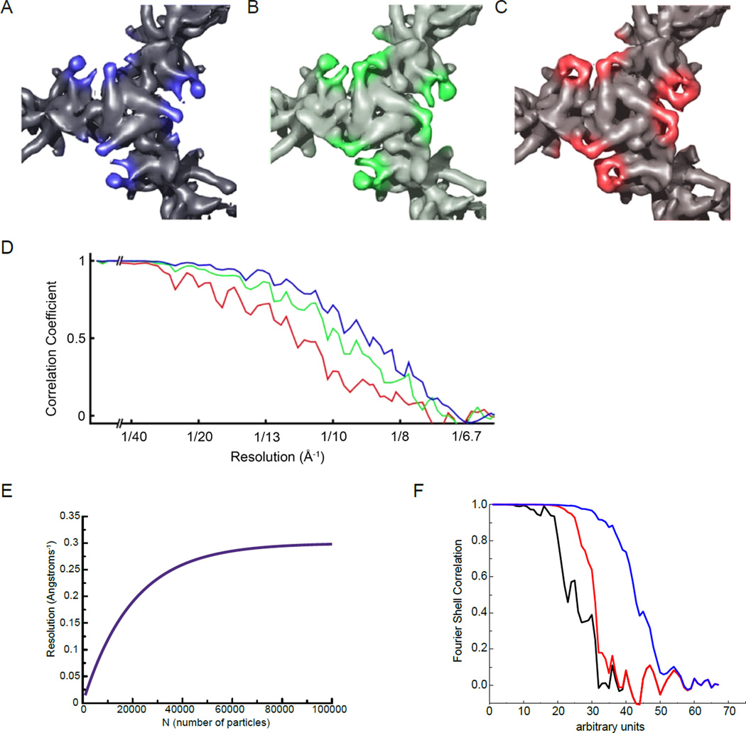
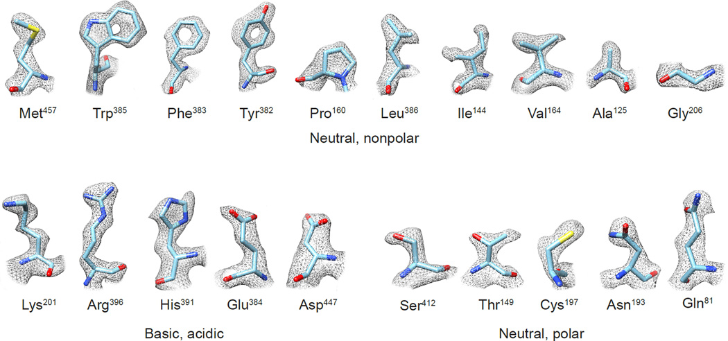
The prospect that the structures of protein assemblies, small and large, can be determined using cryo-electron microscopy (cryo-EM) is beginning to transform the landscape of structural biology and cell biology. Great progress is being made in determining 3D structures of biological assemblies ranging from icosahedral viruses and helical arrays to small membrane proteins and protein complexes. Here, we review recent advances in this field, focusing especially on the emerging use of cryo-EM in mapping the binding of drugs and inhibitors to protein targets, an application that requires structure determination at the highest possible resolutions. We discuss methods used to evaluate the information contained in cryo-EM density maps and consider strengths and weaknesses of approaches currently used to measure map resolution.


Figures

Figure 1
Figure 1
Cryo-EM progress towards drug discovery. (A) Chart showing entries deposited in the Electron Microscopy Data Bank each year since 2012 with reported resolutions better than 4.5 Å for protein complexes smaller than 2.5 MDa. Four select high resolution structures (red circles) determined with bound small molecule drugs (density highlighted in red) are shown in (B–E) with ribbon diagrams of: (B) p97 (EMD-3295, PDB-5ftj) [49]; (C) lactate dehydrogenase (EMD-8191, PDB-5k0z) [48]; (D) TRPV1 (EMD-8117, PDB-5irx) [18]; and (E) Plasmodium falciparum 20S proteasome (EMD-3231, PDB-5fmg) [40].
Figure 2
Figure 2
Visualizing drug density in cryo-EM maps. (A–D) Close-up views of protein (grey mesh) and drug (red solid) densities, fit with the atomic model of the protein. (A) Lactate dehydrogenase bound to the inhibitor GSK2837808A (EMD-8191, PDB-5k0z) [49]. (B) TRPV1 ion channel bound to double-knot toxin (DkTx) and resiniferatoxin (RTX) (EMD-8117, PDB-5irx) [18]. (C) p97 bound to the inhibitor UPCDC30245 (EMD-3295, PDB-5ftj) [49]. (D) Plasmodium falciparum 20S proteasome bound to WLW vinyl sulfone (EMD-3231, PDB-5fmg) [40]. (E, F) LIGPLOT diagrams showing binding pocket interactions of inhibitor bound p97, as shown in (A) and RTX bound to TRPV1, as shown in (B).
Figure 3
Figure 3
Fourier Shell Correlation (FSC) can be an inaccurate measurement of resolution in cryo-EM maps. (A–D) From [53], top views of the pyruvate dehydrogenase E2 catalytic domains, contoured to the same sigma levels, reconstructed using the best 963 (A), 395 (B) or 139 (C) particles. Although features of the map become better resolved as fewer, better particles are used, the reported FSC resolution (D) decreases. Colors of the FSC plots in (D) correspond to map colors in (A–C). (E) An idealized curve for resolution as a function of sample size N, which tends to follow a log-linear relationship [54]. (F) An actual FSC curve between two independent half-map reconstructions of a helical polymer is shown in black (the scale of the abscissa is arbitrary). In red the same FSC is computed, but after adding an artifact to the reconstruction, a Gaussian cylinder of density along the axis with a standard deviation in the radius of the cylinder of 2 pixels. The FSC is improved (it is shifted to the right) and at high resolution the black and red curves are completely superimposed. In the blue curve the Gaussian cylinder is even narrower, with a standard deviation in radius of 1.5 pixels, which greatly improves the overall FSC and still matches the “true” FSC (the black curve) at the highest resolution.
Figure 4
Figure 4
Experimental densities for selected examples of the 20 amino acid types from the 1.8 Å resolution cryo-EM structure of glutamate dehydrogenase (EMDB-8194; PDB-5k12) [48]. Features such as holes in aromatic rings, as well as the “zigzag” structure of extended Arg and Lys sidechains, are visible in the density maps.

References

    1. Egelman EH. The Current Revolution in Cryo-EM. Biophys J. 2016;110:1008–1012. - PMC - PubMed
    1. Kuhlbrandt W. Cryo-EM enters a new era. Elife. 2014;3:e03678. - PMC - PubMed
    1. Li X, Mooney P, Zheng S, Booth CR, Braunfeld MB, Gubbens S, Agard DA, Cheng Y. Electron counting and beam-induced motion correction enable near-atomic-resolution single-particle cryo-EM. Nat Methods. 2013;10:584–590. This paper presents the use of direct detectors and motion correction to achieve near-atomic resolutions of the Thermoplasma acidophilum 20S proteosome, outlining methods that helped drive the subsequent revolution advances in cryo-EM.

    1. Faruqi AR, Cattermole DM, Henderson R, Mikulec B, Raeburn C. Evaluation of a hybrid pixel detector for electron microscopy. Ultramicroscopy. 2003;94:263–276. - PubMed
    1. Cheng Y. Single-Particle Cryo-EM at Crystallographic Resolution. Cell. 2015;161:450–457. - PMC - PubMed

Publication types

LinkOut - more resources