

GEOGRAPHIC ATROPHY: Semantic Considerations and Literature Review
- PMID: 27552292
- PMCID: PMC5115977
- DOI: 10.1097/IAE.0000000000001258
GEOGRAPHIC ATROPHY: Semantic Considerations and Literature Review
Abstract
Purpose: There is a lack of agreement regarding the types of lesions and clinical conditions that should be included in the term "geographic atrophy." Varied and conflicting views prevail throughout the literature and are currently used by retinal experts and other health care professionals.
Methods: We reviewed the nominal definition of the term "geographic atrophy" and conducted a search of the ophthalmologic literature focusing on preceding terminologies and the first citations of the term "geographic atrophy" secondary to age-related macular degeneration.
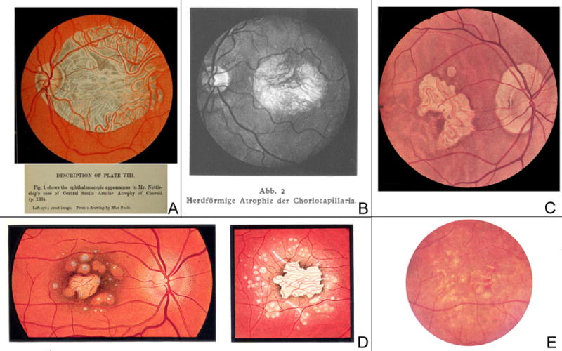
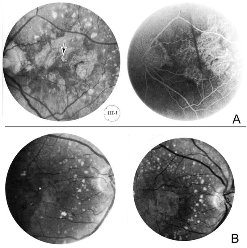
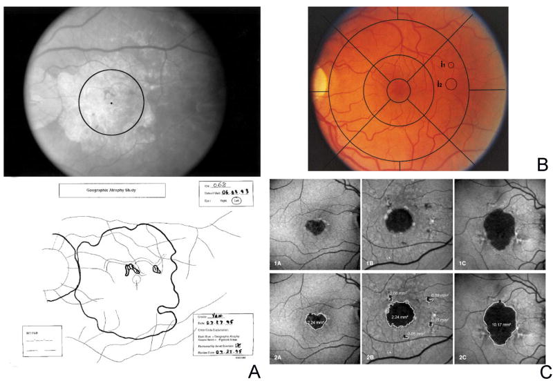
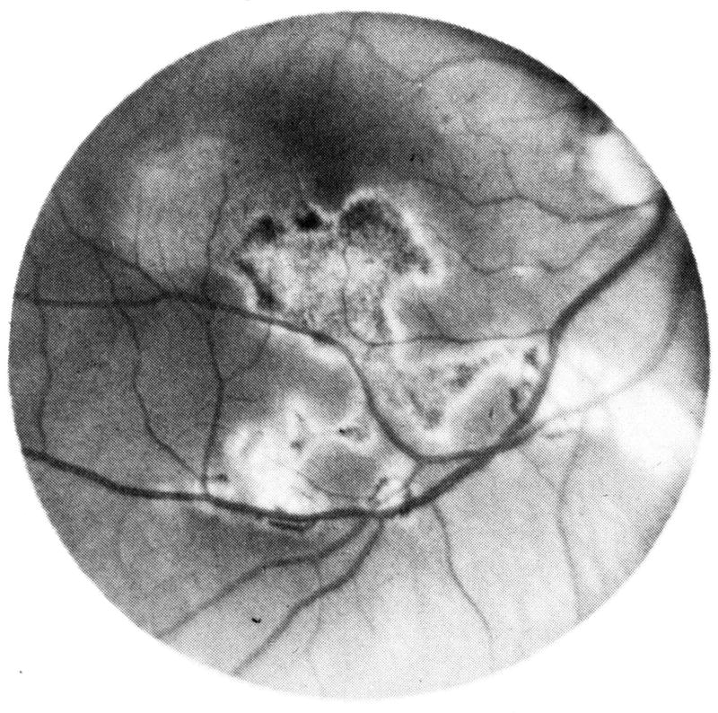
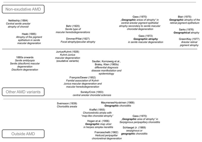
Results: According to the nominal definition, the term "geography" stands for a detailed description of the surface features of a specific region, indicating its relative position. However, it does not necessarily imply that the borders of the region must be sharply demarcated or related to any anatomical structures. The term "geographical areas of atrophy" was initially cited in the 1960s in the ophthalmologic literature in the context of uveitic eye disease and shortly thereafter also for the description of variants of "senile macular degeneration." However, no direct explanation could be found in the literature as to why the terms "geographical" and "geographic" were chosen. Presumably the terms were used as the atrophic regions resembled the map of a continent or well-defined country borders on thematic geographical maps. With the evolution of the terminology, the commonly used adjunct "of the retinal pigment epithelium" was frequently omitted and solely the term "geographic atrophy" prevailed for the nonexudative late-stage of age-related macular degeneration itself. Along with the quantification of atrophic areas, based on different imaging modalities and the use of both manual and semiautomated approaches, various and inconsistent definitions for the minimal lesion diameter or size of atrophic lesions have also emerged.
Conclusion: Reconsideration of the application of the term "geographic atrophy" in the context of age-related macular degeneration seems to be prudent given ongoing advances in multimodal retinal imaging technology with identification of various phenotypic characteristics, and the observation of atrophy development in eyes under antiangiogenic therapy.
Conflict of interest statement
None of the authors has any proprietary interest in the content of the manuscript.
Figures
References
-
- Klein R, Chou CF, Klein BE, et al. Prevalence of age-related macular degeneration in the US population. Arch Ophthalmol. 2011;129:75–80. - PubMed
-
- Chakravarthy U, Augood C, Bentham GC, et al. Cigarette Smoking and Age-Related Macular Degeneration in the EUREYE Study. Ophthalmology. 2007;114:1164–1169. - PubMed
-
- Tomany SC, Wang JJ, Van Leeuwen R, et al. Risk factors for incident age-related macular degeneration: pooled findings from 3 continents. Ophthalmology. 2004;111:1280–1287. - PubMed
-
- Holz FG, Strauss EC, Schmitz-Valckenberg S, et al. Geographic Atrophy: Clinical Features and Potential Therapeutic Approaches. Ophthalmology. 2014;121:1079–1091. - PubMed
-
- Bhisitkul RB, Mendes TS, Rofagha S, et al. Macular atrophy progression and 7-year vision outcomes in subjects from the ANCHOR, MARINA and HORIZON studies (SEVEN-UP Study) Am J Ophthalmol. 2015;159:915–924. - PubMed
Publication types
MeSH terms
Grants and funding
LinkOut - more resources
Full Text Sources
Other Literature Sources
