. 2016 Aug 24:6:32183.
doi: 10.1038/srep32183.

Pathogenicity of duck plague and innate immune responses of the Cherry Valley ducks to duck plague virus

Affiliations

Pathogenicity of duck plague and innate immune responses of the Cherry Valley ducks to duck plague virus

Ning Li et al. Sci Rep. .

Abstract

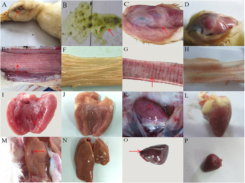
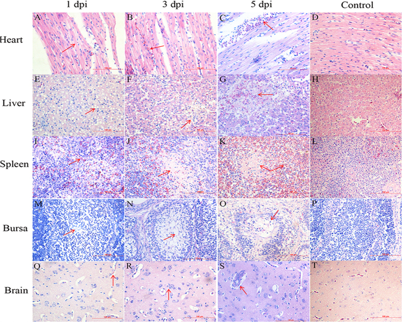
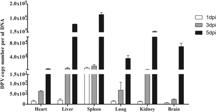
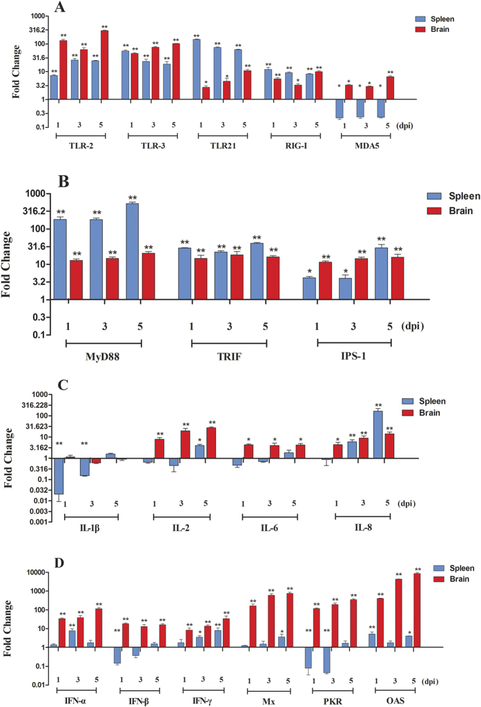
Duck plague caused by duck plague virus (DPV) is an acute and contagious disease. To better understand the pathogenic mechanism of duck plague virus in ducklings, an infection experiment was performed. Our results showed that typical symptoms were observed in the infected ducklings. DPV could replicate quickly in many tissues, leading to pathological lesions, especially on the spleen. Real-time quantitative PCR demonstrated that expression of many innate immune-related genes was mostly up-regulated in the brain, and the antiviral innate immune response was established, but not sufficient to restrict viral replication. In contrast, although the expression of many major pattern recognition receptors (PRRs) increased in the spleen, the expression of most cytokines was declined. Our study indicates that DPV is a pantropic virus that can replicate rapidly in tissues, causing serious pathological lesions but the immune responses are different in the spleen and brain. To our knowledge, this is the first report to systematically explore the expression profiles of the immune genes in the DPV-infected ducks. Our data provide a foundation for further study of the pathogenicity of duck plague.


Figures

Figure 1
Figure 1. Clinical symptoms and gross lesions of the DPV-infected ducks.
(A) Swollen head and neck; (B) Greenish diarrhea; (C) A light-yellow and transparent liquid of the head; (E) Diffuse hemorrhage of the esophageal mucosa; (G) Hemorrhage of the annulus trachealis; (I) Spotted hemorrhage of the endocardium; (K) Petechial hemorrhaging of the epicardium; (M) Liver is enlarged with blood spots; (O) Splenomegaly and hemorrhage. D, F, H, J, L, N, and P represent the head, esophagus mucosa, trachea, endocardium, epicardium, liver, and spleen of ducks from the control group, respectively.
Figure 2
Figure 2. Pathological changes of the DPV-infected ducks at the different time points.
(A) Mild granular degeneration of myocardial fibers at 1 dpi; (B) A small amount of erythrocyte infiltration at 3 dpi; (C) Myocardium haemorrhage at 5 dpi; (E) Fatty degeneration in the liver at 1 dpi; (F) Fatty degeneration and focal necrosis of hepatocyte at 3 dpi; (G) Hepatocyte necrosis with hemorrhage at 5 dpi; (I) Slight congestion in the white pulp of the spleen at 1 dpi; (J) Necrotic foci of lymphocyte at 3 dpi; (K) Lymphocytic necrosis with diffuse hemorrhage at 5 dpi; (M) Slight reduction of lymphocytes in the bursa of Fabricus at 1 dpi; (N) Lymphocytes dissolved and disappeared, the number decreased significantly at 3 dpi; (O) Serious necrosis of lymphocytes at 5 dpi; (Q) Brain edema at 1 dpi; (R) Neuronophagia of the brain at 3 dpi; (S) Perivascular inflammatory infiltrates at 5 dpi. D, H, L, P, and T represent the heart, liver, spleen, bursa of Fabricus, and brain of ducks from the control group, respectively. Magnification, ×400.
Figure 3
Figure 3. DPV replication in the tissues of the infected ducks.
The data were expressed as means ± standard deviation, and five infected ducks were randomly selected for detecting the viral DNA load using the qRT-PCR method.
Figure 4
Figure 4. Expression profiles of PPRs in the spleen and brain of DPV-infected ducks.
The samples of viral infected ducks were collected at 1, 3, and 5 dpi. Total RNA was extracted and cDNA was prepared for detecting the cytokines. The expressions of cytokines tested in this study were measured using 2−ΔΔCt method by relative quantification. Data were expressed as mean fold change (n = 5). (A) The expression of major PRRs contained the TLR2, TLR3, TLR21, RIG-I, and MDA5; (B) The expression of relevant adaptor protein molecules contained the MyD88, TRIF, and IPS-1; (C) The expression of proinflammatory cytokines (IL-1β, IL-2, IL-6, and IL-8); (D) The expression of type I and II IFNs and ISGs (Mx, PKR, and OAS). Differences were analyzed with Student’s t test and were considered significant as follows: *P < 0.05; **P < 0.01.

References

    1. Davison S., Converse K. A., Hamir A. N. & Eckroade R. J. Duck viral enteritis in domestic muscovy ducks in Pennsylvania. Avian Diseases 37, 1142–1146 (1993). - PubMed
    1. Wang G. et al. The comprehensive diagnosis and prevention of duck plague in northwest Shandong province of China. Poultry Science 92, 2892–8 (2013). - PubMed
    1. Salguero F. J. & Sánchez-Cordón P. J. Histopathological and ultrastructural changes associated with herpesvirus infection in waterfowl. Avian Pathology Journal of the W.v.p.a 31, 133–40 (2002). - PubMed
    1. Huang Y. X. Study on duck plague-like disease. J. South China Agric. Univ, 1–12 (1959).
    1. Wei X. X. et al. Isolation and identification of one DPV strain (GM). China Poultry 37, 63–65 (2015).

Publication types

MeSH terms

LinkOut - more resources