

Lymphocytic, cytokine and transcriptomic profiles in peripheral blood of dogs with atopic dermatitis
- PMID: 27553600
- PMCID: PMC4995625
- DOI: 10.1186/s12917-016-0805-6
Lymphocytic, cytokine and transcriptomic profiles in peripheral blood of dogs with atopic dermatitis
Abstract
Background: Canine atopic dermatitis (cAD) is a common chronic and pruritic skin disease in dogs. The development of cAD involves complex interactions between environmental antigens, genetic predisposition and a number of disparate cell types. The aim of the present study was to perform comprehensive analyses of peripheral blood of AD dogs in relation to healthy subjects in order to determine the changes which would be characteristic for cAD.
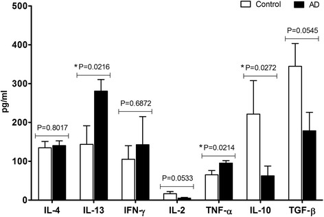
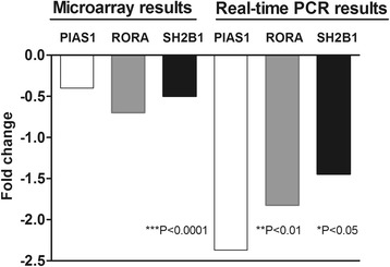
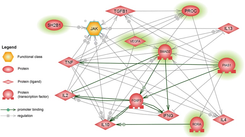
Results: The number of cells in specific subpopulations of lymphocytes was analyzed by flow cytometry, concentration of chosen pro- and anti-inflammatory cytokines (IL-4, IL-10, IL-13, TNF-α, TGF-β1) was determined by ELISA; and microarray analysis was performed on RNA samples isolated from peripheral blood nuclear cells of AD and healthy dogs. The number of Th cells (CD3(+)CD4(+)) in AD and healthy dogs was similar, whereas the percentage of Tc (CD3(+)CD8(+)) and Treg (CD4(+)CD25(+) Foxp3(+)) cells increased significantly in AD dogs. Increased concentrations of IL-13 and TNF-α, and decreased levels of IL-10 and TGF-β1 was observed in AD dogs. The level of IL-4 was similar in both groups of animals. Results of the microarray experiment revealed differentially expressed genes involved in transcriptional regulation (e.g., transcription factors: SMAD2, RORA) or signal transduction pathways (e.g., VEGF, SHB21, PROC) taking part in T lymphocytes lineages differentiation and cytokines synthesis.
Conclusions: Results obtained indicate that CD8(+) T cells, beside CD4(+) T lymphocytes, contribute to the development of the allergic response. Increased IL-13 concentration in AD dogs suggests that this cytokine may play more important role than IL-4 in mediating changes induced by allergic inflammation. Furthermore, observed increase in Treg cells in parallel with high concentrations of TNF-α and low levels of IL-10 and TGF-β1 in the peripheral blood of AD dogs point at the functional insufficiency of Treg cells in patients with AD.
Keywords: Canine atopic dermatitis; Cytokines; Lymphocytes; Microarray; Peripheral blood.
Figures
Similar articles
-
Quantitation of canine regulatory T cell populations, serum interleukin-10 and allergen-specific IgE concentrations in healthy control dogs and canine atopic dermatitis patients receiving allergen-specific immunotherapy.Vet Immunol Immunopathol. 2008 Jun 15;123(3-4):337-44. doi: 10.1016/j.vetimm.2008.02.008. Epub 2008 Feb 17. Vet Immunol Immunopathol. 2008. PMID: 18423890
-
Increased numbers of FoxP3-expressing CD4+ CD25+ regulatory T cells in peripheral blood from dogs with atopic dermatitis and its correlation with disease severity.Vet Dermatol. 2016 Feb;27(1):26-e9. doi: 10.1111/vde.12279. Epub 2015 Dec 21. Vet Dermatol. 2016. PMID: 26748886
-
Comparison of circulating CD4+, CD8+ lymphocytes and cytokine profiles between dogs with atopic dermatitis and healthy dogs.Res Vet Sci. 2022 Jul;145:13-20. doi: 10.1016/j.rvsc.2022.01.018. Epub 2022 Jan 29. Res Vet Sci. 2022. PMID: 35134678
-
Review: Role of genetics and the environment in the pathogenesis of canine atopic dermatitis.Vet Dermatol. 2015 Apr;26(2):95-e26. doi: 10.1111/vde.12198. Epub 2015 Feb 22. Vet Dermatol. 2015. PMID: 25703290 Review.
-
Cytokines and T cells in atopic dermatitis.Eur Cytokine Netw. 2013 Mar;24(1):37-44. doi: 10.1684/ecn.2013.0333. Eur Cytokine Netw. 2013. PMID: 23608610 Review.
Cited by
-
Peculiarities of vascular endothelial growth factor of oral cavity in atopic condition VEGF of oral cavity in atopic condition.Interv Med Appl Sci. 2021 Jul 6;11(4):207-212. doi: 10.1556/1646.2020.00002. eCollection 2021 Aug. Interv Med Appl Sci. 2021. PMID: 36343297 Free PMC article.
-
Serum canine thymus and activation-regulated chemokine (TARC/CCL17) concentrations correlate with disease severity and therapeutic responses in dogs with atopic dermatitis.Vet Dermatol. 2020 Dec;31(6):446-455. doi: 10.1111/vde.12894. Epub 2020 Sep 17. Vet Dermatol. 2020. PMID: 32945018 Free PMC article.
-
Is Vitamin D3 a Worthy Supplement Protecting against Secondary Infections in Dogs with Atopic Dermatitis?Pathogens. 2023 Jan 15;12(1):145. doi: 10.3390/pathogens12010145. Pathogens. 2023. PMID: 36678493 Free PMC article. Review.
-
Cytokine and Lymphocyte Profiles in Dogs with Atopic Dermatitis after Allergen-Specific Immunotherapy.Vaccines (Basel). 2022 Jun 28;10(7):1037. doi: 10.3390/vaccines10071037. Vaccines (Basel). 2022. PMID: 35891200 Free PMC article.
-
Oral treatment with Rosa multiflora fructus extract modulates mast cells in canine atopic dermatitis.Front Vet Sci. 2025 Apr 9;12:1531313. doi: 10.3389/fvets.2025.1531313. eCollection 2025. Front Vet Sci. 2025. PMID: 40271492 Free PMC article.
References
MeSH terms
Substances
LinkOut - more resources
Full Text Sources
Other Literature Sources
Molecular Biology Databases
Research Materials
Miscellaneous
