Comparative Study
. 2016 Aug 24;18(1):50.
doi: 10.1186/s12968-016-0271-0.

Compressed sensing real-time cine cardiovascular magnetic resonance: accurate assessment of left ventricular function in a single-breath-hold

Affiliations
Comparative Study

Compressed sensing real-time cine cardiovascular magnetic resonance: accurate assessment of left ventricular function in a single-breath-hold

Tomoyuki Kido et al. J Cardiovasc Magn Reson. .

Abstract

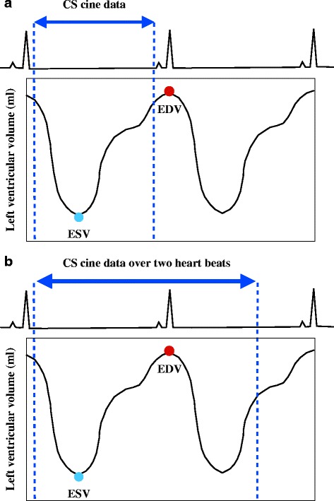
Background: Cardiovascular cine magnetic resonance (CMR) accelerated by compressed sensing (CS) is used to assess left ventricular (LV) function. However, it is difficult for prospective CS cine CMR to capture the complete end-diastolic phase, which can lead to underestimation of the end-diastolic volume (EDV), stroke volume (SV), and ejection fraction (EF), compared to retrospective standard cine CMR. This prospective study aimed to evaluate the diagnostic quality and accuracy of single-breath-hold full cardiac cycle CS cine CMR, acquired over two heart beats, to quantify LV volume in comparison to multi-breath-hold standard cine CMR.

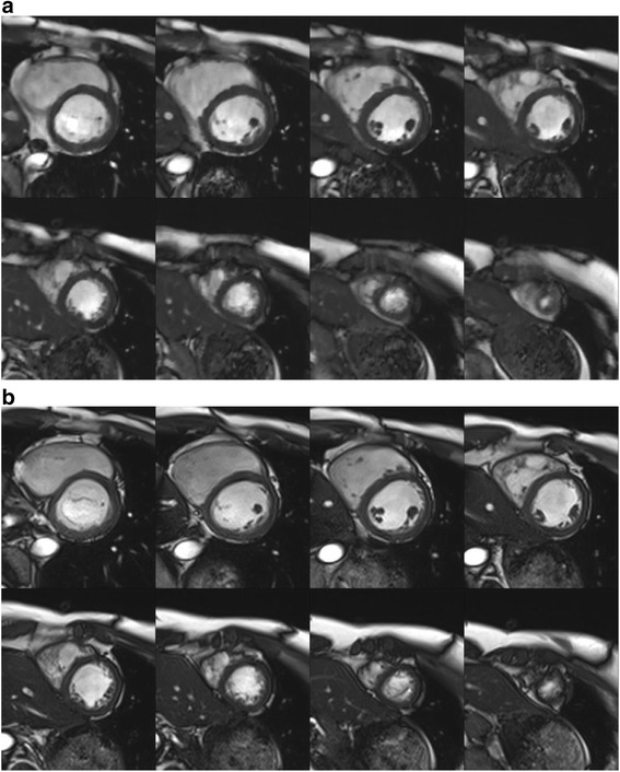
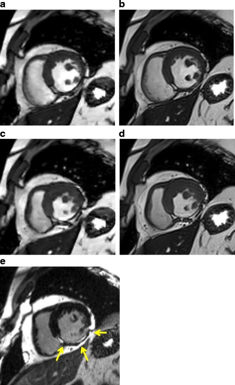
Methods: Eighty-one participants underwent standard segmented breath-hold cine and CS real-time cine CMR examinations to obtain a stack of eight contiguous short-axis images with same high spatial (1.7 × 1.7 mm(2)) and temporal resolution (41 ms). Two radiologists independently performed qualitative analysis of image quality (score, 1 [i.e., "nondiagnostic"] to 5 [i.e., "excellent"]) and quantitative analysis of the LV volume measurements.

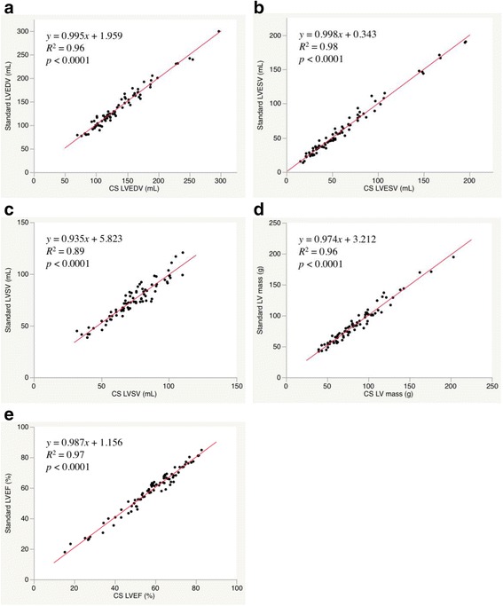
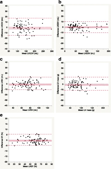
Results: The total examination time was 113 ± 7 s for standard cine CMR and 24 ± 4 s for CS cine CMR (p < 0.0001). The CS cine image quality was slightly lower than standard cine (4.8 ± 0.5 for standard vs. 4.4 ± 0.5 for CS; p < 0.0001). However, all image quality scores for CS cine were above 4 (i.e., good). No significant differences existed between standard and CS cine MR for all quantitative LV measurements. The mean differences with 95 % confidence interval (CI), based on Bland-Altman analysis, were 1.3 mL (95 % CI, -14.6 - 17.2) for LV end-diastolic volume, 0.2 mL (95 % CI, -9.8 to10.3) for LV end-systolic volume, 1.1 mL (95 % CI, -10.5 to 12.7) for LV stroke volume, 1.0 g (95 % CI, -11.2 to 13.3) for LV mass, and 0.4 % (95 % CI, -4.8 - 5.6) for LV ejection fraction. The interobserver and intraobserver variability for CS cine MR ranged from -4.8 - 1.6 % and from -7.3 - 9.3 %, respectively, with slopes of the regressions ranging 0.88-1.0 and 0.86-1.03, respectively.

Conclusions: Single-breath-hold full cardiac cycle CS real-time cine CMR could evaluate LV volume with excellent accuracy. It may replace multi-breath-hold standard cine CMR.

Keywords: Cardiac function; Cardiovascular magnetic resonance; Compressed sensing; Left ventricular ejection fraction; Real-time imaging.


Figures

Fig. 1
Fig. 1
Data acquisition of compressed sensing cine CMR for LV volume measurements. CS, compressed sensing; EDV, end-diastolic volume; ESV, end-systolic volume
Fig. 2
Fig. 2
Sampling pattern of compressed sensing cine CMR
Fig. 3
Fig. 3
Images acquired using compressed sensing cine and standard cine CMR. End-diastolic short-axis views of the left ventricle a by compressed sensing cine CMR and b by standard cine CMR. Both image sets were acquired from a 29-year-old healthy male volunteer. Both observers rated the image quality as excellent (i.e., score 5) for both images
Fig. 4
Fig. 4
Scatter plots for LV volume measurements by standard cine and CS cine. CS, compressed sensing; EF, ejection fraction; LV, left ventricular; LVEDV, left-ventricular end-diastolic volume; LVEF, left ventricular ejection fraction; LVESV, left ventricular end-systolic volume; LVSV, left-ventricular stroke volume; SD, standard deviation
Fig. 5
Fig. 5
Bland–Altman plots for LV volume measurements by standard cine and CS cine. The solid line indicates the difference between two sequences; the long dashed lines indicate the 95 % limits of agreement interval (i.e., the mean ± 1.96 SD); and the short dashed lines indicate the 95 % confidence interval of the mean difference CS, compressed sensing; EF, ejection fraction; LV, left ventricular; LVEDV, left-ventricular end-diastolic volume; LVEF, left-ventricular ejection fraction; LVESV, left-ventricular end-systolic volume; LVSV, left-ventricular stroke volume; SD, standard deviation
Fig. 6
Fig. 6
A 69-year-old patient with an inferior wall infarction. a, c Compressed sensing cine CMR images. b, d Standard cine MR images. e Late Gadolinium Enhancement (LGE) CMR images. The images depict the mid short-axis view a and b in the end-diastolic phase and c and d in the end-systolic phase. Both cine images show thinning and akinesia of the inferior myocardium. The LGE CMR shows a transmural infarction in the inferior myocardium (arrows)

Similar articles

Cited by

References

    1. Curtis JP, Sokol SI, Wang Y, Rathore SS, Ko DT, Jadbabale F, et al. The association of left ventricular ejection fraction, mortality, and cause of death in stable outpatients with heart failure. J Am Coll Cardiol. 2003;42:736–42. doi: 10.1016/S0735-1097(03)00789-7. - DOI - PubMed
    1. Knauth AL, Gauvreau K, Powell AJ, Landzberg MJ, Walsh EP, Lock JE, et al. Ventricular size and function assessed by cardiac MRI predict major adverse clinical outcomes late after tetralogy of Fallot repair. Heart. 2008;94:211–6. doi: 10.1136/hrt.2006.104745. - DOI - PubMed
    1. Bamberg F, Parhofer KG, Lochner E, Marcus RP, Theisen D, Findeisen HM, et al. Diabetes mellitus: long-term prognostic value of whole-body MR imaging for the occurrence of cardiac and cerebrovascular events. Radiology. 2013;269:730–7. doi: 10.1148/radiol.13130371. - DOI - PubMed
    1. White HD, Norris RM, Brown MA, Brandt PW, Whitlock RM, Wild CJ. Left ventricular end-systolic volume as the major determinant of survival after recovery from myocardial infarction. Circulation. 1987;76:44–51. doi: 10.1161/01.CIR.76.1.44. - DOI - PubMed
    1. Rathi VK, Biedermann RW. Imaging of ventricular function by cardiovascular magnetic resonance. Curr Cardiol Rep. 2004;6:55–61. doi: 10.1007/s11886-004-0065-0. - DOI - PubMed

Publication types

MeSH terms