. 2017 Feb;46(1):45-57.
doi: 10.1007/s13744-016-0430-x. Epub 2016 Aug 23.

Investigation of the midgut structure and ultrastructure in Cimex lectularius and Cimex pipistrelli (Hemiptera: Cimicidae)

Affiliations

Investigation of the midgut structure and ultrastructure in Cimex lectularius and Cimex pipistrelli (Hemiptera: Cimicidae)

M M Rost-Roszkowska et al. Neotrop Entomol. 2017 Feb.

Abstract

Cimicidae are temporary ectoparasites, which means that they cannot obtain food continuously. Both Cimex species examined here, Cimex lectularius (Linnaeus 1758) and Cimex pipistrelli (Jenyns 1839), can feed on a non-natal host, C. lectularius from humans on bats, C. pipistrelli on humans, but never naturally. The midgut of C. lectularius and C. pipistrelli is composed of three distinct regions-the anterior midgut (AMG), which has a sack-like shape, the long tube-shaped middle midgut (MMG), and the posterior midgut (PMG). The different ultrastructures of the AMG, MMG, and PMG in both of the species examined suggest that these regions must fulfill different functions in the digestive system. Ultrastructural analysis showed that the AMG fulfills the role of storing food and synthesizing and secreting enzymes, while the MMG is the main organ for the synthesis of enzymes, secretion, and the storage of the reserve material. Additionally, both regions, the AMG and MMG, are involved in water absorption in the digestive system of both Cimex species. The PMG is the part of the midgut in which spherites accumulate. The results of our studies confirm the suggestion of former authors that the structure of the digestive tract of insects is not attributed solely to diet but to the basic adaptation of an ancestor.

Keywords: Midgut epithelium; alimentary tract; digestive cells; secretory cells.


Figures
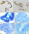
Fig 1
Fig 1
Midgut of Cimex lectularius (A) and Cimex pipistrelli (B) divided into anterior (AMG), middle (MMG), and posterior midgut (PMG). Fg fragment of the foregut, Malpighian tubules. A Stereomicroscope. Bar = 2.7 mm. B Stereomicroscope. Bar = 1.5 mm. C Cimex lectularius. The transverse section through AMG. Light microscope. Bar = 5 μm. D Cimex lectularius. The transverse section through MMG. Light microscope. Bar = 4 μm. E Cimex pipistrelli. The transverse section through PMG. Light microscope. Bar = 4 μm. F Cimex pipistrelli. The transverse section through MMG. Light microscope. Bar = 4 μm. Midgut lumen (l), midgut epithelium (e), visceral muscles (mc), basal lamina (arrowhead), regenerative cells (arrows).
Fig 2
Fig 2
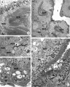
Digestive cells in the epithelium of three regions of the midgut. A Cimex lectularius. AMG. TEM. Bar = 0.7 μm. B Cimex lectularius. MMG. TEM. Bar = 1 μm. C C. pipistrelli. MMG. TEM. Bar = 1 μm. D Cimex pipistrelli. PMG. TEM. Bar = 1.4 μm. Midgut lumen (l), microvilli (mv), mitochondria (m), nucleus (n), basal lamina (bl), basal cell membrane folds (arrows), autophagosomes (au), spherites (sp), cisterns of the smooth endoplasmic reticulum (SER), Golgi complexes (d), vacuoles (v).
Fig 3
Fig 3
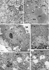
The ultrastructure of the midgut epithelium just before eating. A Cimex lectularius. MMG. TEM. Bar = 0.7 μm. B Cimex pipistrelli. MMG. TEM. Bar = 1 μm. C C. lectularius. AMG. TEM. Bar = 0.4 μm. D Cimex lectularius. AMG. TEM. Bar = 0.7 μm. E C. pipistrelli. PMG. TEM. Bar = 0.8 μm. Midgut lumen (l), mitochondria (m), microvilli (mv), autophagosomes (au), cisterns of the rough (RER) and smooth (SER) endoplasmic reticulum, Golgi complexes (d), nucleus (n), vacuoles (v), smooth septate junction (arrowhead), folds of the basal cell membrane (arrows), basal lamina (bl), visceral muscles (mc).
Fig 4
Fig 4
Ultrastructure of the midgut epithelium during digestion. A Cimex lectularius. MMG. Apical surface of digestive cells with vesicles with medium electron-dense content (arrows). TEM. Bar = 0.2 μm. B Cimex lectularius. AMG. TEM. Bar = 0.5 μm. C Cimex lectularius. MMG. TEM. Bar = 0.5 μm. D Cimex lectularius. AMG. TEM. Bar = 1.5 μm. E Cimex pipistrelli. MMG. TEM. Bar = 0.5 μm. F Cimex pipistrelli. MMG. TEM. Bar = 0.2 μm. Basal lamina (bl), mitochondria (m), microvilli (mv), autophagosomes (au), nucleus (n), vacuoles (v), midgut lumen (l), cisterns of the smooth (SER) and rough (RER) endoplasmic reticulum, visceral muscles (mc), spherites (sp), smooth septate junction (arrowhead), vesicles with electron-medium content (arrows).
Fig 5
Fig 5
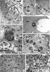
Ultrastructure of the midgut epithelium during digestion. A Cimex lectularius. MMG. TEM. Bar = 0.2 μm. B Cimex lectularius. MMG. TEM. Bar = 2.5 μm. C Cimex pipistrelli. MMG. TEM. Bar = 2.9 μm. D Cimex pipistrelli. MMG. TEM. Bar = 0.5 μm. E Cimex pipistrelli. MMG. TEM. Bar = 0.3 μm. F Cimex pipistrelli. MMG. TEM. Bar = 1.1 μm. G Cimex lectularius. PMG. TEM. Bar = 0.6 μm. H Cimex lectularius. PMG. TEM. Bar = 0.4 μm. Autophagosomes (au), autolysosomes (al), lamellar bodies (lb), residual bodies (rb), mitochondria (m), microvilli (mv), vacuoles (v), cisterns of the rough endoplasmic reticulum (RER), reserve material (rm), midgut lumen (l), exocytosis (arrows), basal lamina (bl), spherites (sp).
Fig 6
Fig 6
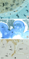
Histochemical staining of the MMG in Cimex lectularius. A Sudan black B—positive reaction. Light microscopy. Bar = 3.5 μm. B BPB—positive reaction. Light microscopy. Bar = 2.5 μm. C PAS—negative reaction. Light microscopy. Bar = 2.5 μm. MMG lumen (l), MMG epithelium (e), reserve material (arrows).
Fig 7
Fig 7
Secretion in AMG and MMG. A Cimex lectularius. AMG. TEM. Bar = 0.1 μm. B Cimex pipistrelli. AMG. TEM. Bar = 1 μm. C Cimex pipistrelli. MMG. TEM. Bar = 0.6 μm. Mitochondria (m), microvilli (mv), midgut lumen (l), reserve material (rm), residual bodies (rb), the protrusions of the apical cell membrane (stars), merocrine vesicles (arrows).
Fig 8
Fig 8
Secretory and regenerative cells. AF Secretory cells in the midgut epithelium. A Cimex lectularius. TEM. Bar = 0.4 μm. B Cimex lectularius. TEM. Bar = 1.3 μm. C Cimex pipistrelli. TEM. Bar = 1.8 μm. D Cimex lectularius. TEM. Bar = 0.7 μm. E Cimex pipistrelli. TEM. Bar = 1 μm. F Cimex pipistrelli. The higher magnification of F. TEM. Bar = 0.2 μm. Secretory cells (sc), regenerative cells (rc), digestive cells (dc), mitochondria (m), nucleus (n), microvilli (mv), basal lamina (bl), midgut lumen (l), cisterns of the rough endoplasmic reticulum (RER), vacuoles (v), electron-dense granules (arrows), cytoskeleton (arrowhead), visceral muscles (mc). G Regenerative cell in Cimex pipistrelli. TEM. Bar = 0.9 μm.

References

    1. Azevedo DO, Neves CA, dos Santos Mallet JR, Monte Gonçalves TC, Zanuncio JC, Serrão JE. Notes on midgut ultrastructure of Cimex hemipterus (Hemiptera: Cimicidae) J Med Entomol. 2009;46:435–441. doi: 10.1603/033.046.0304. - DOI - PubMed
    1. Balvín O, Munclinger P, Kratochvíl L, Vilímová J. Mitochondrial DNA and morphology show independent evolutionary histories of bedbug Cimex lectularius (Heteropera: Cimicidae) on bats and humans. Parasitol Res. 2012;111:457–469. doi: 10.1007/s00436-012-2862-5. - DOI - PubMed
    1. Balvín O, Kratochvíl L, Vilímová J. Batbugs (Cimex pipistrelli group, Heteroptera: Cimicidae) are morphologically, but not genetically differentiated among bat hosts. J Zool Syst Evol Res. 2013;51:287–295. doi: 10.1111/jse.12000. - DOI
    1. Balvín O, Bartonička T, Simov N, Paunović M, Vilímová J. Distribution and host relations of species of the genus Cimex on bats in Europe. Folia Zool. 2014;63:281–289.
    1. Bandani AR, Kazzazi M, Allahyari M. Gut pH and isolation and characterization of digestive α-D-glucosidase of sunn pest. J Agric Sci Technol. 2010;12:265–274.

LinkOut - more resources