. 2016 Nov;113(Pt A):116-124.
doi: 10.1016/j.phrs.2016.08.023. Epub 2016 Aug 20.

S-Sulfhydration of ATP synthase by hydrogen sulfide stimulates mitochondrial bioenergetics

Affiliations

S-Sulfhydration of ATP synthase by hydrogen sulfide stimulates mitochondrial bioenergetics

Katalin Módis et al. Pharmacol Res. 2016 Nov.

Abstract

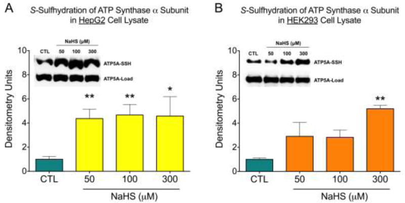
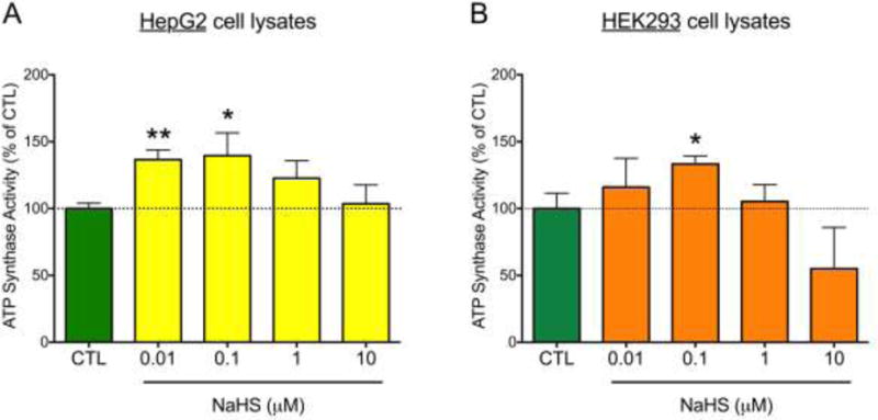
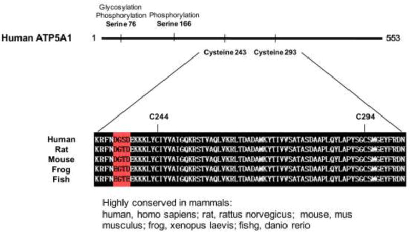
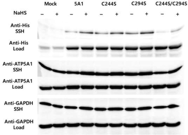
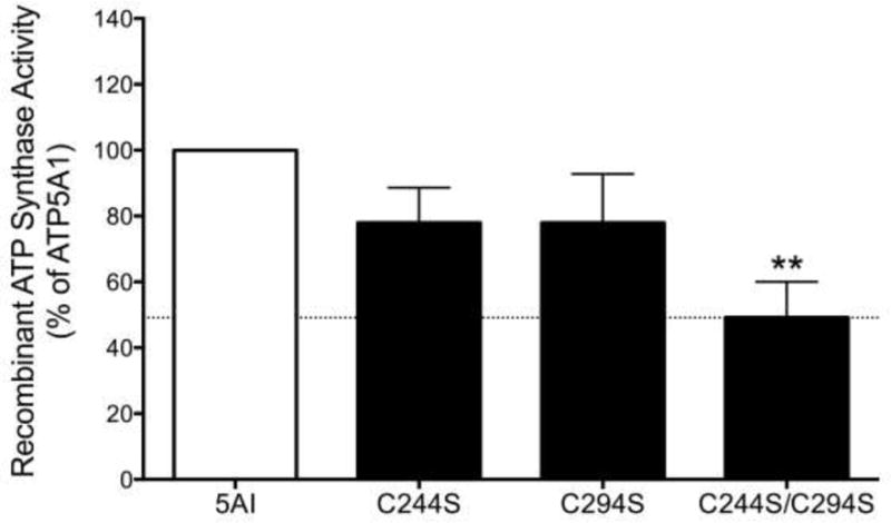
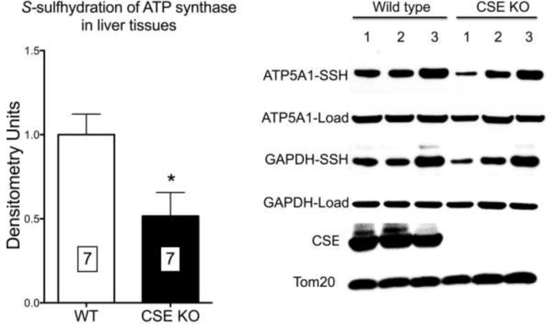
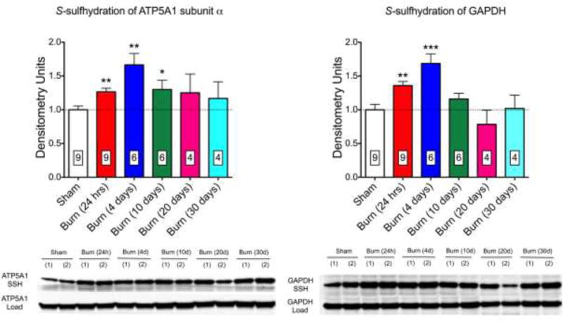
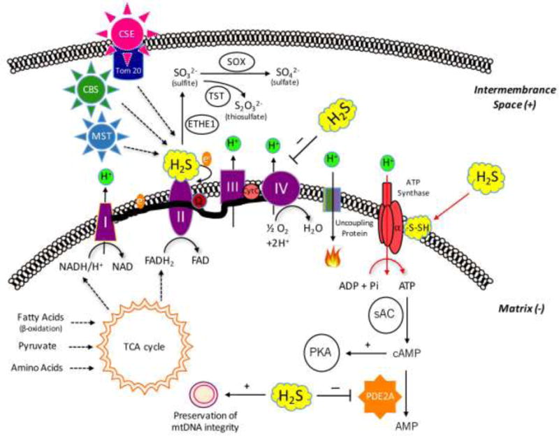
Mammalian cells can utilize hydrogen sulfide (H2S) to support mitochondrial respiration. The aim of our study was to explore the potential role of S-sulfhydration (a H2S-induced posttranslational modification, also known as S-persulfidation) of the mitochondrial inner membrane protein ATP synthase (F1F0 ATP synthase/Complex V) in the regulation of mitochondrial bioenergetics. Using a biotin switch assay, we have detected S-sulfhydration of the α subunit (ATP5A1) of ATP synthase in response to exposure to H2S in vitro. The H2S generator compound NaHS induced S-sulfhydration of ATP5A1 in HepG2 and HEK293 cell lysates in a concentration-dependent manner (50-300μM). The activity of immunocaptured mitochondrial ATP synthase enzyme isolated from HepG2 and HEK293 cells was stimulated by NaHS at low concentrations (10-100nM). Site-directed mutagenesis of ATP5A1 in HEK293 cells demonstrated that cysteine residues at positions 244 and 294 are subject to S-sulfhydration. The double mutant ATP synthase protein (C244S/C294S) showed a significantly reduced enzyme activity compared to control and the single-cysteine-mutated recombinant proteins (C244S or C294S). To determine whether endogenous H2S plays a role in the basal S-sulfhydration of ATP synthase in vivo, we compared liver tissues harvested from wild-type mice and mice deficient in cystathionine-gamma-lyase (CSE, one of the three principal mammalian H2S-producing enzymes). Significantly reduced S-sulfhydration of ATP5A1 was observed in liver homogenates of CSE-/- mice, compared to wild-type mice, suggesting a physiological role for CSE-derived endogenous H2S production in the S-sulfhydration of ATP synthase. Various forms of critical illness (including burn injury) upregulate H2S-producing enzymes and stimulate H2S biosynthesis. In liver tissues collected from mice subjected to burn injury, we detected an increased S-sulfhydration of ATP5A1 at the early time points post-burn. At later time points (when systemic H2S levels decrease) S-sulfhydration of ATP5A1 decreased as well. In conclusion, H2S induces S-sulfhydration of ATP5A1 at C244 and C294. This post-translational modification may be a physiological mechanism to maintain ATP synthase in a physiologically activated state, thereby supporting mitochondrial bioenergetics. The sulfhydration of ATP synthase may be a dynamic process, which may be regulated by endogenous H2S levels under various pathophysiological conditions.

Keywords: ATP synthase; Bioenergetics; Burn; Burn injury; Cysteine; H(2)S; Hydrogen sulfide; Mitochondria; S-Sulfhydration.


Conflict of interest statement

The authors declare no conflicts of interest in relationship to this study.

Figures

Figure 1
Figure 1
H2S induced S-sulfhydration of ATP5A1. HepG2 (A) and HEK 293 (B) cell lysates were incubated with NaHS at various concentrations (50–300 μM) for 30 min at 37°C. S-sulfhydration of ATP5A1 was detected by the biotin switch assay using anti-ATP5A1 antibody. Densitometry data show mean±SEM of n=5 independent experiments, *p<0.05 and **p<0.01 indicate significantly increased S-sulfhydration after NaHS, compared to the vehicle control (CTL) group.
Figure 2
Figure 2
NaHS stimulates ATP5A1 enzyme activity in HepG2 (A) and HEK293 (B) cell lysates. ATP synthase protein, immunocaptured from the cell homogenates, was incubated with NaHS for 30 min at 37°C, and enzyme activity was determined by a kinetic, spectrophotometric assay which measures the consumption of ATP and the consequent production of ADP (which is coupled to the oxidation of NADH to NAD+). The oligomycin-inhibitable component of the ATP consumption was considered specific ATP synthase activity. Data were expressed as percent values of control (CTL) the ATP synthase activity (immunocaptured ATP synthase treated with NaHS vehicle). Data show mean±SEM values from n=3 independent experiments. *p<0.05 and **p<0.01 indicate the stimulatory effect of NaHS on ATP synthase.
Figure 3
Figure 3
Domains of ATP5A1, indicating the sites of highly conserved cysteine residues across different species.
Figure 4
Figure 4
Western blot analysis of HEK-293 cells transfected either with empty vector, or native human ATP5A1 (5A1), or cysteine 244 mutant (C244S) human ATP5A1, or cysteine 294 mutant (C294S) human ATP5A1, or the double mutant C244S/C294S human ATP5A1 plasmids for 48 hrs. Cell lysates were incubated with NaHS (100 μM) for an additional 30 min at 37°C. The biotin switch assay using anti-tetraHis antibody was used to differentiate the recombinant ATP5A1 from the endogenous ATP5A1.
Figure 5
Figure 5
The double mutant recombinant protein (C244S/C294S) exhibits a significantly reduced enzyme activity compared to the intact human ATP5A1 protein. Mean±SEM values are shown from n=3 independent experiments. **p<0.01 indicates significant suppression of the catalytic activity of the double mutant protein, compared to wild-type protein.
Figure 6
Figure 6
ATP5A1 prepared from livers of CSE−/− mice exhibits reduced S-sulfhydration compared to ATP synthase prepared from livers of wild-type mice. Densitometry analysis represents mean±SEM values of n=7 wild-type or n=7 CSE−/− livers. *p<0.05 shows significant difference between the CSE−/− and the wild-type group.
Figure 7
Figure 7
S-sulfhydration of ATP5A1 (A) and GAPDH (B) is increased at early time points after burn injury in livers of mice. A representative western blot and densitometry analysis (mean±SEM) of n=4–9 liver tissues is shown. *p<0.05 and **p<0.01 represent significant increases in the sulfhydration of the target enzyme after burn injury compared to the sham group.
Figure 8
Figure 8
Summary of the various effects of H2S on mitochondrial bioenergetics. (a) The novel findings contained in the current report (S-sulfhydration of ATP synthase) are indicated with red arrows. H2S can be produced in the mitochondrion constitutively by two distinct H2S-producing enzymes, 3-MST, and CBS. Moreover, CSE is capable of translocating to the mitochondrial outer membrane under certain stress condition (e.g.: increased intracellular calcium signal), contributing the growth of intramitochondrial H2S level. Lower concentrations of H2S exert stimulatory effects on mitochondrial function via several different mechanisms: (b) H2S can donate electrons to mitochondrial electron transport chain. (c) H2S can act as an antioxidant neutralizing mitochondrial-derived reactive oxygen and nitrogen species, stabilizing the electron transport chain proteins, as well as preventing mitochondrial DNA damage. (d) H2S oxidization can results in sulfite, sulfate and thiosulfate; some of these species can also act as “pools” or sources of biologically active H2S. (e) H2S can inhibit mitochondrial PDE2A enzyme, which increases intramitochondrial cAMP levels, and stimulates mitochondrial function via activation of the cAMP-dependent protein kinase (PKA). Higher concentrations of H2S can also exert marked inhibitory effects on mitochondrial function: (f) H2S can inhibits cytochrome c oxidase (complex IV) resulting in a reversible inhibition of mitochondrial electron transport and ATP production.

Similar articles

Cited by

References

    1. Szabo C. Hydrogen sulphide and its therapeutic potential. Nat Rev Drug Discov. 2007;6:917–35. - PubMed
    1. Calvert JW, Coetzee WA, Lefer DJ. Novel insights into hydrogen sulfide–mediated cytoprotection. Antioxid Redox Signal. 2010;12:1203–17. - PMC - PubMed
    1. Zhao W, Zhang J, Lu Y, Wang R. The vasorelaxant effect of H2S as a novel endogenous gaseous KATP channel opener. EMBO J. 2001;20:6008–16. - PMC - PubMed
    1. Li L, Rose P, Moore PK. Hydrogen sulfide and cell signaling. Annu Rev Pharmacol Toxicol. 2011;51:169–87. - PubMed
    1. Mustafa AK, Sikka G, Gazi SK, Steppan J, Jung SM, Bhunia AK, Barodka VM, Gazi FK, Barrow RK, Wang R, Amzel LM, Berkowitz DE, Snyder SH. Hydrogen sulfide as endothelium-derived hyperpolarizing factor sulfhydrates potassium channels. Circ Res. 2011;109:1259–68. - PMC - PubMed

Publication types

MeSH terms