. 2016 Dec 5;55(49):15250-15253.
doi: 10.1002/anie.201606060. Epub 2016 Aug 24.

Paper-Origami-Based Multiplexed Malaria Diagnostics from Whole Blood

Affiliations

Paper-Origami-Based Multiplexed Malaria Diagnostics from Whole Blood

Gaolian Xu et al. Angew Chem Int Ed Engl. .

Abstract

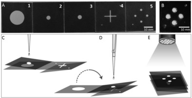
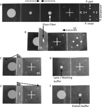
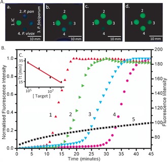
We demonstrate, for the first time, the multiplexed determination of microbial species from whole blood using the paper-folding technique of origami to enable the sequential steps of DNA extraction, loop-mediated isothermal amplification (LAMP), and array-based fluorescence detection. A low-cost handheld flashlight reveals the presence of the final DNA amplicon to the naked eye, providing a "sample-to-answer" diagnosis from a finger-prick volume of human blood, within 45 min, with minimal user intervention. To demonstrate the method, we showed the identification of three species of Plasmodium, analyzing 80 patient samples benchmarked against the gold-standard polymerase chain reaction (PCR) assay in an operator-blinded study. We also show that the test retains its diagnostic accuracy when using stored or fixed reference samples.

Keywords: diagnostics; malaria; microfluidics; nucleic acid based test; paper origami.


Figures

Figure 1
Figure 1
Paper‐based multiplexed LAMP detection of malaria in blood. A) Foldable paper devices: Dark areas are printed with hydrophobic wax. The device consists of five panels (1–5) folding onto each other, and a plastic cover for LAMP processing to avoid evaporation (B). The design also incorporates alignment marks on two corners (in the bottom left and top right corners) to assign the results. C) Illustrates the extraction process. Panels 2 and 3 are folded together and onto Panel 1. The sample is dispensed onto the device (panel 3) and extracted using capillary flows vertically (flow of liquid from Panel 3 to Panel 1). By folding the device (flipping the Panel 2–3 fold onto Panels 4 and 5), the sample is transferred to the LAMP spots (D) where the reaction is carried out. The signal is read out using a UV flashlight (365 nm; E).
Figure 2
Figure 2
Paper‐folding steps for fluidic manipulation of assay steps. The broken arrows indicate folding direction. Panels numbered as in Figure 1. A) A hole from the center of the third Panel has a glass‐fiber disc onto which the sample is dispensed; Numbers in the last panel indicate the different reagents placed onto the four different spots for amplification of different species. 1. Internal control (IC); 2. Plasmodium pan; 3. P. falciparum; 4. P. vivax. B) The second/third Panel are folded and clamped to form structure 1 (S1); C) The fifth Panel is folded onto the back of the fourth frame to form structure 2 (S2); D) S1 is folded onto the first Panel before adding lysis/washing buffer for DNA extraction and purification; E–F) S1 is folded onto S2 for elution and elution buffer added.
Figure 3
Figure 3
Results of multiplex LAMP amplification under UV light. Under UV excitation, green calcein emission occurs in the presence of pyrophosphate A) Numbers denote different species‐specific LAMP reaction. 1. Internal control (IC); 2. Plasmodium pan; 3. P. falciparum; 4. P. vivax. Letters denote the different positive results: a. 1 positive; b. 1 and 2 positive; c. 1, 2 and 3 positive; d. all positive. B) Real‐time amplification curve of Plasmodium pan LAMP with the 10‐fold serially diluted target (1–4, normalized real‐time amplification curves), and ddH2O as a negative control (5): 1. 108 IU/mL (red up‐triangle); 2. 107 IU/mL (green right‐triangle); 3. 106 IU/mL (cyan down‐triangle); 4. 105 IU/mL (magenta circle). 5. Negative control (black square: no target DNA). As the concentration increases, the amplification is initiated earlier, evidenced by the exponential increase in the fluorescence. C) Threshold time (defined as the time corresponding to 50 % of the maximum fluorescence intensity, Tt) as a function of target concentration. This Figure of merit is analogous to the cycle threshold (Ct) of real‐time PCR.23 Data is the average of three repeats and error bars represent the standard deviation. The data was fitted with linear regression (R 2=0.98).

References

    1. Kettelhut M. M., Chiodini P. L., Edwards H., Moody A., J. Clin. Pathol. 2003, 56, 927–932. - PMC - PubMed
    1. Lindblade K. A., Steinhardt L., Samuels A., Kachur S. P., Slutsker L., Expert Rev. Anti-Infect. Ther. 2013, 11, 623–639. - PubMed
    1. Snounou G., Viriyakosol S., Zhu X. P., Jarra W., Pinheiro L., do Rosario V. E., Thaithong S., Brown K. N., Mol. Biochem. Parasitol. 1993, 61, 315–320. - PubMed
    1. Rougemont M., Saanen M. V., Sahli R., Hinrikson H. P., Bille J., Jaton K., J. Clin. Microbiol. 2004, 42, 5636–5643. - PMC - PubMed
    1. Reyburn H., Mbatia R., Drakeley C., Carneiro I., Mwakasungula E., Mwerinde O., Saganda K., Shao J., Kitua A., Olomi R., et al., BMJ 2004, 329, 1212–1215. - PMC - PubMed

Publication types