. 2016 Oct 14;291(42):21963-21970.
doi: 10.1074/jbc.M116.737601. Epub 2016 Aug 23.

Substrate Affinity Differentially Influences Protein Kinase C Regulation and Inhibitor Potency

Affiliations

Substrate Affinity Differentially Influences Protein Kinase C Regulation and Inhibitor Potency

Ruth F Sommese et al. J Biol Chem. .

Abstract

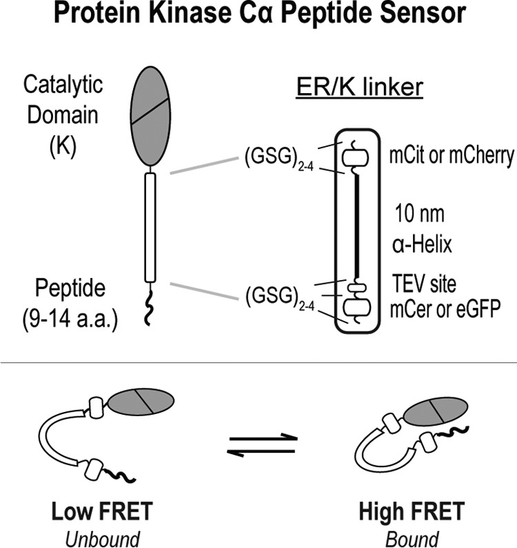
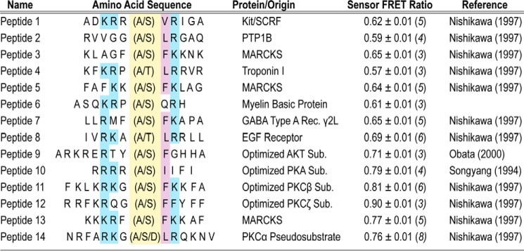
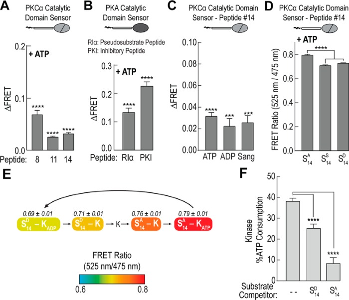
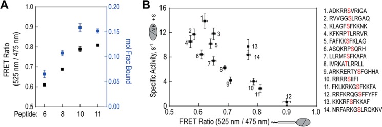
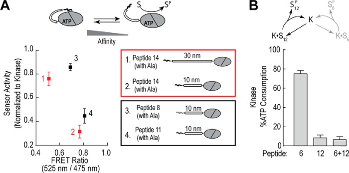
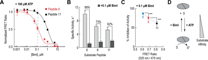
The overlapping network of kinase-substrate interactions provides exquisite specificity in cell signaling pathways, but also presents challenges to our ability to understand the mechanistic basis of biological processes. Efforts to dissect kinase-substrate interactions have been particularly limited by their inherently transient nature. Here, we use a library of FRET sensors to monitor these transient complexes, specifically examining weak interactions between the catalytic domain of protein kinase Cα and 14 substrate peptides. Combining results from this assay platform with those from standard kinase activity assays yields four novel insights into the kinase-substrate interaction. First, preferential binding of non-phosphorylated versus phosphorylated substrates leads to enhanced kinase-specific activity. Second, kinase-specific activity is inversely correlated with substrate binding affinity. Third, high affinity substrates can suppress phosphorylation of their low affinity counterparts. Finally, the substrate-competitive inhibitor bisindolylmaleimide I displaces low affinity substrates more potently leading to substrate selective inhibition of kinase activity. Overall, our approach complements existing structural and biophysical approaches to provide generalizable insights into the regulation of kinase activity.

Keywords: fluorescence resonance energy transfer (FRET); inhibitor; phosphorylation; protein kinase C (PKC); substrate specificity.


Figures

FIGURE 1.
FIGURE 1.
Schematic of PKCα peptide FRET sensors. PKCα peptide sensors are based around an ER/K FRET linker (14, 15), which separates the catalytic domain of PKCα and a variety of short peptides ≤14 residues in length (Fig. 2). In the majority of the experiments, the fluorophores used in the ER/K linker are monomeric Cerulean (mCer, FRET donor) and monomeric Citrine (mCit, FRET acceptor). For the time-resolved measurements in Fig. 4A, monomeric eGFP and monomeric Cherry (mCherry) were used instead.
FIGURE 2.
FIGURE 2.
PKCα FRET sensors display a range of basal FRET interactions. Peptides used in activity assays (Ser- or Thr-containing) or FRET-based sensors (Ala- or Asp-containing) are shown. In the amino acid sequence, the phosphorylation site is highlighted in yellow. Blue (hydrophobic) or purple (basic) shading indicates consensus site residues important for phosphorylation (18). The basal FRET ratio is shown for each alanine-containing sensor and are derived from at least three protein preparations (n ≥ 3 preparations).
FIGURE 3.
FIGURE 3.
Loss of affinity for phosphorylated substrate enhances kinase specific activity. A–C, increase in the FRET ratio (ΔFRET) upon addition of 100 μm ATP, 100 μm ADP, or 100 μm sangivamycin (Sang) for sensors containing short alanine-substituted substrate peptides and either (A and C) the catalytic domain of PKCα or (B) the catalytic domain of PKA. For PKC peptide sequence information see Fig. 2. D, FRET ratios for PKCα peptide number 14 sensors containing an alanine (S14A), serine (S14S), or aspartic acid (S14D, phosphomimetic) with 100 μm ATP. E, schematic of a subset of steps in the catalytic cycle of PKCα. Coloring denotes steady-state FRET ratios of PKCα peptide number 14 sensors containing either an aspartic acid (S14D) or an alanine residue (S14A) with 100 μm ATP or 100 μm ADP. States are arranged based on progression of FRET through the catalytic cycle and do not indicate the relative prevalence of all possible states. F, ATP consumption of the catalytic domain of PKCα for serine-containing peptide number 14 upon addition of equal concentrations of either aspartic acid-substituted (S14D) or alanine-substituted (S14A) peptide. For all FRET and activity readings, data are derived from at least three independent protein preparations with at least two replicated measurements for each condition per experiment (mean ± S.E., n ≥ 3 experiments).
FIGURE 4.
FIGURE 4.
Inverse correlation between substrate affinity and kinase activity. A, comparison between steady-state FRET and fractional binding for four alanine-containing peptide sensors. For fractional binding, fluorescence decay single photon counting data were fit to I(t) = A1et/τ1 + A2et/τ2, where τ1 was set to the lifetime of a 10-nm ER/K linker control (see Table 1). Molecular fraction of the bound state(s) was calculated as A2/(A1 + A2). B, steady-state FRET of PKCα catalytic domain sensors with 14 short peptides (Fig. 2) compared with activity of the catalytic domain with corresponding substrate peptides. Activity assays were performed at 21–22 °C with equimolar peptide concentrations (500 μm) and kinase (50 nm). Specific activity is reported as mole of ATP consumed per mol of kinase per s (s−1). In all experiments, data are derived from at least three protein preparations (mean ± S.E., n ≥ 3 experiments).
FIGURE 5.
FIGURE 5.
High affinity substrates suppress phosphorylation of low affinity counterparts. A, relative activity of sensors containing either the alanine-substituted peptide number 14 with different linker distances (10 versus 30 nm; 1 versus 2; red) or containing different alanine-substituted peptides (peptide number 8 versus number 11; 3 versus 4; black) with the same 10-nm linker. Activity data are plotted versus the steady-state FRET ratio of the peptide sensors. B, activity of the isolated catalytic domain for peptide number 6, peptide number 12, and equimolar concentrations of peptide numbers 6 and 12. For both FRET and activity experiments, data are derived from at least three protein preparations with at least two replicates for each condition per independent experiment (mean ± S.E., n ≥ 3 experiments).
FIGURE 6.
FIGURE 6.
Kinase-substrate binding affinity impacts inhibitor potency. A, titration of peptide numbers 8 and 11 FRET sensors in the presence of 100 μm ATP by the substrate competitive inhibitor BimI. The FRET ratio for each sensor was normalized to the FRET ratio of that sensor in the absence of any inhibitor. B, inhibition of PKCα catalytic domain activity by 0.1 μm BimI for three different substrate peptides (order of peptide affinity: 11 > 8 > 6). Activity assays were performed with equimolar peptide concentrations (500 μm) and kinase (50 nm) and specific activity is reported as mole of ATP consumed per mol of kinase per s (s−1). C, percent inhibition of PKCα catalytic domain activity by 0.1 μm BimI for substrate peptides in B. Percent inhibition is graphed versus the steady-state FRET ratio for the corresponding alanine-substituted FRET sensors. For all FRET and activity readings, data are derived from at least three protein preparations with at least two replicates per independent experiment (mean ± S.E., n ≥ 3 experiments). D, for PKCα, varying substrate affinity reduces the potency of substrate-competitive inhibitors like BimI.

References

    1. Hornbeck P. V., Zhang B., Murray B., Kornhauser J. M., Latham V., and Skrzypek E. (2015) PhosphoSitePlus, 2014: mutations, PTMs and recalibrations. Nucleic Acids Res. 43, D512–520 - PMC - PubMed
    1. Pearce L. R., Komander D., and Alessi D. R. (2010) The nuts and bolts of AGC protein kinases. Nat. Rev. Mol. Cell. Biol. 11, 9–22 - PubMed
    1. Manning G., Whyte D. B., Martinez R., Hunter T., and Sudarsanam S. (2002) The protein kinase complement of the human genome. Science 298, 1912–1934 - PubMed
    1. de Oliveira P. S., Ferraz F. A., Pena D. A., Pramio D. T., Morais F. A., and Schechtman D. (2016) Revisiting protein kinase-substrate interactions: toward therapeutic development. Sci. Signal. 9, re3. - PubMed
    1. Hanks S. K., and Hunter T. (1995) Protein kinases 6. The eukaryotic protein kinase superfamily: kinase (catalytic) domain structure and classification. FASEB J. 9, 576–596 - PubMed

MeSH terms