. 2015 Jul 29;4(3):59-68.
doi: 10.1016/j.jbo.2015.06.002. eCollection 2015 Sep.

RANK and RANK ligand expression in primary human osteosarcoma

Affiliations

RANK and RANK ligand expression in primary human osteosarcoma

Daniel Branstetter et al. J Bone Oncol. .

Abstract

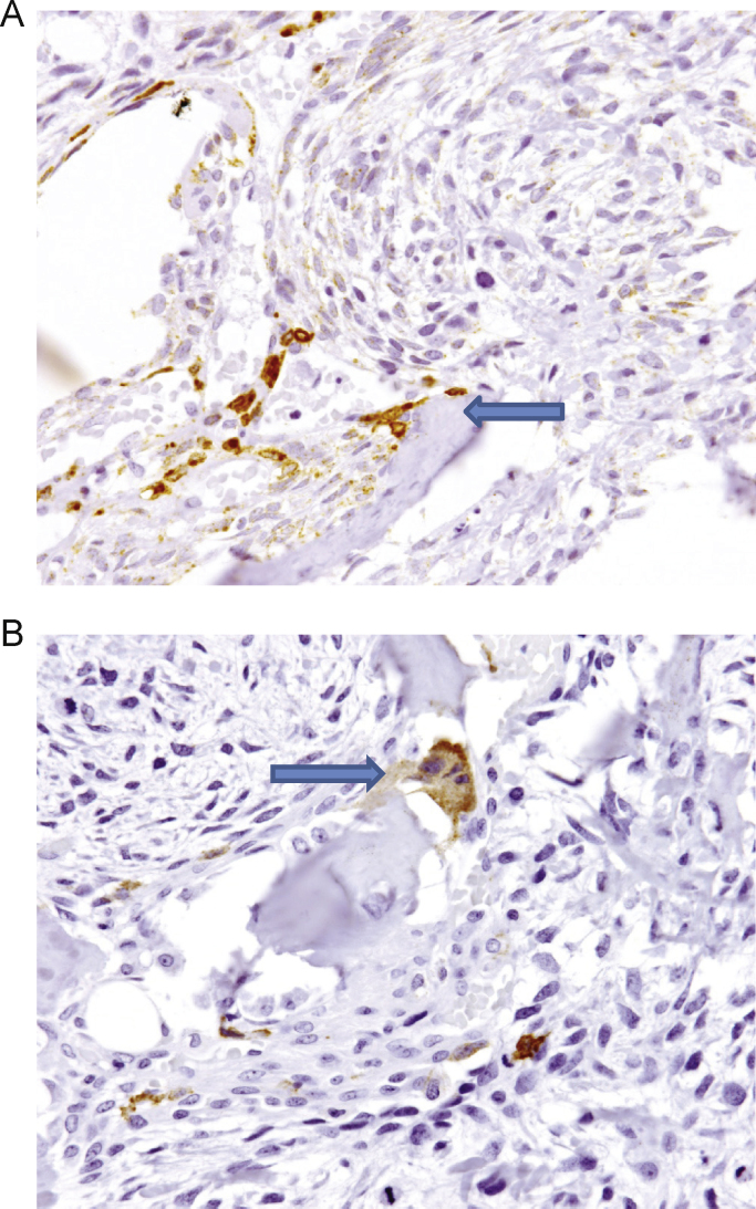
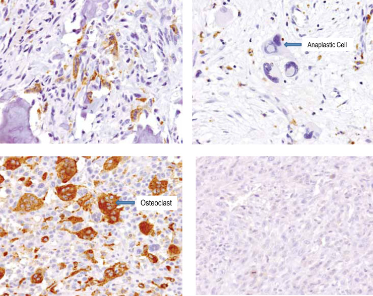
Receptor activator of nuclear factor kappa-B ligand (RANKL) is an essential mediator of osteoclast formation, function and survival. In patients with solid tumor metastasis to the bone, targeting the bone microenvironment by inhibition of RANKL using denosumab, a fully human monoclonal antibody (mAb) specific to RANKL, has been demonstrated to prevent tumor-induced osteolysis and subsequent skeletal complications. Recently, a prominent functional role for the RANKL pathway has emerged in the primary bone tumor giant cell tumor of bone (GCTB). Expression of both RANKL and RANK is extremely high in GCTB tumors and denosumab treatment was associated with tumor regression and reduced tumor-associated bone lysis in GCTB patients. In order to address the potential role of the RANKL pathway in another primary bone tumor, this study assessed human RANKL and RANK expression in human primary osteosarcoma (OS) using specific mAbs, validated and optimized for immunohistochemistry (IHC) or flow cytometry. Our results demonstrate RANKL expression was observed in the tumor element in 68% of human OS using IHC. However, the staining intensity was relatively low and only 37% (29/79) of samples exhibited≥10% RANKL positive tumor cells. RANK expression was not observed in OS tumor cells. In contrast, RANK expression was clearly observed in other cells within OS samples, including the myeloid osteoclast precursor compartment, osteoclasts and in giant osteoclast cells. The intensity and frequency of RANKL and RANK staining in OS samples were substantially less than that observed in GCTB samples. The observation that RANKL is expressed in OS cells themselves suggests that these tumors may mediate an osteoclastic response, and anti-RANKL therapy may potentially be protective against bone pathologies in OS. However, the absence of RANK expression in primary human OS cells suggests that any autocrine RANKL/RANK signaling in human OS tumor cells is not operative, and anti-RANKL therapy would not directly affect the tumor.

Keywords: APC, allophycocyanin; ATCC, American Type Culture Collection; Antibodies; ELISA, enzyme linked immunosorbent assay; FACS, fluorescence-activated cell sorting; FBS, fetal bovine serum; FFPE, formalin-fixed, paraffin-embedded; GCTB, giant cell tumor of bone; Human osteosarcoma; IHC, immunohistochemistry; ISH, in situ hybridization; IgG1, immunoglobulin G1; Immunohistochemistry; LN, lymph node; OS, osteosarcoma; Protein expression; RANK; RANK, receptor activator of nuclear factor kappa-B; RANKL; RANKL, receptor activator of nuclear factor kappa-B ligand; RNA, ribonucleic acid; RT-PCR, reverse transcriptase polymerase chain reaction; cDNA, complementary deoxyribonucleic acid; mAb, monoclonal antibody; mRNA, messenger ribonucleic acid.


Figures

Fig. 1
Fig. 1
huRANKL antibody validation for IHC methods. (A) Anti-huRANKL mAb M366 IHC reveals specific signal in mouse L929 cells transduced with huRANKL cDNA (L929 huRANKL), but not parental L929 cells (L929 control). (B) Similarly, analysis of dissociated proteins on western blot using mAb 366 of L929 huRANKL cells detected a protein of approximately 45 kDa, the predicted size for full-length human RANKL . Positions of molecular weight markers are illustrated on left (kDa). (C) Determination of RANKL cell surface protein expression on L929 cells was performed using flow cytometry. Expression of RANKL was detected using the M366 mAb and a goat antimouse secondary antibody conjugated to APC. M366 staining on huRANKL transduced L929 cells is indicated with the red line and on parental L929 cells (L929 control) a solid black line. The M366 anti-huRANKL antibody detects a signal by IHC, western blot, and flow cytometry specifically in L929 cells transduced with huRANKL and not parental L929 cells. APC, allophycocyanin; cDNA, complementary deoxyribonucleic acid; IHC, immunohistochemistry; mAb, monoclonal antibody; RANKL, receptor activator of nuclear factor kappa-B ligand.
Fig. 2
Fig. 2
RANKL and RANK IHC of GCTB. IHC performed on FFPE section of GCTB, which serves as an internal positive and negative control since RANKL and RANK are expressed in distinct compartments . GCTB is composed of osteoclast-like giant cells and myeloid giant cell precursors that express RANK and stromal tumor cells that express RANKL. (A) The RANKL IHC using M366 clearly indicates stromal tumor cells but is excluded from the giant cell component. (B) Conversely, the RANK IHC using N-2B10 recognizes giant cells and myeloid osteoclast precursors but is excluded from the stromal compartment. FFPE, formalin-fixed, paraffin-embedded; GCTB, giant cell tumor of bone; IHC, immunohistochemistry; RANK, receptor activator of nuclear factor kappa-B; RANKL, RANK ligand.
Fig. 3
Fig. 3
huRANK antibody validation for IHC methods. (A) RANK IHC was performed on FFPE samples of xenografts of the specified human cancer cells. Parental cells did not detectably express RANK as confirmed by multiple independent methods. There was no detectable signal with the isotype control antibody. Both antibodies against huRANK (N-1H8 and N-2B10) provide a similar staining pattern, essentially cross-validating one another. (B) The same cells used for the above tumor xenografts were grown in vitro and processed for flow cytometry. For anti-RANK staining, the pattern of staining by both test antibodies N-1H8 and N-2B10 was compared with a previously identified mAb (M331) useful for flow cytometry applications (Armstrong [31]). Solid grey line: Unstained; Red line: Secondary control=Goat antimouse APC; Blue line: Isotype control 4D2 (anti-AGP3 muIgG1), 1 μg/mL; Purple line: M331 (anti-huRANK muIgG1), 1 μg/mL; Green line: N-1H8 (anti-huRANK muIgG1), 1 μg/mL; Black line: N-2B10 (anti-huRANK muIgG1), 1 μg/mL. The RANK signal by IHC is concordant with the signal by flow cytometry. APC, allophycocyanin; FFPE, formalin-fixed, paraffin-embedded; IHC, immunohistochemistry; mAb, monoclonal antibody; RANK, receptor activator of nuclear factor kappa-B.
Fig. 4
Fig. 4
RANKL and RANK H-score distribution in OS. The H score incorporates intensity (scale of 0–3) and percentage of tumor cells stained positive, giving a range of 0–300. RANK IHC was performed using N-1H8 mAb on the small core TMA samples and confirmed using N-2B10 mAb on the larger OS tumor sections. RANKL IHC was performed using M366 mAb on both the TMA and large tissue sections. The H-score distribution for both analytes are depicted for both (A) the small TMA core samples and (B) the larger tissue sections. IHC, immunohistochemistry; OS, osteosarcoma; RANK, receptor activator of nuclear factor kappa-B; RANKL, RANK ligand.
Fig. 5
Fig. 5
Range of RANKL protein expression in OS. RANKL IHC performed with mAb M366. Representative IHC images from four separate OS tumor samples are shown. Arrows indicate RANKL-positive tumor cells and tumor anaplastic cells. In addition, RANKL-negative osteoclasts are also clearly delineated (arrow). The four images demonstrate a range of RANKL positivity, as indicated by the different H-scores. The H-score for each image was as follows: upper left panel, H-score=260; upper right panel, H-score=210; lower left panel, H-score=180; lower right panel, H-score=20. IHC, immunohistochemistry; OS, osteosarcoma; RANK, receptor activator of nuclear factor kappa-B ligand.
Fig. 6
Fig. 6
RANKL and RANK expression within adjacent bone tissue. RANKL and RANK IHC performed with mAb N-1H8 and M366, respectively. (A) RANKL-expressing stroma at bone interface. (B) RANK expression in bone osteoclast. IHC, immunohistochemistry; mAb, monoclonal antibody; RANK, receptor activator of nuclear factor kappa-B; RANKL, RANK ligand.
Fig. 7
Fig. 7
RANK protein expression in OS is limited to myeloid osteoclasts and osteoclast precursors. RANK IHC performed with mAb N-1H8. Representative IHC images from four separate OS tumor samples are shown. Upper left panel shows RANK-positive normal osteoclasts at the tumor–bone interface. Upper right panel shows tumor anaplastic cells were RANK-negative. Lower left panel shows abundant RANK-positive tumor-associated osteoclasts were present within the tumor in some tumors. Lower right panel shows no RANK-positive cells were present in some tumors. The H-score was zero for all images for RANK. RANK expression was observed in osteoclasts and myeloid osteoclast precursors in a majority of samples but not in the sarcoma component. IHC, immunohistochemistry; mAb, monoclonal antibody; OS, osteosarcoma; RANK, receptor activator of nuclear factor kappa-B.

References

    1. Klein M.J., Siegal G.P. Osteosarcoma: anatomic and histologic variants. Am J Clin Pathol. 2006;125:555–581. - PubMed
    1. Mutsaers A.J., Walkley C.R. Cells of origin in osteosarcoma: mesenchymal stem cells or osteoblast committed cells? Bone. 2014;62:56–63. - PubMed
    1. Botter S.M., Neri D., Fuchs B. Recent advances in osteosarcoma. Curr Opin Pharmacol. 2014;16:15–23. - PubMed
    1. Lacey D.L., Boyle W.J., Simonet W.S., Kostenuik P.J., Dougall W.C., Sullivan J.K. Bench to bedside: elucidation of the OPG-RANK-RANKL pathway and the development of denosumab. Nat Rev Drug Discov. 2012;11:401–419. - PubMed
    1. Stopeck A.T., Lipton A., Body J.J., Steger G.G., Tonkin K., de Boer R.H. Denosumab compared with zoledronic acid for the treatment of bone metastases in patients with advanced breast cancer: a randomized, double-blind study. J Clin Oncol. 2010;28:5132–5139. - PubMed