. 2016 Sep 20;7(38):61458-61468.
doi: 10.18632/oncotarget.11369.

Ets2 knockdown inhibits tumorigenesis in esophageal squamous cell carcinoma in vivo and in vitro

Affiliations

Ets2 knockdown inhibits tumorigenesis in esophageal squamous cell carcinoma in vivo and in vitro

Qinghua Li et al. Oncotarget. .

Abstract

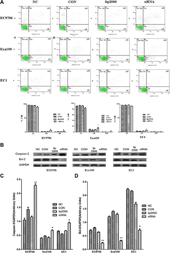
Increased expression of Ets2 is reported upregulated in esophageal squamous cell carcinoma tissue. However, the function of Ets2 in carcinogenesis of ESCC is poorly understood. Here, the rise of Ets2 was confirmed in ESCC cells and Ets2 depletion by RNA interference promotes cell apoptosis, inhibits cell proliferation, attenuates cell invasion and induces cell cycle G0/G1 arrest in vitro. Moreover, in vivo, Xenograft mouse model studies showed Ets2 knockdown inhibits tumor formation and metastasis significantly. Furthermore, Ets2 depletion inactivates the mTOR/p70S6K signaling pathway both in vitro and in vivo. Taken together, these findings strongly suggest that a critical role of Ets2 in human ESCC pathogenesis via the inactivation of the mTOR/p70S6K signaling pathway.

Keywords: Ets2; apoptosis; esophageal squamous cell carcinoma; mTOR/p70S6K signaling pathway; proliferation.


Conflict of interest statement

These authors declare that there is no conflicts of interests concerning this paper.

Figures

Figure 1
Figure 1. Expression of Ets2 protein was notably increased in ESCC cells and knocked down by siRNA
(A) Western Blot analysis of Ets2 in Het-1A, EC1, EC9706 and Eca109 cells. (B) Semi-quantitative analysis showed that Ets2 was increased by 3.2-fold in EC9706 cells (P < 0.01), 2.2-fold in Eca109 cells (P < 0.05), and 1.93-fold in EC1 cells (P < 0.05) respectively compared with that in Het-1A cells. *P < 0.05, **P < 0.01 versus that in Het-1A cells. (C) and (D) Western blotting analysis of the expression of Ets2 in EC9706 cells at 48 h and 72 h after transfection with 3 candidate siRNA sequences (marked as siRNA1, siRNA2, siRNA3). (E) Quantitative results of the Western blotting analysis obtained via densitometric analysis. Ets2 protein expression was obviously inhibited by interference only with siRNA1 and siRNA2 (P < 0.01) sequences at 48 h after transfection and the interference efficiency of siRNA1 sequence was higher than that of siRNA2 (P < 0.05). *P < 0.05, **P < 0.01 versus the NC groups. CON, ESCC cells were cultured normally; lip2000, ESCC cells were transfected with equal amounts of Lipofectamine® 2000 Reagent; NC, ESCC cell were transfected with non-targeting control siRNA as negative control.
Figure 2
Figure 2. Proliferative rates of ESCC cells when Ets2 was knocked down
(A) the numbers of proliferative rates of siEts2-EC9706, siEts2-Eca109 and siEts2-EC1 cells were 19.97%, 29.2% and 22.3% respectively, reducing by 40%, 45% and 57% compared with NC cells measured with EdU assay. (B) the cell survival rates of EC9706, Eca109 and EC1 cells were 69%, 58% and 68% after transfection with siEts2, reducing by 31%, 42% and 32% compared with NC (Figure 3B), respectively. *P < 0.05.
Figure 3
Figure 3. Ets2 knockdown induced apoptosis of ESCC cells in vitro
(A) apoptosis of ESCC cells was detected using annexin V-FITC and PI dual staining. Quadrants were designed as follows, LL: non-stained cells indicating viable cells; LR: annexin V-FITC stained cells indicating early apoptosis; UR: annexin V-FITC and PI stained cells indicating late apoptosis; and UL: PI stained cells indicating secondary necrosis. All dot plots are a representation of equal cell populations (n = 10,000). (B) apoptosis factor proteins caspase-3 and Bcl-2 were analyzed by Western blotting. (C) and (D) semi-quantitative analysis of caspase-3 and Bcl-2 expression. Caspase-3 was increased by 120%, 75% and 30% roughly in EC9706, Eca109 and EC1 cells compared with NC, while anti-apoptotic Bcl-2 protein was decreased by 70%, 73% and 68% in EC9706, Eca109 and EC1 cells compared with NC. *P < 0.05, **P < 0.01.
Figure 4
Figure 4. Effects of Ets2 knockdown on cell cycle
The number of siEts2-EC9706 cells at each cell cycle phase had no change compared with that of NC cells. The numbers of siEts2-Eca109 and siEts2-EC1 cells at G0/G1 phase were more than that of CON cells (P < 0.05). *P < 0.05.
Figure 5
Figure 5. Inhibitory effects of Ets2 depletion on the invasive capacity of ESCC cells
(A) The results of Transwell chamber assay. The numbers of invaded siEts2-EC9706, siEts2-Eca109 and siEts2-EC1 cells were significantly reduced compared with that of invaded NC cells. (B) E-cadherin expression was detected using Western blotting and the results revealed that E-cadherin protein was significantly increased by 54%, 130% and 67% respectively compared with that in NC. *P < 0.05, **P < 0.01.
Figure 6
Figure 6. Effect of Ets2 knockdown on mTOR/p70S6K signaling pathway
When Ets2 was knocked down, the expression of Prdx1 protein was reduced concomitantly with the reduction of Ets2 in EC9706, Eca109 and EC1 cells (P < 0.05). Both p-mTOR and p-p70S6K proteins were decreased in siEts2-EC9706, siEts2-Eca109 and siEts2-EC1 cells compared with that in NC groups (P < 0.05). *P < 0.05, **P < 0.01.
Figure 7
Figure 7. Effects of Ets2 silence on tumor size and protein expressions in vivo
(A) the tumor growth curve of xenograft mice. The tumor volume in LV-shEts2-Eca109 cell injected into nude mice was significantly smaller than that in LV-Eca109 and Eca109 cell-bearing mice (P < 0.001). (B) the tumor weight in xenograft mice. The average tumor weight in LV-shEts2-Eca109 cell-bearing mice was much lighter than that in LV-Eca109 and Eca109 cell-bearing mice (P < 0.05). (C) and (D), the protein expressions were analyzed by Western blotting. Protein caspase-3 and E-cadherin were significantly enhanced in LV-shEts2-Eca109 injected mice compared with that in Eca109 and LV-Eca109 injected mice (P < 0.001), while the proteins of Bcl-2, p-mTOR, p-p70S6K and Prdx1 were significantly reduced as the reduction of Ets2 in xenograft mice (P < 0.001). *P < 0.05, **P < 0.01.
Figure 8
Figure 8. Ets2 silence promotes ESCC cells apoptosis in vivo
Tunel assay demonstrated a 17.4% apoptotic rate in Ets2 silence tissue.

References

    1. Hou G, Zhang Q, Wang L, Liu M, Wang J, Xue L. mTOR inhibitor rapamycin alone or combined with cisplatin inhibits growth of esophageal squamous cell carcinoma in nude mice. Cancer Lett. 2010;290:248–54. doi: 10.1016/j.canlet.2009.09.015. - DOI - PubMed
    1. Kim T, Grobmyer SR, Smith R, Ben-David K, Ang D, Vogel SB, Hochwald SN. Esophageal cancer-the five year survivors. J Surg Oncol. 2011;103:179–83. doi: 10.1002/jso.21784.. - DOI - PubMed
    1. Seth A, Watson DK. ETS transcription factors and their emerging roles in human cancer. Eur J Cancer. 2005;41:2462–78. - PubMed
    1. Sharrocks AD. The ETS-domain transcription factor family. Nat Rev Mol Cell Biol. 2001;2:827–37. - PubMed
    1. Kasten M, Giordano A. Cdk10, a Cdc2-related kinase, associates with the Ets2 transcription factor and modulates its transactivation activity. Oncogene. 2001;20:1832–38. - PubMed

MeSH terms

Substances