

Effects of low-level laser therapy on stem cells from human exfoliated deciduous teeth
- PMID: 27556203
- PMCID: PMC4990361
- DOI: 10.1590/1678-775720150275
Effects of low-level laser therapy on stem cells from human exfoliated deciduous teeth
Abstract
Objective: This study aimed to evaluate the influence of different laser therapy energy densities on SHED viability and proliferation.
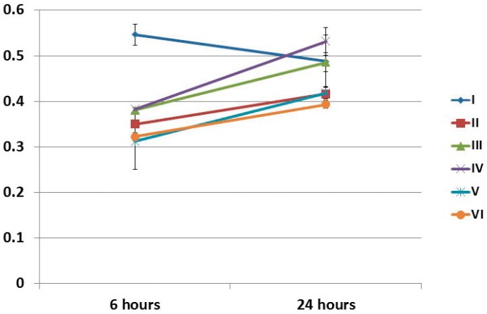
Material and methods: SHED were irradiated according to the groups: I (1.2 J/cm2 - 0.5 mW - 10 s), II (2.5 J/cm2 - 10 mW - 10 s), III (3.7 J/cm2 - 15 mW - 10 s), IV (5.0 J/cm2 - 20 mW - 10 s), V (6.2 J/cm2 - 25 mW - 10 s), and VI (not irradiated - control group). Cell viability was assessed 6 and 24 h after irradiation measuring the mitochondrial activity and using the Crystal Violet assay. Cell proliferation was assessed after 24, 48, and 72 h of irradiation by SRB assay.
Results: MTT assay demonstrated differences from 6 to 24 hours after irradiation. After 24 h, groups I and IV showed higher absorbance values than those of control group. Crystal Violet assay showed statistically differences in the absorbance rate from 6 to 24 h after irradiation for groups III and VI. At 24 h after irradiation, Group III absorbance rate was greater than that of groups I, II, and IV. Group VI absorbance rate was greater than that of groups I and IV. SRB assay showed that the group I had higher rates than those of groups II, III, V, and VI, at 24 h after irradiation. After 48 h, group I exhibited the greatest cell proliferation rate followed by groups III, V, and VI. After 72 h, group III exhibited the lowest cell proliferation rate than those of groups II, IV, and V.
Conclusions: The Low-Level Laser Therapy energy densities used in this study did not cause loss of cell viability and stimulated SHED proliferation within the parameters described in this study.
Figures


References
-
- AlGhamdi KM, Kumar A, Moussa NA. Low-level laser therapy: a useful technique for enhancing the proliferation of various cultured cells. Lasers Med Sci. 2012;27(1):237–249. - PubMed
-
- Almeida-Lopes L, Rigau J, Zângaro RA, Guidugli J, Neto, Jaeger MM. Comparison of the low level laser therapy effects on cultured human gingival fibroblasts proliferation using different irradiance and same fluence. Lasers Surg Med. 2001;29:179–184. - PubMed
-
- Azevedo LH, Paula Eduardo F, Moreira MS, Paula Eduardo C, Marques MM. Influence of different power densities of LILT on cultured human fibroblast growth: a pilot study. Lasers Med Sci. 2006;21:86–89. - PubMed
Publication types
MeSH terms
Substances
LinkOut - more resources
Full Text Sources
Other Literature Sources
Medical
