. 2016 Aug 22;17(8):1375.
doi: 10.3390/ijms17081375.

Development of a Patient-Derived Xenograft (PDX) of Breast Cancer Bone Metastasis in a Zebrafish Model

Affiliations

Development of a Patient-Derived Xenograft (PDX) of Breast Cancer Bone Metastasis in a Zebrafish Model

Laura Mercatali et al. Int J Mol Sci. .

Abstract

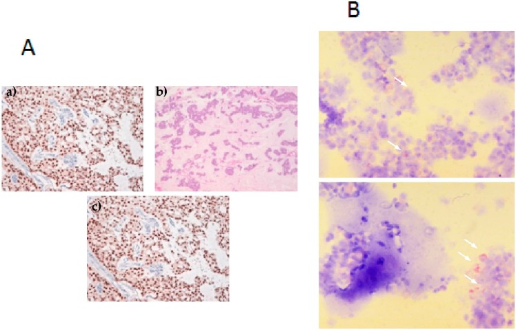
Bone metastasis is a complex process that needs to be better understood in order to help clinicians prevent and treat it. Xenografts using patient-derived material (PDX) rather than cancer cell lines are a novel approach that guarantees more clinically realistic results. A primary culture of bone metastasis derived from a 67-year-old patient with breast cancer was cultured and then injected into zebrafish (ZF) embryos to study its metastatic potential. In vivo behavior and results of gene expression analyses of the primary culture were compared with those of cancer cell lines with different metastatic potential (MCF7 and MDA-MB-231). The MCF7 cell line, which has the same hormonal receptor status as the bone metastasis primary culture, did not survive in the in vivo model. Conversely, MDA-MB-231 disseminated and colonized different parts of the ZF, including caudal hematopoietic tissues (CHT), revealing a migratory phenotype. Primary culture cells disseminated and in later stages extravasated from the vessels, engrafting into ZF tissues and reaching the CHT. Primary cell behavior reflected the clinical course of the patient's medical history. Our results underline the potential for using PDX models in bone metastasis research and outline new methods for the clinical application of this in vivo model.

Keywords: bone metastasis; breast cancer; patient-derived xenograft; zebrafish model.


Figures

Figure 1
Figure 1
Breast cancer bone metastasis. (A): (a) Histological hematoxylin and eosin (H & E) staining of bone metastasis (BM) primary culture (5× magnification). Mucinous areas show low cellularity; (b) H & E staining of BM primary culture (20× magnification). Monomorphic cells with round nucleus and nucleolus are seen in nests with a minor mucus quantity; (c) ER staining showing positivity; (B): Cytospin of BM primary cells. White arrows show pancytokeratin-positive cells.
Figure 2
Figure 2
Representative image of MDA-MB-231 cell line five days after injection into the duct of Couvier of 2 day post fertilization (dpf), Tg(fli1:GFP) ZF embryo. MDA-MB-231, labeled in red, were monitored on a daily basis for the duration of the experiment (five days) and showed progressive and extensive dissemination throughout the developing embryo (25× magnification).
Figure 3
Figure 3
Images of carboxyfluorescein succinimidyl ester (CFSE)-labeled, patient-derived breast cancer bone metastasis (primary cells) xenografted in Tg(kdrl:mcherry) ZF embryos. (A) Whole-body image of the zebrafish embryo at 5 dpi (25× magnification). Primary cells disseminated predominantly in the caudal hematopoietic tissues (CHT) of the embryo; (B) Details of (A), showing the interactions of primary cells with the zebrafish vessels in the CHT. Cells extravasated in the CHT of the ZF embryo and engrafted in close proximity of the vessels. Fluorescent images merged with brightfield image, 63× magnification; (C) Combined picture of the CHT of embryo in (A,B). Individual images were taken at 63× magnification.
Figure 4
Figure 4
Gene expression analysis of aggressiveness and osteomimicry markers. (A) Heatmap configuration of gene expression analysis in BM primary culture and breast cancer cell lines; green/red bars refer to high/low gene expression, respectively; black bars refer to intermediate levels of expression; (B) Gene expression quantification by comparative threshold cycle (Ct) value method (∆∆Ct). The primary culture was chosen as calibrator. See the Experimental Section for selected genes.

References

    1. Weidle U.H., Birzele F., Kollmorgen G., Rüger R. Molecular mechanisms of bone metastasis. Cancer Genom. Proteom. 2016;13:1–12. - PubMed
    1. Guan X. Cancer metastasis: Challenges and opportunitis. Acta Pharm. Sin. B. 2015;5:402–418. doi: 10.1016/j.apsb.2015.07.005. - DOI - PMC - PubMed
    1. Roodman G.D. Mechanisms of bone metastasis. N. Engl. J. Med. 2004;350:1655–1664. doi: 10.1056/NEJMra030831. - DOI - PubMed
    1. Coleman R.E. Metastatic bone disease: Clinical features, pathophysiology and treatment strategies. Cancer Treat. Rev. 2001;27:165–176. doi: 10.1053/ctrv.2000.0210. - DOI - PubMed
    1. Mundy G.R. Metastasis to bone: Causes, consequences and therapeutic opportunities. Nat. Rev. Cancer. 2002;2:584–593. doi: 10.1038/nrc867. - DOI - PubMed

LinkOut - more resources