. 2016 Aug 24;10(8):e0004921.
doi: 10.1371/journal.pntd.0004921. eCollection 2016 Aug.

Identification of RNA Binding Proteins Associated with Dengue Virus RNA in Infected Cells Reveals Temporally Distinct Host Factor Requirements

Affiliations

Identification of RNA Binding Proteins Associated with Dengue Virus RNA in Infected Cells Reveals Temporally Distinct Host Factor Requirements

Olga V Viktorovskaya et al. PLoS Negl Trop Dis. .

Abstract

Background: There are currently no vaccines or antivirals available for dengue virus infection, which can cause dengue hemorrhagic fever and death. A better understanding of the host pathogen interaction is required to develop effective therapies to treat DENV. In particular, very little is known about how cellular RNA binding proteins interact with viral RNAs. RNAs within cells are not naked; rather they are coated with proteins that affect localization, stability, translation and (for viruses) replication.

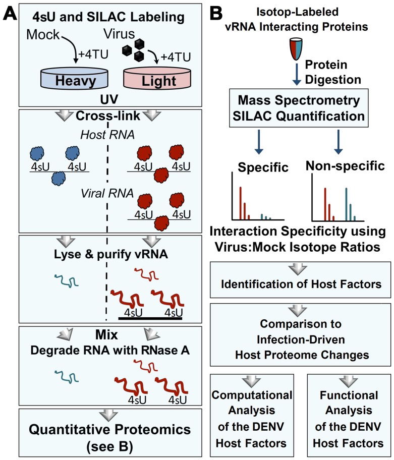
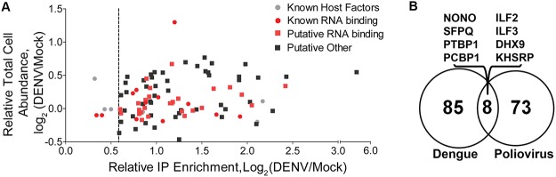
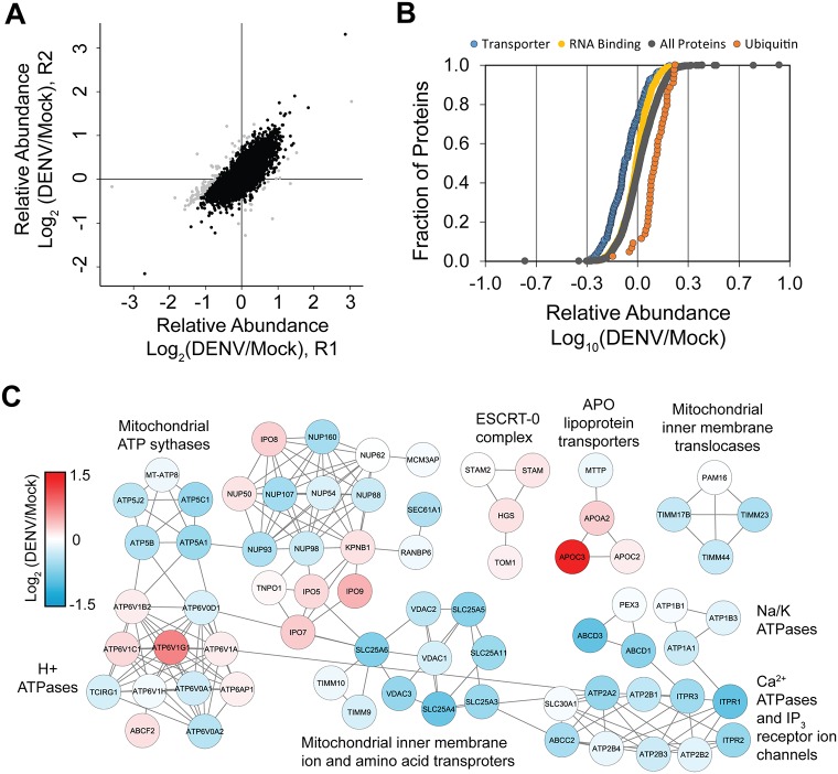
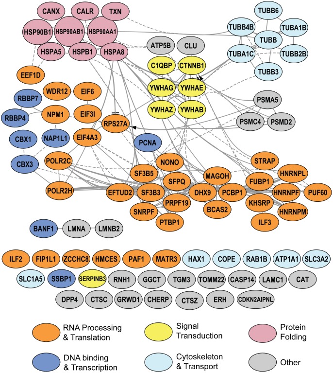
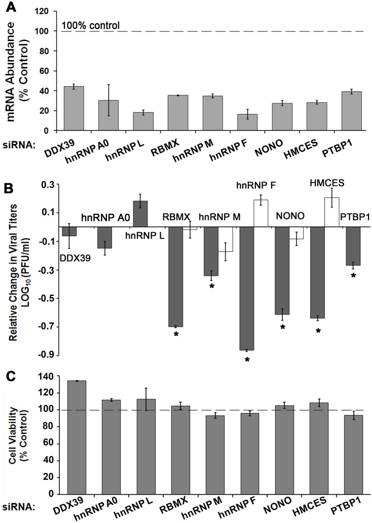
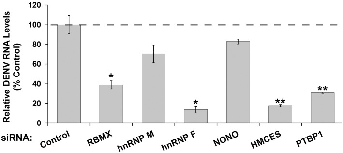
Methodology/principal findings: Seventy-nine novel RNA binding proteins for dengue virus (DENV) were identified by cross-linking proteins to dengue viral RNA during a live infection in human cells. These cellular proteins were specific and distinct from those previously identified for poliovirus, suggesting a specialized role for these factors in DENV amplification. Knockdown of these proteins demonstrated their function as viral host factors, with evidence for some factors acting early, while others late in infection. Their requirement by DENV for efficient amplification is likely specific, since protein knockdown did not impair the cell fitness for viral amplification of an unrelated virus. The protein abundances of these host factors were not significantly altered during DENV infection, suggesting their interaction with DENV RNA was due to specific recruitment mechanisms. However, at the global proteome level, DENV altered the abundances of proteins in particular classes, including transporter proteins, which were down regulated, and proteins in the ubiquitin proteasome pathway, which were up regulated.

Conclusions/significance: The method for identification of host factors described here is robust and broadly applicable to all RNA viruses, providing an avenue to determine the conserved or distinct mechanisms through which diverse viruses manage the viral RNA within cells. This study significantly increases the number of cellular factors known to interact with DENV and reveals how DENV modulates and usurps cellular proteins for efficient amplification.


Conflict of interest statement

The authors have declared that no competing interests exist.

Figures

Fig 1
Fig 1. Isolation and identification of cellular proteins that associate with DENV RNA by qTUX-MS.
(A) Diagram of the qTUX-MS method and SILAC labelling. Huh7.5UPRT cells labelled with the ‘light’ (red) or ‘heavy’ (blue) amino acids are infected with DENV or treated with mock, respectively, in the presence of 4TU. 4sU is incorporated into cellular and DENV RNAs and proteins are UV cross-linked to the contacting thio-containing RNA (represented as either balls to indicate native conformation or curved lines to indicate denatured proteins) in living cells at 28 hpi prior to cell lysis under denaturing conditions. Viral ribonucleoprotein complexes were isolated using DNA molecules complementary to DENV RNA bound to magnetic beads, the RNA was degraded with RNase A and the proteins were identified by mass spectrometry. (B) Workflow for quantitative proteomic analysis of RNA-bound host factors isolated in (A). Isolated proteins were mixed between mock and virus-infected samples, digested into peptides, and analysed by mass spectrometry. Relative ‘light’ and ‘heavy’ peptide abundances were quantified to determine the specificity of interaction. Host factor candidates were identified and subjected to functional validation.
Fig 2
Fig 2. Comparison of DENV/MOCK qTUX-MS protein enrichment and whole cell expression.
(A) Scatter plot comparison of the relative log2 total cell protein abundance versus the relative log2 qTUX-MS enrichment for 93 proteins quantified by qTUX-MS. Grey and red circles, known pro- & anti-viral factors and known vRNA-binding proteins, respectively; red and black squares, putative factors with RNA binding annotation and putative host factors with other annotation, respectively. (B) Venn diagram representing the overlap between the cellular factors identified to bind DENV RNA (this study) and the proteins previously described to bind poliovirus RNA [15]. The proteins found in both studies are listed above the diagram.
Fig 3
Fig 3. DENV virus infection induces differential expression of selected protein classes.
(A) Comparison of 4,907 log2 DENV/Mock host protein abundance ratios between replicate experiments (R1 and R2). Black dots, proteins that were quantified with < 50% variance between replicates. (B) Comparison of log10 relative abundances of all host proteins (grey circles) to proteins annotated in the Transporter (blue circles, N = 209), RNA binding (yellow circles, N = 522), and ubiquitin proteasome pathway (orange circles, N = 44). Abundance ratios are plotted in increasing magnitude and expressed as a fraction of the total number of proteins. (C) Reactome functional interaction networks of proteins annotated in the transporter PANTHER protein class ontology. Four sub-networks (≥ 3 proteins per network) containing 71 proteins were formed. Nodes labeled with gene symbols and colored by relative log2 protein abundance. Clusters are labeled with representative transporter classes.
Fig 4
Fig 4. Functional network of known and putative host factors interacting with DENV RNA identified by qTUX-MS.
Ninety-three host proteins identified by qTUX-MS were analyzed by the Reactome FI Cytoscape plugin. Network nodes are labeled with gene symbols and color-coded by broad functional classes
Fig 5
Fig 5. Depletion of cellular proteins identified by qTUX-MS affect DENV titer.
HeLaUPRT cells were transfected with either control or specific siRNAs and 48 hours post transfection were infected with DENV2, Ad5 or tested for cell viability (C). 40 hours post infection knockdown efficiencies were assessed at the mRNA levels (A), and DENV and Ad5 titers were determined (B). (A) mRNA levels were measured by RT-qPCR and quantified using the 2-ΔΔCt approach and normalized to β-actin mRNA levels. (B) DENV and Ad5 were collected at either 40 hpi or 30 hpi, respectively, and titers were determined by plaque assays. Average viral titers for DENV (dark grey) and Ad5 (white) are reported relative to the titer obtained from the control siRNA knockdown (Log10 = 0). Error bars represent ± standard error, p<0.05 (*) are shown. Absolute values for DENV and Ad5 titers are shown in S6 Fig. (C) Relative viability of non-infected cells was measured using an MTT assay (Invitrogen) 48 hours following knockdown by the indicated siRNAs.
Fig 6
Fig 6. Knockdown of qTUX-MS host factors reduces DENV RNA levels.
Cells were infected with DENV 48 hours following siRNA transfections and collected 40 hpi. Total RNA was extracted from the cells and viral RNA levels were determined using qRT-PCR analysis. Each bar represents an average value of the viral RNA normalized to β-actin mRNA levels from at three independent experiments in the indicated host factor depleted cells relative to control siRNA knockdown. Error bars represent ± standard deviation, p<0.05 (*), p<0.01 (**) are shown.

References

    1. Bhatt S, Gething PW, Brady OJ, Messina JP, Farlow AW, Moyes CL, et al. The global distribution and burden of dengue. Nature. 2013;496(7446):504–7. 10.1038/nature12060 - DOI - PMC - PubMed
    1. Leyssen P, De Clercq E, Neyts J. Perspectives for the treatment of infections with Flaviviridae. Clinical microbiology reviews. 2000;13(1):67–82, table of contents. - PMC - PubMed
    1. Rodenhuis-Zybert IA, Wilschut J, Smit JM. Dengue virus life cycle: viral and host factors modulating infectivity. Cellular and molecular life sciences: CMLS. 2010;67(16):2773–86. 10.1007/s00018-010-0357-z . - DOI - PMC - PubMed
    1. Welsch S, Miller S, Romero-Brey I, Merz A, Bleck CK, Walther P, et al. Composition and three-dimensional architecture of the dengue virus replication and assembly sites. Cell host & microbe. 2009;5(4):365–75. 10.1016/j.chom.2009.03.007 . - DOI - PMC - PubMed
    1. Sayce AC, Miller JL, Zitzmann N. Targeting a host process as an antiviral approach against dengue virus. Trends Microbiol. 2010;18(7):323–30. 10.1016/j.tim.2010.04.003 . - DOI - PubMed

Publication types

LinkOut - more resources