. 2016 Sep 20;7(38):62292-62304.
doi: 10.18632/oncotarget.11410.

Induction of exportin-5 expression during melanoma development supports the cellular behavior of human malignant melanoma cells

Affiliations

Induction of exportin-5 expression during melanoma development supports the cellular behavior of human malignant melanoma cells

Corinna Anna Ott et al. Oncotarget. .

Abstract

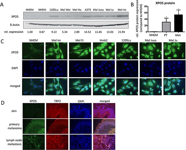
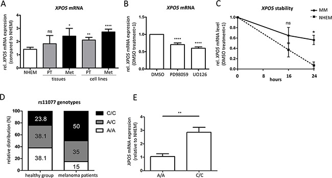
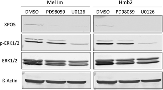
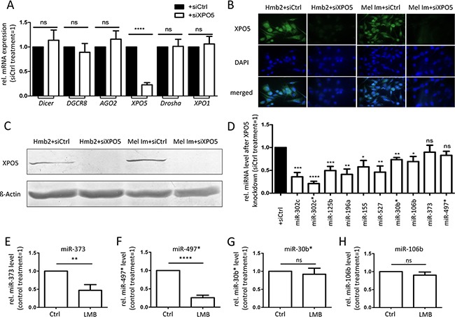
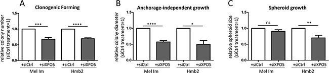
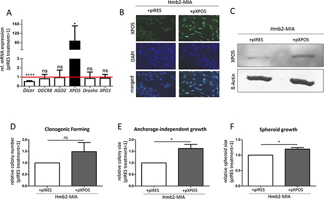
Regulation of gene expression via microRNAs is known to promote the development of many types of cancer. In melanoma, miRNAs are globally up-regulated, and alterations of miRNA-processing enzymes have already been identified. However, mis-regulation of miRNA transport has not been analyzed in melanoma yet. We hypothesized that alterations in miRNA transport disrupt miRNA processing. Therefore, we investigated whether the pre-miRNA transporter Exportin-5 (XPO5) was involved in altered miRNA maturation and functional consequences in melanoma. We found that XPO5 is significantly over-expressed in melanoma compared with melanocytes. We showed enhanced XPO5 mRNA stability in melanoma cell lines which likely contributes to up-regulated XPO5 protein expression. In addition, we identified MEK signaling as a regulator of XPO5 expression in melanoma. Knockdown of XPO5 expression in melanoma cells led to decreased mature miRNA levels and drastic functional changes. Our data revealed that aberrant XPO5 expression is important for the maturation of miRNAs and the malignant behavior of melanoma cells. We suggest that the high abundance of XPO5 in melanoma leads to enhanced survival, proliferation and metastasis and thereby supports the aggressiveness of melanoma.

Keywords: XPO5; mRNA stability; malignant melanoma; miR-SNP; microRNA.


Conflict of interest statement

The authors declare no conflicts of interest.

Figures

Figure 1
Figure 1. XPO5 protein expression in NHEMs, melanoma cell lines and tissue samples
A. Western blot results showing XPO5 protein expression in normal human epidermal melanocytes (NHEMs) and primary (PT) and metastatic (Met) melanoma cell lines. ß-Actin was used as a loading control. The relative densitometric quantifications of XPO5 protein expression are indicated under the blot and show a strong induction of XPO5 protein expression in melanoma cells compared with NHEMs. B. Relative densitometric quantification of three independent Western blot analyses compared with ß-Actin. XPO5 expression in NHEMs was set as 1. C. Protein expression of XPO5 (as detected by immunofluorescence staining) was enhanced in melanoma cell lines compared with NHEMs. D. Protein expression of XPO5 (as detected by immunofluorescence staining) was enhanced in primary melanoma tissue and tissue of a lymph node metastasis compared with healthy skin. TRP2 staining was used to stain melanocytes.
Figure 2
Figure 2. Elevated XPO5 mRNA expression and stability in malignant melanoma
A. qRT-PCRs showed an increase in expression of XPO5 mRNA during melanoma progression in primary and metastatic melanoma tissues (n=4) and cell lines (n=13) compared with NHEMs (n=15). B. XPO5 mRNA expression decreased after treatment with the MEK inhibitors PD98059 and U0126 in comparison to DMSO-treated cells (n=10). C. The stability of XPO5 mRNA was determined by qRT-PCR after treatment of NHEMs and melanoma cell lines (MM) with alpha-amanitin for 0, 16 and 24 hours. The remaining XPO5 mRNA level in the melanoma cell lines (solid line) was significantly different to that of the NHEMs (dashed line) after the 24 h treatment (n=3). D. The relative distribution of the rs11077 genotypes in melanoma patients (n=20) versus the healthy control group (n-21). In the control group, the values are equivalent to the distribution in the Caucasian population. In melanoma patients, half of the patients had the C/C variant SNP (50%). E. Relative XPO5 mRNA expression compared to NHEM in the homozygous rs11077 genotypes A/A and C/C.
Figure 3
Figure 3. Analysis of potential regulators of XPO5 protein expression
Western blot analyses after the treatment of the melanoma cell lines Mel Im and Hmb2 with the MEK inhibitors PD98059 and U0126 showed a strong downregulation of XPO5 expression. The p-ERK1/2 staining reflects inhibition of ERK phosphorylation by U0126 treatment in both cell lines compared with the control treatment (DMSO). ERK1/2 antibody staining was used to show overall ERK level. ß-Actin was analyzed in the same protein samples on a second blot (due to similar molecular weights) and served as a loading control.
Figure 4
Figure 4. Consequences of siRNA-induced XPO5 downregulation in the metastatic melanoma cell lines Mel Im and Hmb2
A. Relative expression of genes involved in miRNA processing and mRNA transport. A significant knockdown of XPO5 in melanoma cell lines did not alter the gene expression of Dicer, DGCR8, AGO2, Drosha or XPO1 compared with control treatment (siCtrl) (n=7). B. Immunofluorescence staining and C. Western blot analysis of XPO5 showed a strong downregulation of XPO5in Mel Im and Hmb2 cells after siXPO5 treatment. D. Relative levels of mature miRNAs after XPO5 knockdown compared with siCtrl (n=4). Except for miR-373 and miR-497*, the levels of all tested miRNAs were significantly lower compared with those of the siCtrl treatment. E-H. Relative miRNA levels after XPO1 inhibition (n=4). The levels of (E) miR-373 and (F) miR-497* were significantly lower after XPO1 inhibition via leptomycin B (LMB) compared with control-treated cells (Ctrl). The levels of (G) miR-30b* and (H) miR-106b were not affected by XPO1 inhibition.
Figure 5
Figure 5. Functional consequences of XPO5 knockdown in melanoma cell lines
A. siRNA-induced XPO5 knockdown in Mel Im and Hmb2 significantly decreased the number of colonies formed in the clonogenic forming assay compared with that of siCtrl-treated cells (n=7). B. XPO5 knockdown resulted in significantly smaller colonies of Mel Im and Hmb2 cells in the anchorage-independent growth assay using soft agar (n=3). C. Spheroid growth of siXPO5-treated Hmb2 cells was significantly decreased compared with siCtrl-treated cells (n=5).
Figure 6
Figure 6. Over-expression of XPO5 in MIA-deficient Hmb2 cell clones and its functional consequences
A. Significant over-expression of XPO5 in Hmb2-MIA cells did not alter the gene expression of DGCR8, AGO2, Drosha or XPO1 compared with that in the control treatment (pIRES) but significantly decreased Dicer gene expression (n=3). B. Immunofluorescence staining and C. Western blot analysis of XPO5 showed strong XPO5 over-expression in Hmb2-MIA cells after pXPO5 treatment compared with pIRES transfection. D-F. Functional consequences of XPO5 over-expression in Hmb2-MIA cells. (D) XPO5 over-expression in Hmb2-MIA increased the number of colonies formed in the clonogenic forming assay compared with pIRES-treated cells, but the increase was not statistically significant (n=4). (E) XPO5 over-expression resulted in significantly larger colonies of in the anchorage-independent growth assay (n=3). (F) Spheroid growth of pXPO5 transfected Hmb2-MIA cells was significantly increased compared with that of pIRES-treated cells (n=4).

References

    1. Haass NK, Schumacher U. Melanoma never says die. Experimental dermatology. 2014;23:471–2. doi: 10.1111/exd.12400. - DOI - PubMed
    1. Gebauer F, Hentze MW. Molecular mechanisms of translational control. Nature reviews Molecular cell biology. 2004;5:827–835. doi: 10.1038/nrm1488. - DOI - PMC - PubMed
    1. Meister G, Landthaler M, Dorsett Y, Tuschl T. Sequence-specific inhibition of microRNA- and siRNA-induced RNA silencing. RNA (New York NY) 2004;10:544–50. - PMC - PubMed
    1. Dalmay T, Edwards DR. MicroRNAs and the hallmarks of cancer. Oncogene. 2006;25:6170–6175. doi: 10.1038/sj.onc.1209911. - DOI - PubMed
    1. Bonazzi VF, Stark MS, Hayward NK. MicroRNA regulation of melanoma progression. Melanoma Research. 2012;22:101–113. doi: 10.1097/CMR.0b013e32834f6fbb. - DOI - PubMed

MeSH terms