. 2016 Oct 6;538(7623):79-83.
doi: 10.1038/nature19089. Epub 2016 Aug 24.

Surface patterning of nanoparticles with polymer patches

Affiliations

Surface patterning of nanoparticles with polymer patches

Rachelle M Choueiri et al. Nature. .

Abstract

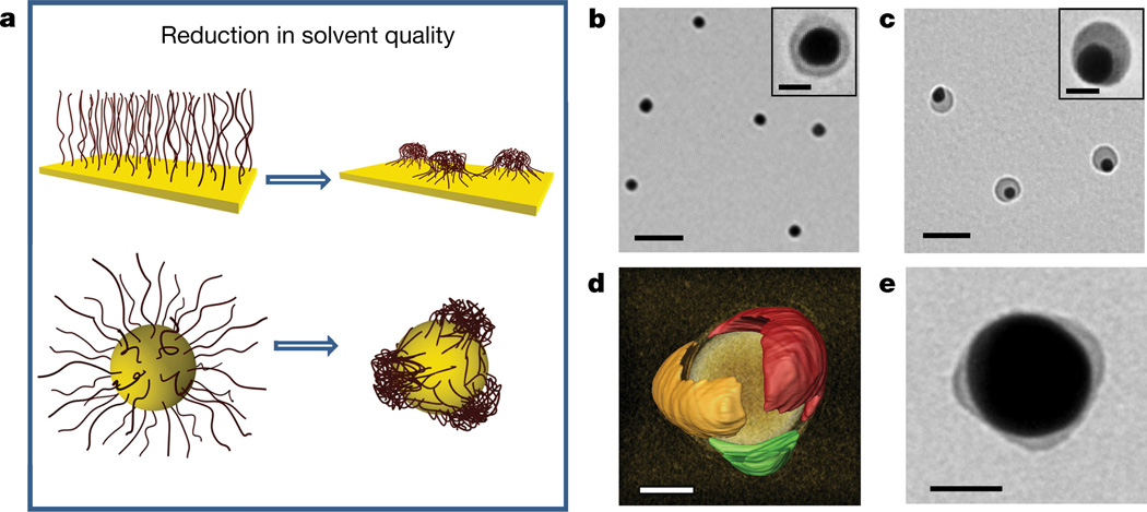
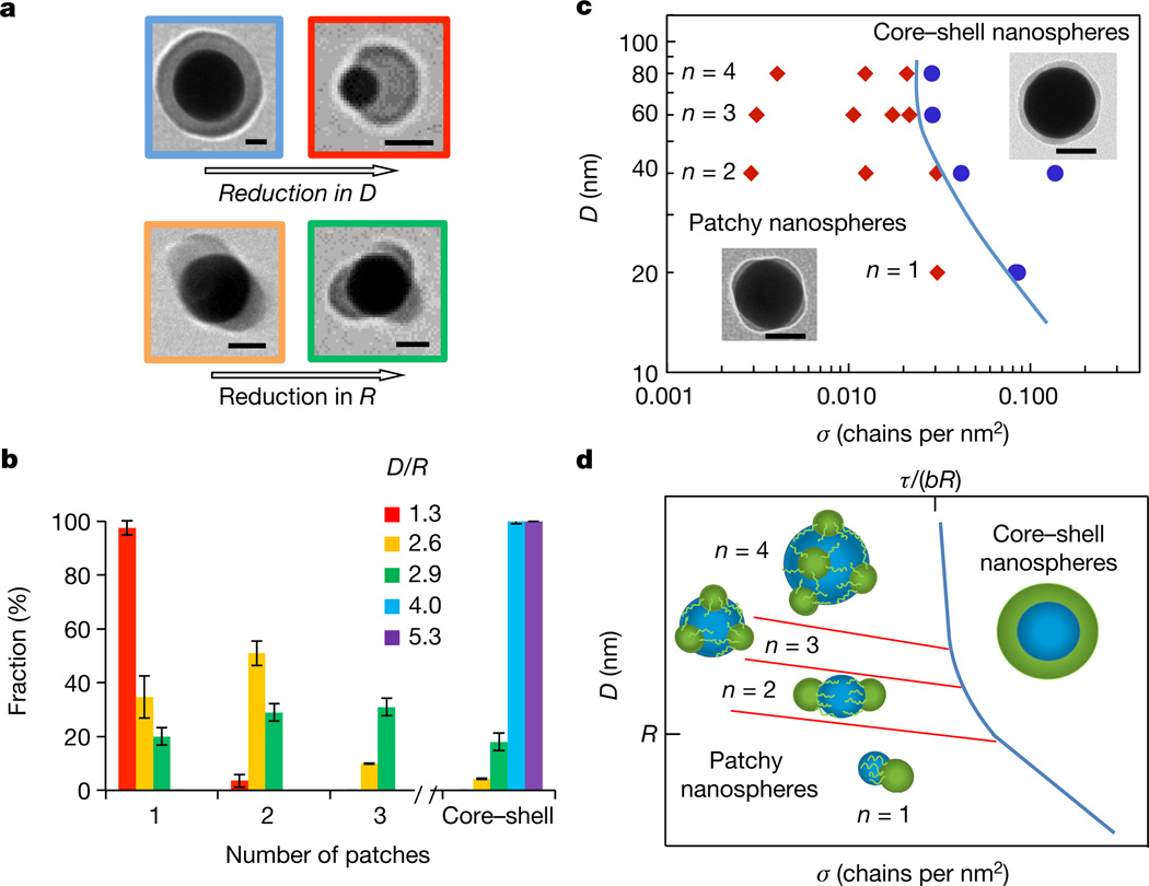
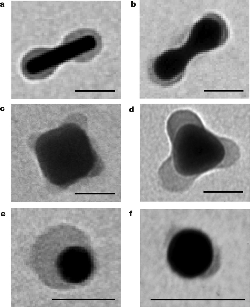
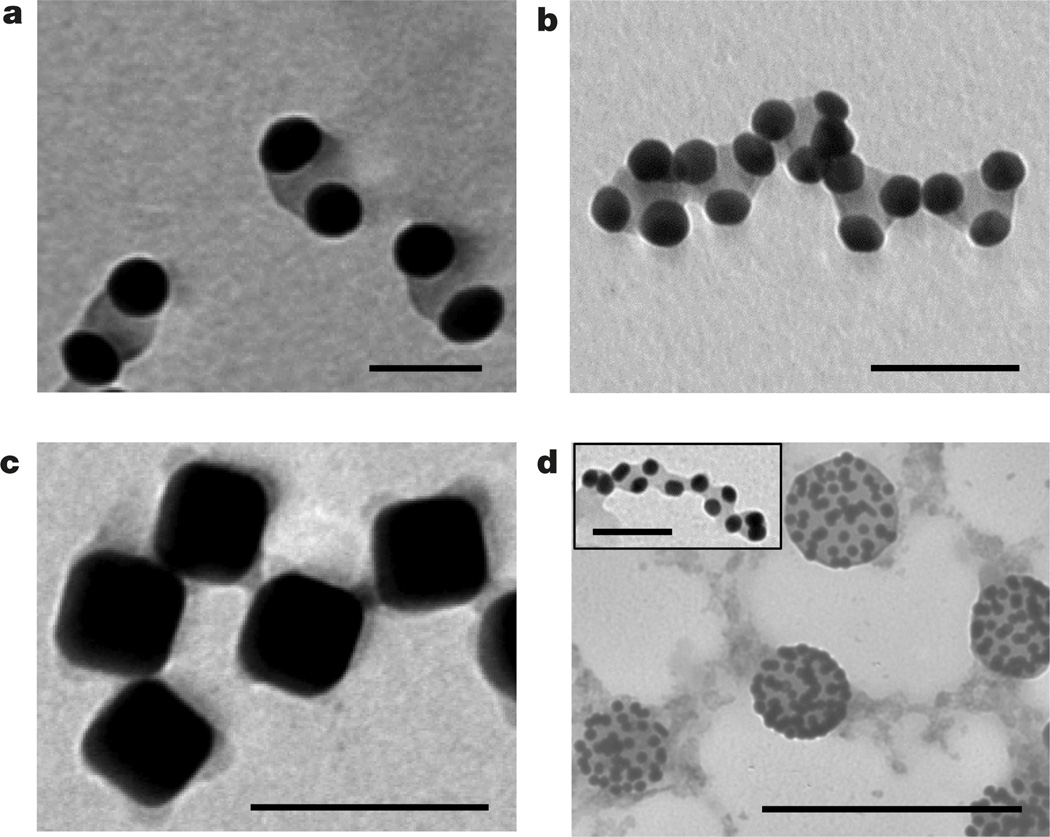
Patterning of colloidal particles with chemically or topographically distinct surface domains (patches) has attracted intense research interest. Surface-patterned particles act as colloidal analogues of atoms and molecules, serve as model systems in studies of phase transitions in liquid systems, behave as 'colloidal surfactants' and function as templates for the synthesis of hybrid particles. The generation of micrometre- and submicrometre-sized patchy colloids is now efficient, but surface patterning of inorganic colloidal nanoparticles with dimensions of the order of tens of nanometres is uncommon. Such nanoparticles exhibit size- and shape-dependent optical, electronic and magnetic properties, and their assemblies show new collective properties. At present, nanoparticle patterning is limited to the generation of two-patch nanoparticles, and nanoparticles with surface ripples or a 'raspberry' surface morphology. Here we demonstrate nanoparticle surface patterning, which utilizes thermodynamically driven segregation of polymer ligands from a uniform polymer brush into surface-pinned micelles following a change in solvent quality. Patch formation is reversible but can be permanently preserved using a photocrosslinking step. The methodology offers the ability to control the dimensions of patches, their spatial distribution and the number of patches per nanoparticle, in agreement with a theoretical model. The versatility of the strategy is demonstrated by patterning nanoparticles with different dimensions, shapes and compositions, tethered with various types of polymers and subjected to different external stimuli. These patchy nanocolloids have potential applications in fundamental research, the self-assembly of nanomaterials, diagnostics, sensing and colloidal stabilization.


Figures

Figure 1
Figure 1. Polymer segregation on the nanoparticle surface
a, Schematics of solvent-mediated formation of pinned polymer micelles (surface patches) on a planar macroscopic surface (top) and on the nanoparticle surface (bottom). b, c, TEM images of gold nanospheres capped with polystyrene-50K at the grafting density of 0.03 chains per square nanometre and deposited on the grid from the 0.3 nM nanosphere solution in DMF (b) and from the DMF/water mixture at Cw = 4 vol% after 24 h incubation at 40 °C (c). Scale bars in b and c are 100 nm. Insets in b and c show the corresponding images of individual nanospheres. Inset scale bars are 20 nm. d, Electron tomography reconstruction image of the 60-nm-diameter nanosphere with three polystyrene-50K patches, each shown for clarity with a different arbitrary colour. The image of a gold core is removed to highlight the structure of polymer patches. The estimated resolution is 2–3 nm. Patchy nanospheres were formed as described in c. The grafting density of polystyrene-50K is 0.02 chains per square nanometre. e, TEM image of the gold nanosphere carrying photocrosslinked thiol-terminated polystyrene-co-polyisoprene patches preserved after 24 h incubation in tetrahydrofuran (a good solvent for polystyrene-co-polyisoprene). Original patchy nanoparticles were formed and crosslinked in the DMF/water mixture at Cw = 1 vol%. Scale bars in d and e are 20 nm.
Figure 2
Figure 2. Structural transitions in the polymer layer on the surface of gold nanospheres
a, Effect of nanosphere size (top) and polymer dimensions (bottom) on patch formation. The nanospheres are functionalized with polystyrene-50K (top row and bottom left) and polystyrene-30K (bottom right) at σ = 0.03 chains per square nanometre. Scale bars are 25 nm. b, Distribution of populations of nanospheres with a different patch number. The red, yellow, blue and violet bars correspond to the 20-, 40-, 60- and 80-nm-diameter nanospheres capped with polystyrene-50K, respectively; the green bar represents 32-nmdiameter nanospheres functionalized with polystyrene-30K. σ = 0.03 chains per square nanometre. The error bars represent the standard deviations. Each experiment was run in triplicate. The inset shows theD/R ratios, with colours corresponding to the colours of bars and the frames of the images in a. c, Experimental diagram of nanosphere states. The blue line separates the regions of core–shell and patchy nanospheres with a different average patch number n. The insets illustrate patchy and a core– shell nanospheres with σ of 0.012 and 0.03 chains per square nanometre, respectively. Scale bars are 50 nm. In b and c, 200–300 nanospheres were analysed for each population. d, Theoretical diagram of nanosphere states. Transitions between nanospheres with different values of n begin at DR (red lines). The blue line shows the boundary between the smooth and patchy polymer layer, approaching the grafting density σ = τ/(bR) for large nanospheres, similar to c.
Figure 3
Figure 3. Generality of polymer patterning of nanoparticle surface
a–d, TEM images of polystyrene-50K-coated gold spherocylindrical nanorods (a), gold dumbbell-shaped nanorods (b), silver nanocubes (c) and silver triangular prisms (d), all in the DMF/water mixture at Cw = 4 vol%. e, f, TEM images of thiol-terminated poly(4-vinyl pyridine) (Mn = 22,000 g mol−1) in water at pH = 10.5 (e) and thiol-terminated poly(N-vinylcarbazole) (Mn = 19,800 g mol−1) in the DMF/water mixture at Cw = 4 vol% (f), both on the surface of gold nanospheres. All scale bars are 40 nm.
Figure 4
Figure 4. Self-assembly of patterned nanoparticles
a, Dimers of single-patch gold nanospheres. b, Self-assembly of trimers of single-patch gold nanospheres in chains. In a and b the nanospheres were capped with polystyrene-50K and incubated for 15 days in the DMF/water solution at Cw = 4 vol% at 40 °C. Scale bars in a and b are 40 nm. c, Self-assembly of patchy silver nanocubes functionalized with polystyrene-50K in the DMF/water mixture at Cw = 20 vol%; scale bar is 100 nm. d, Self-assembly of gold nanospheres on the surface of droplets enriched with free nonthiolated polystyrene. The self-assembly was induced by adding water at Cw = 4 vol% to the mixed solution of free non-thiolated polystyrene (Mn = 50,000 g mol−1) and gold nanospheres tethered with polystyrene- 50K in DMF; scale bar is 250 nm. The inset to d shows self-assembly of patchy polystyrene-50K-capped gold nanospheres in the DMF/water mixture at Cw = 4 vol% in the presence of 0.625 nM of non-thiolated polystyrene, following 5 min sonication of the solution. Scale bar is 40 nm.

References

    1. Bianchi E, Blaak R, Likos CN. Patchy colloids: state of the art and perspectives. Phys. Chem. Chem. Phys. 2011;13:6397–6410. - PubMed
    1. Preisler Z, Vissers T, Munaŏ G, Smallenburg F, Sciortino F. Equilibrium phases of one-patch colloids with short-range attractions. Soft Matter. 2014;10:5121–5128. - PubMed
    1. Chen Q, Bae SC, Granick S. Directed self-assembly of a colloidal kagome lattice. Nature. 2011;469:381–384. - PubMed
    1. Glotzer SC, Solomon MJ. Anisotropy of building blocks and their assembly into complex structures. Nat. Mater. 2007;6:557–562. - PubMed
    1. Gröschel AH, et al. Guided hierarchical co-assembly of soft patchy nanoparticles. Nature. 2013;503:247–251. - PubMed