. 2016 Oct 15;129(20):3948-3957.
doi: 10.1242/jcs.184424. Epub 2016 Aug 24.

Exosomes bind to autotaxin and act as a physiological delivery mechanism to stimulate LPA receptor signalling in cells

Affiliations

Exosomes bind to autotaxin and act as a physiological delivery mechanism to stimulate LPA receptor signalling in cells

Susanna A Jethwa et al. J Cell Sci. .

Abstract

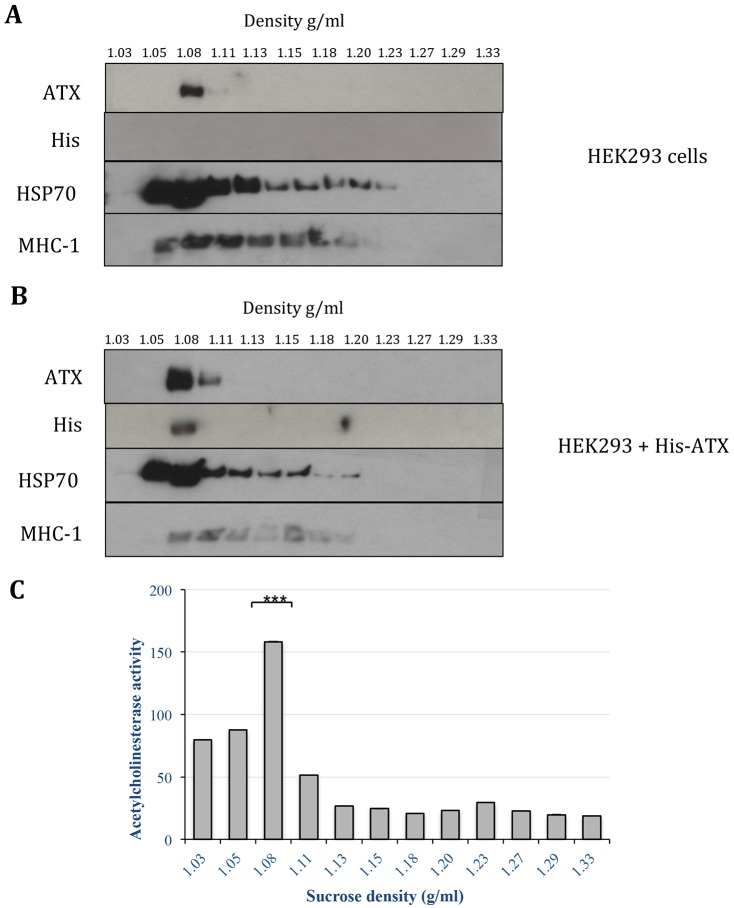
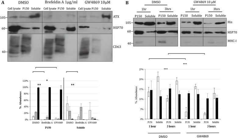
Autotaxin (ATX; also known as ENPP2), the lysophospholipase responsible for generating the lipid receptor agonist lysophosphatidic acid (LPA), is a secreted enzyme. Here we show that, once secreted, ATX can bind to the surface of cell-secreted exosomes. Exosome-bound ATX is catalytically active and carries generated LPA. Once bound to a cell, through specific integrin interactions, ATX releases the LPA to activate cell surface G-protein-coupled receptors of LPA; inhibition of signalling by the receptor antagonist Ki1642 suggests that these receptors are LPAR1 and LPAR3. The binding stimulates downstream signalling, including phosphorylation of AKT and mitogen-activated protein kinases, the release of intracellular stored Ca2+ and cell migration. We propose that exosomal binding of LPA-loaded ATX provides a means of efficiently delivering the lipid agonist to cell surface receptors to promote signalling. We further propose that this is a means by which ATX-LPA signalling operates physiologically.

Keywords: Autotaxin; Exosome; Integrin; LPA.


Conflict of interest statement

The authors declare no competing or financial interests.

Figures

Fig. 1.
Fig. 1.
Detection of ATX with vesicular fractions of conditioned serum-free medium. (A) His–ATX was expressed in HEK293 cells from which conditioned media were generated and fractionated. Proteins were separated by using SDS-PAGE and were immunoblotted with antibodies against His, ATX and proteins commonly detected in exosomes. Immunoblots are from the same membrane. P15 is the pellet generated from the 1500 g centrifugation step. P150 and S150 are the pellet and supernatant fractions generated from the 150,000 g ultracentrifugation step. (B) Vesicular ATX is also present in serum. Fractionated conditioned media samples were separated by using SDS-PAGE and were analysed for the presence of ATX by immunoblotting, and the band density was quantified (black bars, P15; grey bars, P150; white bars, S150). The different molecular mass bands reflect distinct glycosylation patterns. (C) P150 pellets are rich in exosome-like vesicles, as observed by performing electron microscopy. P150 pellets isolated by ultracentrifugation were fixed in 2% paraformaldehyde with 2.5% glutaraldehyde in 0.1 M sodium cacodylate buffer (pH 7.2). Fixed pellets were resin-embedded and imaged by using transmission electron microscopy (TEM). Arrows point to vesicles displaying the characteristics of exsomes.
Fig. 2.
Fig. 2.
Fractionation of vesicular ATX with sucrose density centrifugation. P150 samples were purified by performing ultracentrifugation through sucrose density gradients from (A) untransfected HEK293 cells and (B) HEK293 cells that had been transfected with His–ATX. Proteins were precipitated from each fraction with TCA in acetone and fractionated by performing SDS-PAGE. ATX, His–ATX, HSP70 and MHC-1 were detected by immunoblotting, and (C) acetylcholinesterase activity was assayed in triplicate using the 5,5′-dithiobis(2-nitrobenzoic acid) (DNTB) system. The mean data shown are from three technical repeats (representative of at least three separate experiments). The immunoblots shown in A,B are from the same membranes. ***P≤0.01, significance relates to activity comparison between density of 1.08 g/ml and 1.05g/ml sucrose. (Student's t-test).
Fig. 3.
Fig. 3.
Autotaxin associates with the outer exosome membrane. Exosomes were isolated and incubated with 0.1% Triton X-100, with 0.05% trypsin, or a combination of both for 20 min at room temperature. Samples were fractionated using SDS-PAGE and then analysed by immunoblotting with antibodies against ATX, His and HSP70. The immunoblots shown are from the same membranes. Data shown are representative of at least four experiments.
Fig. 4.
Fig. 4.
Extracellular ATX does not traffic through the MVB pathway. HEK293 cells were cultured for 16 h in the presence of (A) DMSO control, 1 μg/ml brefeldin A or 20 μM GW4869. CD63 was used as the marker for exosomes. Fractionated culture media were immunoblotted to observe ATX and HSP70 (white bars, ATX; grey bars, HSP70; black bars, CD63). The graph labelled % Abundance reports the distribution of ATX, HSP70 and CD63 between the P150 and S150 fractions in percent. **P≤0.01, *P≤0.05 (Student's t-test). (B) Soluble His–ATX was isolated and added to HEK293 cells in the presence or absence of 20 μM GW4869 for 1 or 3 h. ATX binding to exosomes was detected by immunoblotting with an anti-His antibody. Exosome abundance was determined by co-immunoblotting for HSP70 and MHC-I. The immunoblots shown in A,B are from the same membranes; MHC-1 was used as a marker for exosomes. Data shown are representative of at least three experiments (white bars, ATX; black bars, HSP70). The graph labelled % Abundance reports the distribution of ATX and HSP70 between the P150 and S150 fractions in percent. Mean±s.d., ***P≤0.001 (Student's t-test). P150 and S150 are the pellet and supernatant fractions, respectively, generated from the 150, 000 g ultracentrifugation step. Soluble His-ATX was purified from the S150 fraction.
Fig. 5.
Fig. 5.
ATX is detected when exosomes are immunopurified with an antibody against laminin γ 1. (A) Laminin γ1 (LAMC1) was immunoprecipitated from lysed exosomes with an anti-laminin-γ1 antibody conjugated to protein-G agarose beads. Laminin γ1 and ATX were detected by performing SDS-PAGE followed by immunoblotting. Results are representative of five experiments. (B) His–ATX was immunoprecipitated from exosomes and used to immunopurify purified recombinant laminin α2,β1,γ1 (211) and α5,β1,γ1 (511) complexes. Binding was assessed by immunoblotting with antibodies against ATX and laminin γ1 (LAMC1). Results are representative of at least two experiments. Control, exosomes isolated from cells that were not transfected with any vector. Vector, exosomes isolated from cells that were transfected with a vector that does not contain ATX cDNA. (C) Soluble His–ATX was isolated from His–ATX-transfected HEK293 conditioned medium and incubated with untransfected HEK293 cells with or without recombinant laminin α2,β1,γ1 and α5,β1,γ1 complexes. Exosomes and soluble proteins were blotted for ATX. Results are representative of two experiments. P150 and S150 are the pellet and supernatant fractions, respectively, generated from the 150,000 g ultracentrifugation step. Soluble refers to the S150 fraction. Control, isolated soluble His-ATX incubated with untransfected HEK293 cells in the absence of recombinant laminin. The immunoblots shown in A–C are from the same membranes.
Fig. 6.
Fig. 6.
Mass spectrometry analysis of LPA and LPC. (A) LPA and (B) LPC species were determined by mass spectrometry analysis of extracted exosomes from the conditioned serum-free media of control (white bars) and His–ATX-overexpressing (black bars) HEK293 cells. The amount of each LPA and LPC species was expressed as a ratio of the total phosphatidylcholine (PC) detected. Results are representative of two experiments performed in triplicate (i.e. shown are results from three technical repeats from a single experiment). (C) Mass spectrometry analysis of exosomes isolated from serum-free conditioned media of HEK293 cells treated with DMSO (white bars) or 10 µM HA-130 (black bars). Results are representative of three experiments performed in triplicate (D). Purified His–ATX incubated with C17 LPC or C17 LPA±10 µM HA-130. Results are representative of three experiments performed in triplicate. ns, not significant, *P=<0.05, **P≤0.01, ***P≤0.001, ****P≤0.0001; (Student's t-test). Data are mean±s.d.
Fig. 7.
Fig. 7.
Vesicular ATX stimulation of signalling is dependent of LPA receptors. Exosomes were added to serum-starved HEK293 cells that had been pre-treated with either EtOH or 10 μM Ki16425. His ATX T210A is a catalytically inactive mutant. (A) Phosphorylated (pERK) and ERK2 in lysates were detected by immunoblotting. (B) Phosphorylated (pAKT) and total AKT in lysates were detected by immunoblotting. Blots were quantified by using scanning densitometry analysis. (C) Exosomes induce Ca2+ mobilisation in NIH3T3 cells in an LPA-like manner. Quiescent cells were seeded onto glass coverslips and loaded with Fura-2 AM, which was alternately excited at 340 nm and 380 nm wavelengths, and the Fura-2 emission at >450 nm was sampled at 2 Hz. The y-axis represents the ratiometric Fura-2 emission, and is proportional to cytosolic Ca2+ concentration. Cells were stimulated with exosomes followed by a threshold and a maximal dose of LPA. The cytosolic Ca2+ concentration in four individual cells is shown. The arrows indicate the series of low-amplitude exosome-induced transient Ca2+ elevations, which were not seen in untreated cells, or in cells that had been co-incubated with exosomes and Ki16425 (n=40 cells imaged on four separate days). (D) HEK293 cells were transfected with His–ATX, His–ATX-RGE, His–ATX-LNV, His–ATX-RGE–LNV, His–ATX-H119A or His–ATX-ΔSMB and cultured in OptiMEM for 36 h to condition media. Exosomes were isolated by differential centrifugation and added to serum-starved NIH3T3 cells. Phosphorylated and total ERK were detected by immunoblotting of lysates. Bands were quantified by performing scanning densitometry analysis and normalised to levels of total ERK. The shown immunoblot is representative of three experiments, and quantification is mean±s.d. of three experiments. The immunoblots shown in A,B,D are from the same membranes. *P≤0.05, **P≤0.01, ***P≤0.0001 (Student's t-test). Control, OptiMEM without LPA or exosomes.
Fig. 8.
Fig. 8.
ATX induces migration but not DNA synthesis in manner that is dependent on LPA receptors. (A) Exosomes isolated from media conditioned by either untransfected or His–ATX-transfected HEK293 cells or LPA were added to quiescent NIH3T3 cells that had been pre-treated with EtOH (white bars) or 10 μM Ki16425 (black bars) for 24 h. The cells were then pulsed with [3H] thymidine for 6 h, fixed and lysed, and the radioactivity was determined. Results are mean±s.d. of three experiments performed in triplicate. (B) Exosomes±10 µM Ki16425 were added to cultures of serum-starved HEK293 cells that had been scratched to leave a clear region into which cells migrated in response to the addition of exosomes±Ki16425. Results are mean±s.d. of two experiments each performed in triplicate. x-axis in B,C indicates the percentage change in the area of the wound covered. (C) Exosomes±10 µM HA-130 were added to cultures of serum-starved HEK293 cells that had been scratched to leave a clear region into which cells migrated in response to the addition of exosomes±HA-130. Results are the mean±s.d. of two experiments each performed in triplicate. *P≤0.05, **P≤0.01, ***P≤0.0001; ns, not significant (Student's t-test). Control, OptiMEM without exosomes. Additional chemicals were added as indicated in B and C.

Similar articles

Cited by

References

    1. Baumforth K. R. N., Flavell J. R., Reynolds G. M., Davies G., Pettit T. R., Wei W., Morgan S., Stankovic T., Kishi Y., Arai H. et al. (2005). Induction of autotaxin by the Epstein-Barr virus promotes the growth and survival of Hodgkin lymphoma cells. Blood 106, 2138-2146. 10.1182/blood-2005-02-0471 - DOI - PubMed
    1. Bootman M. D., Berridge M. J. and Lipp, P. (1997). Cooking with calcium: the recipes for composing global signals from elementary events. Cell 91, 367-373. 10.1016/S0092-8674(00)80420-1 - DOI - PubMed
    1. Fulkerson Z., Wu T., Sunkara M., Kooi C. V., Morris A. J. and Smyth, S. S. (2011). Binding of autotaxin to integrins localizes lysophosphatidic acid production to platelets and mammalian cells. J. Biol. Chem. 286, 34654-34663. 10.1074/jbc.M111.276725 - DOI - PMC - PubMed
    1. Hausmann J., Kamtekar S., Christodoulou E., Day J. E., Wu T., Fulkerson Z., Albers H. M. H. G., van Meeteren L. A., Houben A. J. S., van Zeijl L. et al. (2011). Structural basis of substrate discrimination and integrin binding by autotaxin. Nat. Struct. Mol. Biol. 18, 198-204. 10.1038/nsmb.1980 - DOI - PMC - PubMed
    1. Hohenester E. and Yurchenco, P. D. (2013). Laminins in basement membrane assembly. Cell Adh. Migr. 7, 56-63. 10.4161/cam.21831 - DOI - PMC - PubMed

Publication types

MeSH terms