. 2016 Sep 30;291(40):21123-21136.
doi: 10.1074/jbc.M116.745372. Epub 2016 Aug 24.

Phosphorylation of PP1 Regulator Sds22 by PLK1 Ensures Accurate Chromosome Segregation

Affiliations

Phosphorylation of PP1 Regulator Sds22 by PLK1 Ensures Accurate Chromosome Segregation

Hequan Duan et al. J Biol Chem. .

Erratum in

Abstract

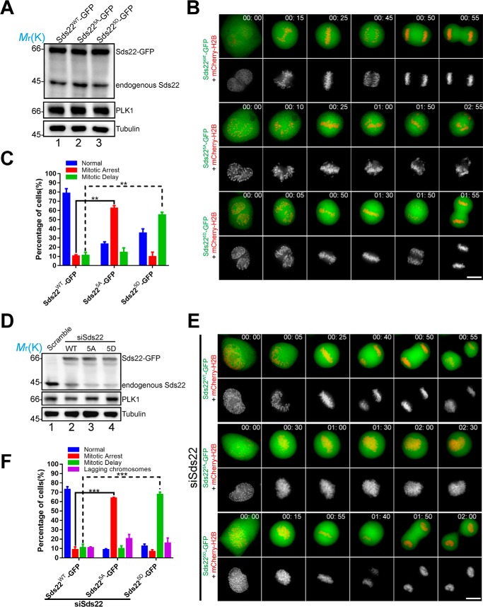
During cell division, accurate chromosome segregation is tightly regulated by Polo-like kinase 1 (PLK1) and opposing activities of Aurora B kinase and protein phosphatase 1 (PP1). However, the regulatory mechanisms underlying the aforementioned hierarchical signaling cascade during mitotic chromosome segregation have remained elusive. Sds22 is a conserved regulator of PP1 activity, but how it regulates PP1 activity in space and time during mitosis remains elusive. Here we show that Sds22 is a novel and cognate substrate of PLK1 in mitosis, and the phosphorylation of Sds22 by PLK1 elicited an inhibition of PP1-mediated dephosphorylation of Aurora B at threonine 232 (Thr232) in a dose-dependent manner. Overexpression of a phosphomimetic mutant of Sds22 causes a dramatic increase in mitotic delay, whereas overexpression of a non-phosphorylatable mutant of Sds22 results in mitotic arrest. Mechanistically, the phosphorylation of Sds22 by PLK1 strengthens the binding of Sds22 to PP1 and inhibits the dephosphorylation of Thr232 of Aurora B to ensure a robust, error-free metaphase-anaphase transition. These findings delineate a conserved signaling hierarchy that orchestrates dynamic protein phosphorylation and dephosphorylation of critical mitotic regulators during chromosome segregation to guard chromosome stability.

Keywords: cell cycle; centromere; cyclin-dependent kinase (CDK); cytoskeleton; kinetochore; phosphatase; phosphorylation; tubulin.


Figures

FIGURE 1.
FIGURE 1.
PLK1 interacts with and phosphorylates Sds22. A, yeast cells were co-transformed with an Sds22 prey construct and the indicated kinetochore protein bait constructs (PLK1, Mad2, Mad1, Sgo1, and CENP-H). An example of such an experiment in which cells were selected on supplemented minimal plates lacking adenine, tryptophan, leucine, and histidine is presented. B, aliquots of HEK293T cells were transiently transfected to express Sds22 with wild type PLK1 (PLK1WT) and kinase-deficient mutant of PLK1 (PLK1KD). 36 h after transfection, the cells were lysed and precipitated with FLAG-agarose. The beads were washed and boiled in 1× SDS-PAGE sample buffer prior to electrophoresis. Western blotting analyses confirmed the co-precipitation of PLK1 with Sds22. C, schematic illustration of human PLK1 functional domain. D, the co-precipitation assay revealed that Sds22 binds to PLK1 full-length (PLK-FL), kinase domain (PLK1-N), and PBD (PLK1-C). Note that the PBD domain showed higher binding efficiency than the kinase domain. E, PLK1 immunoprecipitates from nocodazole-synchronized HeLa cells were immunoblotted with antibodies against PLK1, Sds22, and BubR1. Note that PLK1 immunoprecipitation brought down Sds22 and BubR1. F, Sds22 is a substrate of PLK1 in vivo. HeLa cells transfected with FLAG-Sds22 were arrested at prometaphase using nocodazole. The mitotic cells were collected and divided into two groups: one was treated with DMSO, and the other was treated with the PLK1 inhibitor BI 2536 (10 nm) for 30 min in the presence of 20 μm MG132. The cells were lysed after being pelleted and washed with PBS. The cell lysates clarified by centrifugation were incubated with FLAG M2 beads at 4 °C with an end to end mixing for 3 h. After incubation, the FLAG M2 beads immunoprecipitated with FLAG-Sds22 proteins were washed and then boiled in 1× SDS-PAGE sample buffer. The bound FLAG-Sds22 proteins were separated on an SDS-PAGE for CBB staining. The indicated two FLAG-Sds22 bands were cut from the SDS-PAGE gel for phosphorylation sites identification using two-dimensional LC/MS techniques. G, Sds22 is a substrate of PLK1 in vitro. Bacterial expression of recombinant MBP-Sds22 fusion proteins were phosphorylated in vitro using [γ-32P]ATP and active PLK1 as described under “Materials and Methods.” The samples were separated by SDS-PAGE. Left panel, CBB-stained gel of samples of MBP-Sds22WT plus PLK1, and MBP-Sds225A plus PLK1. Right panel, the same gel was dried and subsequently exposed with x-ray film. Note that there was dramatic incorporation of 32P into wild type Sds22 but little into the mutant Sds22 or MBP.
FIGURE 2.
FIGURE 2.
Phosphorylation of Sds22 is critical for mitotic progression. A, the expression levels of Sds22WT-GFP, Sds225A-GFP, and Sds225D-GFP in HeLa cells co-transfected with mCherry-H2B. Cells were collected at 48 h after transfection and analyzed by Western blots using indicated antibodies. Tubulin served as loading control. B, live cell imaging of chromosome movements in HeLa cells expressing wild type and mutant Sds22. Aliquots of HeLa cells were transiently transfected to express various GFP-tagged Sds22 plasmids with mCherry-H2B. 36 h after transfection; images were acquired at the indicated time points. Note that expression of Sds225A-GFP resulted in mitotic arrest phenotype, and expression of Sds225D-GFP resulted in mitotic delay phenotype. Scale bar, 10 μm. C, statistical analyses of mitotic phenotypes as in B. Error bars represent means ± S.E.; >20 cells of each category from three independent preparations. Student's t test was used to calculate p value for comparison. **, p < 0.01. D, the expression levels of Sds22WT-GFP and its mutants in endogenous Sds22-depleted HeLa cells. Cells were collected at 48 h after transfection and analyzed by Western blots using indicated antibodies. Tubulin served as loading control. E, live cell imaging of chromosome movements in Sds22 depleted HeLa cells expressing wild type and mutant Sds22 proteins. Note that in Sds22-depleted HeLa cells, expression of Sds225A-GFP resulted in mitotic arrest phenotype, and expression of Sds225D-GFP resulted in mitotic delay phenotype. Scale bar, 10 μm. F, statistical analyses of mitotic phenotypes seen in live cell imaging as in E. Error bars represent means ± S.E.; >20 cells of each category from three independent preparations. Student's t test was used to calculate p value for comparison. ***, p < 0.001.
FIGURE 3.
FIGURE 3.
Phosphorylation of Sds22 promotes Aurora B kinase activity. A, phosphorylation of a kinetochore Aurora B substrate CENP-A is decreased by expression of Sds225A. Compared with Sds225A-GFP, overexpression of Sds22WT-GFP and Sds225D-GFP produces stronger CENP-A Ser(P)7 staining on the kinetochores. Scale bar, 10 μm. B, statistical analyses of CENP-A Ser(P)7 immunofluorescence intensity at the kinetochores as in A indicate that Sds225A decreases Aurora B activity. Error bars represent means ± S.E.; >20 cells of each categories from three independent preparations. Student's t test was used to calculate p value for comparison. *, p < 0.05; **, p < 0.01. C, phosphorylation of CENP-A at Ser7 (a kinetochore substrate of Aurora B) is increased by Sds225D expression in PLK1-depleted HeLa cells. Compared with Sds225D-GFP, overexpression of Sds22WT-GFP and Sds225A-GFP produces weaker CENP-A Ser(P)7 staining on the kinetochores. Scale bar, 10 μm. D, statistical analyses of CENP-A Ser(P)7 immunofluorescence intensity at the kinetochores as in C indicatethat Sds225D expression increases Aurora B activity in PLK1-depleted HeLa cells. Error bars represent means ± S.E.; >20 cells of each categories from three different independent preparations. Student's t test was used to calculate p value for comparison. *, p < 0.05; ***, p < 0.001. E, the expression levels of Sds22WT-GFP and its mutants in endogenous PLK1-depleted HeLa cells. Cells were collected at 48 h after transfection and analyzed by Western blotting analyses using indicated antibodies. Tubulin served as loading control. F, phosphorylation of CENP-A at Ser7 is decreased by expression of Sds225A in Sds22-depleted HeLa cells. Compared with Sds225A-GFP, in Sds22 depleted HeLa cells, overexpression of Sds22WT-GFP and Sds225D-GFP produces stronger CENP-A Ser(P)7 staining on the kinetochores. Scale bar, 10 μm. G, statistical analyses of CENP-A Ser(P)7 immunofluorescence intensity at the kinetochores as in F indicate that in Sds22-depleted HeLa cells, Sds225A decreases Aurora B activity (Error bars represent means ± S.E.; >20 cells of each categories from three different preparations). Student's t test was used to calculate p value for comparison. **, p < 0.01; ***, p < 0.001. H, phosphorylation of CENP-A at Ser7 is decreased by kinetochore targeted Sds225A in Sds22-depleted HeLa cells. Compared with Hec1-mCherry-Sds225A, in Sds22-depleted HeLa cells, overexpression of Hec1-mCherry-Sds22WT and Hec1-mCherry-Sds225D produced stronger CENP-A Ser(P)7 staining on the kinetochores. Scale bar, 10 μm. I, statistical analyses of CENP-A Ser(P)7 immunofluorescence intensity at the kinetochores as in H indicate that in Sds22-depleted HeLa cells, kinetochore targeted Sds225A decreases Aurora B activity. Error bars represent means ± S.E.; >20 cells of each categories from three different preparations. Student's t test was used to calculate p value for comparison. ***, p < 0.001. J, the expression levels of Hec1-mCherry-Sds22WT and its mutants in endogenous Sds22-depleted HeLa cells. Cells were collected at 48 h after transfection and analyzed by Western blots using indicated antibodies. Tubulin served as loading control. ACA, anti-centromere antibody.
FIGURE 4.
FIGURE 4.
Phosphorylation of Sds22 by PLK1 is essential for Aurora B kinase activity. A, schematic illustration of experimental design. B, HeLa cells expressing the centromere-targeted Aurora B kinase sensor were imaged at metaphase. Aurora B kinase sensor is responsive to Aurora B inhibitor Hesperadin. PLK1 inhibitor BI 2536 also modulates Aurora B kinase activity in mitosis. Scale bar, 10 μm. C, HeLa cells expressing the centromere-targeted Aurora B kinase sensor and mCherry-Sds22WT and its mutants were imaged at metaphase. Aurora B kinase sensor is responsive to mCherry-Sds225A. Scale bar, 10 μm. D, statistical analyses of the FRET/CFP emission ratio as in B and C indicate that mCherry-Sds225A decreases Aurora B activity. Error bars represent means ± S.E.; >20 cells of each categories from three different preparations. Student's t test was used to calculate p value for comparison. *, p < 0.05; **, p < 0.01. E, Western blotting analysis of the level of Ser(P)7-CENP-A indicates that expression of Sds225A suppresses Aurora B kinase activity. F, statistical analyses of band intensity of CENP-A Ser(P)7 as in E indicate that expression of Sds225A decreases Aurora B activity. Western blot signal intensities were quantified using ImageJ software. The average protein band intensities were measured, and the background intensities were subtracted. The CENP-A Ser(P)7 intensities were then normalized against CENP-A values to account for any variations in immunoblot acquisition. Error bars represent means ± S.E. from three different preparations. Student's t test was used to calculate p value for comparison. *, p < 0.05; **, p < 0.01; ***, p < 0.001.
FIGURE 5.
FIGURE 5.
Phosphorylation of Sds22 inhibits PP1 activity. A, in vitro pulldown assay of PP1γ with Sds22WT and its mutants. The gel was stained with CBB (upper panel). Western blotting analysis with an anti-GST antibody (lower panel) confirmed the interaction between PP1γ with Sds22. Note that compared with MBP-Sds225A, MBP-Sds22WT, and MBP-Sds225D showed higher binding efficiency with GST-PP1γ (lane 10). B, association of PP1γ with Sds22. Aliquots of HEK293T cells were transiently transfected to express various GFP-tagged Sds22 plasmids with FLAG-PP1γ. 36 h after the transfection, the cells were lysed and precipitated with FLAG-M2 agarose beads as described under “Materials and Methods.” Note that Sds225A-GFP association with PP1γ is weak. C, Sds22 inhibits PP1-mediated dephosphorylation of Aurora B Thr(P)232 in a dose-dependent manner. PP1 specifically dephosphorylates Aurora B Thr(P)232. Recombinant, autophosphorylated Aurora B was incubated either alone or with PP1 plus purified Sds22 at indicated concentrations. λ-Phosphatase (λ-PPase) was used as a positive control. Phosphorylation of Aurora B Thr(P)232 was detected by Western blot with phospho-specific antibodies. Equal loading was monitored by GST blot. D, enzymatic kinetics of PP1-mediated dephosphorylation of Aurora B Thr(P)232 in response to increased concentrations of Sds22. PP1 concentration is 40 nm. The Michaelis-Menten data of PP1 activity at various concentrations of Sds22 were shown. Error bars represent means ± S.E. from three different preparations. E, Lineweaver-Burk plot of Lineweaver-Burk analysis (D) is an efficient method of linearizing substrate velocity data so as to determine the kinetic constants Km and Vmax. Based on Lineweaver-Burk analysis, the calculated Km was derived for PP1/Sds22WT (0.04 ± 0.003), PP1/Sds225A (0.013 ± 0.006), and PP1/Sds225D (0.03 ± 0.002). The kinetic analyses indicate that phosphorylation of Sds22 inhibits PP1-mediated dephosphorylation of Thr(P)232 of Aurora B. F, a proposed working model accounting for hierarchy signaling regulation of Aurora B by PP1/Sds22 and PLK1 during mitosis. See text for details.

Similar articles

Cited by

References

    1. Holland A. J., and Cleveland D. W. (2009) Boveri revisited: chromosomal instability, aneuploidy and tumorigenesis. Nat. Rev. Mol. Cell Biol. 10, 478–487 - PMC - PubMed
    1. Lampson M. A., Renduchitala K., Khodjakov A., and Kapoor T. M. (2004) Correcting improper chromosome-spindle attachments during cell division. Nat. Cell Biol. 6, 232–237 - PubMed
    1. Welburn J. P., Vleugel M., Liu D., Yates J. R. 3rd, Lampson M. A., Fukagawa T., and Cheeseman I. M. (2010) Aurora B phosphorylates spatially distinct targets to differentially regulate the kinetochore-microtubule interface. Mol. Cell 38, 383–392 - PMC - PubMed
    1. Zannini L., Delia D., and Buscemi G. (2014) CHK2 kinase in the DNA damage response and beyond. J. Mol. Cell Biol. 6, 442–457 - PMC - PubMed
    1. Pinsky B. A., Kotwaliwale C. V., Tatsutani S. Y., Breed C. A., and Biggins S. (2006) Glc7/protein phosphatase 1 regulatory subunits can oppose the Ipl1/Aurora protein kinase by redistributing Glc7. Mol. Cell Biol. 26, 2648–2660 - PMC - PubMed

MeSH terms

LinkOut - more resources