Review
. 2017 Feb;74(4):663-682.
doi: 10.1007/s00018-016-2344-5. Epub 2016 Aug 24.

Convergent evolution of defensin sequence, structure and function

Affiliations
Review

Convergent evolution of defensin sequence, structure and function

Thomas M A Shafee et al. Cell Mol Life Sci. 2017 Feb.

Abstract

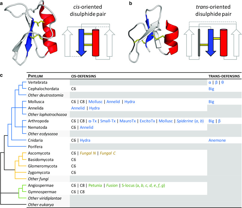
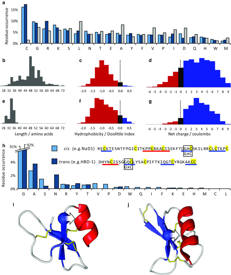
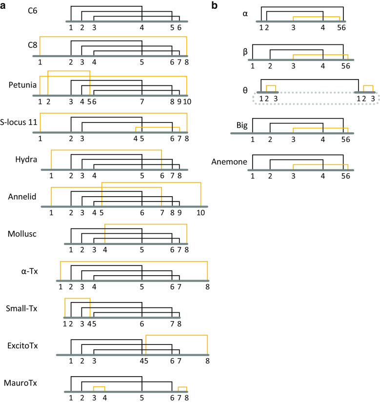
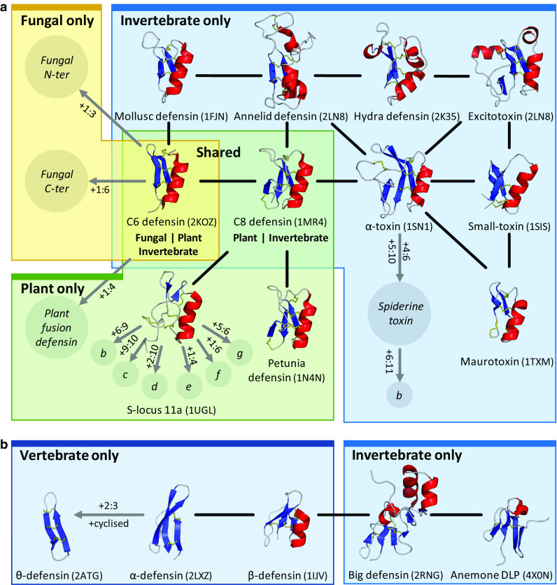
Defensins are a well-characterised group of small, disulphide-rich, cationic peptides that are produced by essentially all eukaryotes and are highly diverse in their sequences and structures. Most display broad range antimicrobial activity at low micromolar concentrations, whereas others have other diverse roles, including cell signalling (e.g. immune cell recruitment, self/non-self-recognition), ion channel perturbation, toxic functions, and enzyme inhibition. The defensins consist of two superfamilies, each derived from an independent evolutionary origin, which have subsequently undergone extensive divergent evolution in their sequence, structure and function. Referred to as the cis- and trans-defensin superfamilies, they are classified based on their secondary structure orientation, cysteine motifs and disulphide bond connectivities, tertiary structure similarities and precursor gene sequence. The utility of displaying loops on a stable, compact, disulphide-rich core has been exploited by evolution on multiple occasions. The defensin superfamilies represent a case where the ensuing convergent evolution of sequence, structure and function has been particularly extreme. Here, we discuss the extent, causes and significance of these convergent features, drawing examples from across the eukaryotes.

Keywords: Antimicrobial peptide; Disulphide-rich protein; Divergent evolution; Evolutionary constraint; Evolvability; Protein superfamily.


Figures

Fig. 1
Fig. 1
Architecture and taxonomic distribution of cis- and trans-defensins. a The plant defensin NaD1 (PDB:1MR4) is a typical cis-defensin in which both of the most conserved disulphides (yellow) from the final β-strand (blue) point in the same direction and bond to the same α-helix (red). b The human β-defensin HBD-1 (PDB:1IJV) is a typical trans-defensin in which the disulphides from the final β-strand point in opposite directions, therefore, bonding to different secondary structure elements. Non-conserved disulphides are represented as dashed lines in the secondary structure diagrams. Adapted from [13]. c A simplified phylogeny of eukaryotic phyla, annotated with the occurrence of different structural classes and cysteine motifs (in italics) from each defensin superfamily. Classes specific to a kingdom are coloured as in the phylogeny. Classes are described in more detail in Figs. 4 and 5. Phyla with no known defensins from each of the superfamilies are filled in grey
Fig. 2
Fig. 2
Organisation of defensin precursor proteins. All defensins are produced with N-terminal endoplasmic reticulum (ER) signal sequences (to direct them to the ER for disulphide bond formation) in addition to the mature defensin domain (Def). Examples of defensins that adopt this structure include a scorpion C6 and plant C8 class I defensins. Other defensins are produced with additional prodomains (Pro) that can be positioned b C-terminally (e.g. mussel, and plant C8 class II defensins) or c N-terminally (e.g. insect C6 and vertebrate α- and β-defensins) of the mature domain. d θ-defensin precursors are truncated α-defensin prologues with a premature stop codon after the first 12 residues, from which a 9-mer fragment is excised, dimerised, and ligated to create the backbone-cyclised θ-defensin. The sequence after the stop codon is still highly similar to the α-defensin (Pseudo). Domain lengths not to scale
Fig. 3
Fig. 3
Amino acid sequence properties of cis- and trans-defensins. a Average amino acid residue occurrence for the cis-defensins (light blue), trans-defensins (dark blue) and whole Uniprot database (grey). Distributions of length, hydrophobicity and charge for bd 1820 cis-defensins and eg 894 trans-defensins. The common GxC motif occurs in both cis-defensins (e.g. NaD1) and trans-defensins (e.g. HBD-1). h Residue bias in the first position of the GxC motif in the cis-defensins (excluding S-locus and spiderines, which have an additional disulphide at this location) and the trans-defensins (excluding α- and θ-defensins, which lack an α-helix and so are unconstrained at this location). In both i cis-defensins (PDB:1MR4) and j trans-defensins (PDB:1IJV), the glycine (sphere) is oriented such that a non-hydrogen R-group on any other amino acid in this position (arrow) would clash with the α-helix. β-Strands in blue, α-helices in red, disulphide bonds in yellow
Fig. 4
Fig. 4
Defensin disulphide connectivities. Disulphide connectivities for the a cis-defensins and b trans-defensins. The most highly conserved disulphides are indicated in black and disulphides that are unique to each class are indicated in yellow. The dashed line indicates cyclisation of the θ-defensin
Fig. 5
Fig. 5
Relatedness within the cis- and trans-defensins. Evidence for common origin in the a cis-defensins and b trans-defensins. Structures are shown for cysteine patterns with solved structures, classes with unresolved structures are represented by italicised names in circles. Putative disulphides unique to a class are denoted as x:y where x and y are the additional cysteines involved in the disulphide. Uncharacterised variants with additional disulphides are denoted by single letters (e.g. S-locus 11b, etc.). Black lines indicate homology evidence from structural similarity, grey lines indicate evidence from gene structure and organisation. The PDB codes for the proteins are given in parentheses. Structures are organised by kingdom, with a fungal representative as an example of the shared C6 defensins and a plant representative for the shared C8 defensins (colours as used in Fig. 1)
Fig. 6
Fig. 6
Defensin dimerisation and lipid-mediated oligomerisation. a The plant C8 defensin NaD1 (PDB:4CQK) forms a homodimer that binds negatively charged phospholipid head groups via a cationic grip [94]. b The human β-defensin HBD-2 (PDB:1FD4) forms a structurally similar dimer [102]. Protein surface charge is indicated by blue (positive) and red (negative). Lipids are shown as sticks with phosphate in white and oxygen in red. c NaD1 dimers assemble into an arching oligomeric structure after interaction with the anionic head groups of PIP2 within an extended cationic groove on the surface of the NaD1 oligomer (PDB:4CQK). Alternating dimers in white and blue
Fig. 7
Fig. 7
Blocking of ion channels by defensin-like peptides. a The common cis-defensin C6 fold is adapted in some scorpion toxins, along with four toxin-specific structural classes with distinct additional disulphides. A structurally uncharacterised cis-defensin is also present in lynx spider venom (spiderine). b The trans-defensin fold has been recruited to toxic function such as crotamine in snakes, OvDLP from platypus and helofensin from bearded lizards. The anemone fold is also used in sea anemone neurotoxins. c Comparison of the C6 defensin fold with different functions. Toxins contain a conserved KCφN motif, whereas antimicrobial defensins contain the broader ζCxx motif at the same location, in addition to a large, flexible loop (φ = hydrophobe, ζ = hydrophile). d Charybdotoxin binds to the tetrameric K v channel (white surface) and inserts a lysine residue into the first of the channel’s four K+ binding sites, blocking the transport of K+ ions (blue spheres) through the cell membrane (blue) (PDB:4JTA) [93]
Fig. 8
Fig. 8
Enzyme inhibition by defensin-like peptides. a The C8 cis-defensins fold has been adapted to enzyme inhibitory function in the Arabidopsis thaliana trypsin inhibitor (ATT) (PDB:1JXC) [11], and b the trans-defensins contain an α-amylase inhibitor, helianthamide, from sea anemones (PDB:4X0N) [12]. Inhibitory loop highlighted in green (putative for ATT) [11]. c The enzyme α-amylase (white surface) uses an aspartate-glutamate dyad in its active site for hydrolysis (green), which is competitively inhibited by the bound helianthamide (PDB:4X0N)

References

    1. Brogden KA. Antimicrobial peptides: pore formers or metabolic inhibitors in bacteria? Nat Rev Microbiol. 2005;3(3):238–250. doi: 10.1038/nrmicro1098. - DOI - PubMed
    1. Ganz T. Defensins: antimicrobial peptides of innate immunity. Nat Rev Immunol. 2003;3(9):710–720. doi: 10.1038/nri1180. - DOI - PubMed
    1. Boulanger N, et al. Characterization of a defensin from the sand fly Phlebotomus duboscqi induced by challenge with bacteria or the protozoan parasite Leishmania major . Infect Immun. 2004;72(12):7140–7146. doi: 10.1128/IAI.72.12.7140-7146.2004. - DOI - PMC - PubMed
    1. Pushpanathan M, Gunasekaran P, Rajendhran J. Antimicrobial peptides: versatile biological properties. Int J Pept. 2013;2013:675391. doi: 10.1155/2013/675391. - DOI - PMC - PubMed
    1. Assadi-Porter FM, et al. Key amino acid residues involved in multi-point binding interactions between brazzein, a sweet protein, and the T1R2-T1R3 human sweet receptor. J Mol Biol. 2010;398(4):584–599. doi: 10.1016/j.jmb.2010.03.017. - DOI - PMC - PubMed

Publication types