. 2016 Sep 26;8(9):2896-2913.
doi: 10.1093/gbe/evw195.

Animal Mitochondrial DNA as We Do Not Know It: mt-Genome Organization and Evolution in Nonbilaterian Lineages

Affiliations

Animal Mitochondrial DNA as We Do Not Know It: mt-Genome Organization and Evolution in Nonbilaterian Lineages

Dennis V Lavrov et al. Genome Biol Evol. .

Abstract

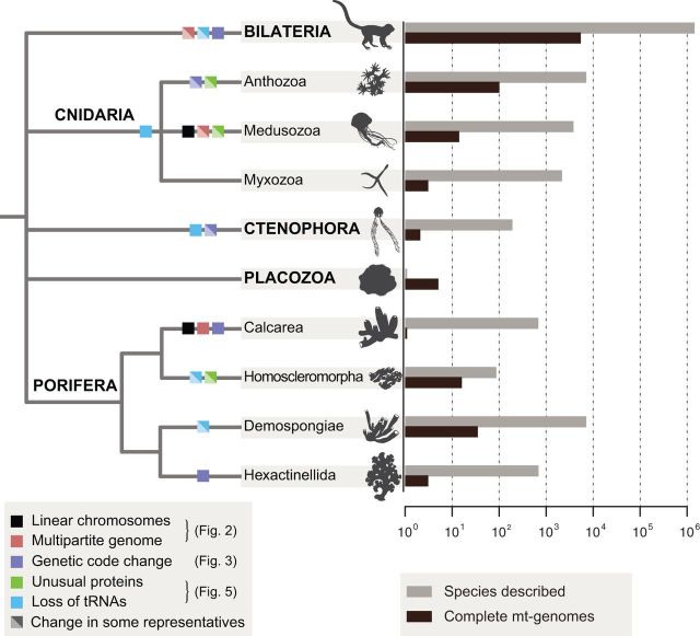
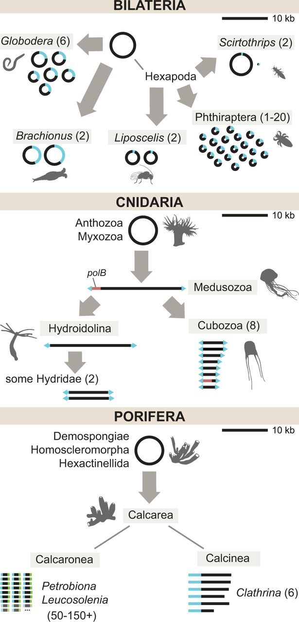
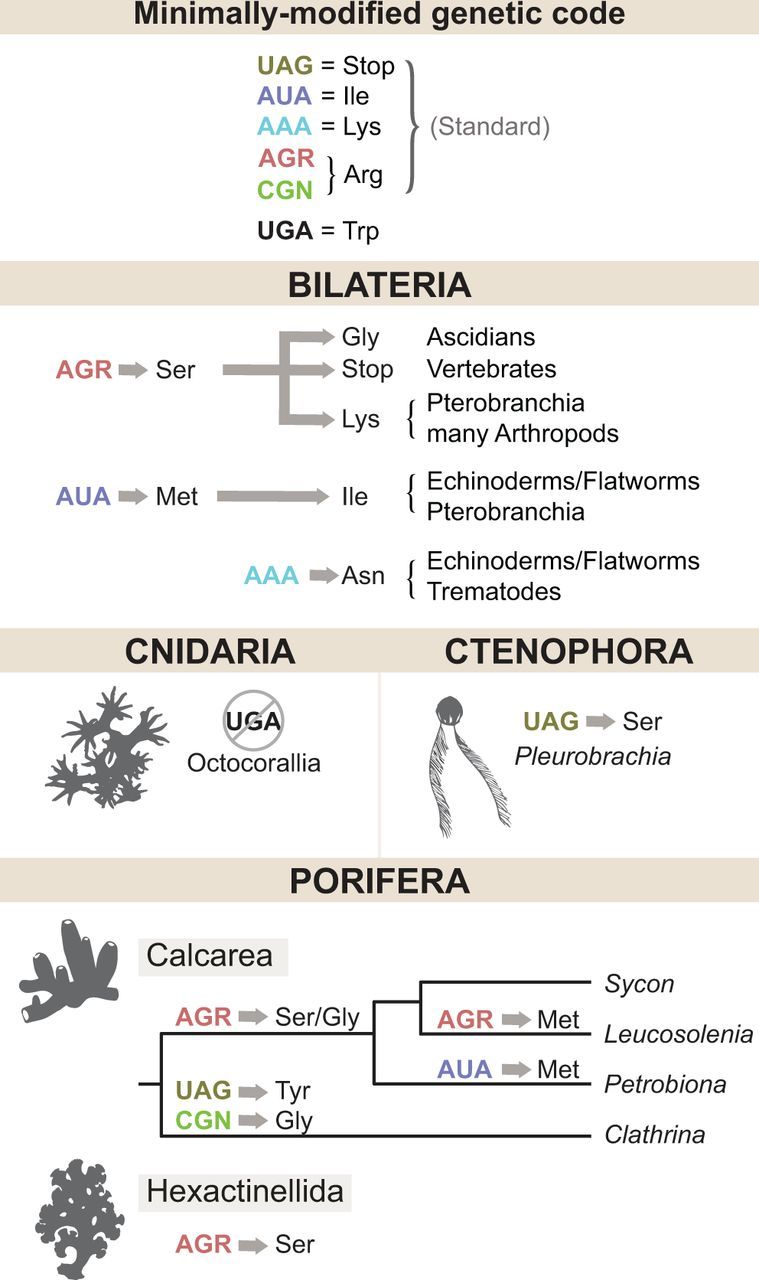
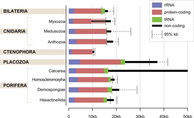
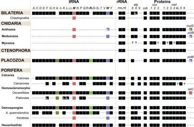
Animal mitochondrial DNA (mtDNA) is commonly described as a small, circular molecule that is conserved in size, gene content, and organization. Data collected in the last decade have challenged this view by revealing considerable diversity in animal mitochondrial genome organization. Much of this diversity has been found in nonbilaterian animals (phyla Cnidaria, Ctenophora, Placozoa, and Porifera), which, from a phylogenetic perspective, form the main branches of the animal tree along with Bilateria. Within these groups, mt-genomes are characterized by varying numbers of both linear and circular chromosomes, extra genes (e.g. atp9, polB, tatC), large variation in the number of encoded mitochondrial transfer RNAs (tRNAs) (0-25), at least seven different genetic codes, presence/absence of introns, tRNA and mRNA editing, fragmented ribosomal RNA genes, translational frameshifting, highly variable substitution rates, and a large range of genome sizes. This newly discovered diversity allows a better understanding of the evolutionary plasticity and conservation of animal mtDNA and provides insights into the molecular and evolutionary mechanisms shaping mitochondrial genomes.

Keywords: Cnidaria; Ctenophora; Metazoa; Placozoa; Porifera; mitochondrial DNA.


Figures

F<sc>ig</sc>. 1.—
Fig. 1.—
Mitochondrial genomes in Metazoa. Consensus view of animal relationships with mapped changes in mt-genome architecture and gene content. Diagonally split boxes indicate changes that occur only in some representatives of the group. Black bars to the right indicate numbers of complete mtDNA sequences in the NCBI RefSeq database as of April 9, 2016 (for data on nuclear genomes, see [Dunn and Ryan 2015]). Gray bars indicate estimated numbers of described species and were obtained from the Global Invertebrate Genomics Alliance (GIGA Community of Scientists 2014). Only the taxa that are mentioned in the text are included. See associated figures for a more detailed depiction of each of these changes. Branch lengths were chosen for illustration purposes only.
F<sc>ig</sc>. 2.—
Fig. 2.—
Evolution of linear and multipartite mitochondrial DNA in animals. Gray arrows indicate independent reorganization events in the common ancestor of the indicated group. Blue segments indicate regions of homology shared across chromosomes. Numbers in parentheses indicate the range of chromosome numbers (greater than 1) found in the group. Top: Multipartite mtDNA in Bilateria. Not shown: An unknown number of mini-circle chromosomes in Dicyema (Watanabe et al. 1999). Middle: Linear and multipartite mtDNA in Cnidaria. The red segment denotes the presence of a polB coding sequence in some Medusozoa. Arrowheads at the ends of linear chromosomes signify inverted terminal repeat sequences. Bottom: Multipartite linear mtDNA in Porifera. Faded chromosomes and ellipses indicate the inferred presence of an uncertain number of additional chromosomes. Blue and green segments in Leucosolenia and Petrobiona show distinct end sequences shared across chromosomes, but which are not homologous between the two genera. Genomes are drawn to scale with respect to the 10 kb scale bar in the upper right part of each panel.
F<sc>ig</sc>. 3.—
Fig. 3.—
Changes in the animal mitochondrial genetic code. Inferred changes in the identities of six codons (listed in the top panel) are shown by arrows. Consecutive changes are shown by a sequence of arrows. Amino-acid identities are designated by the IUPAC three-letter code. A crossed out circle indicates a codon that is no longer used.
F<sc>ig</sc>. 4.—
Fig. 4.—
Size and coding content of animal mitochondrial DNA. Colored bars indicate the average number of nucleotides coding for ribosomal RNA (blue), protein (red), transfer RNA (green), or noncoding (black) computed using all available complete mt-genome sequences published in the NCBI RefSeq database as of April 9, 2016. Gray error bars indicate the upper and lower boundaries of the 95% interquantile range for genome sizes (2.5th and 97.5th percentiles). The shaded portion of the red bar in Myxozoa indicates additional protein-coding genes likely present in these genomes, but not recognizable due to an extreme rate of sequence evolution.
F<sc>ig</sc>. 5.—
Fig. 5.—
Variation in RNA and protein-coding gene content in animal mitochondrial DNA. Left: taxonomic positions of groups with variable gene content. Central: variation in tRNA gene content. Middle: the presence of rRNA genes. Right: the presence/absence of protein-coding genes. Colored boxes highlight the identities of specific genes discussed in the text. Diagonally split boxes indicate genes that are absent in some lineages of the indicated group. Question marks denote genes for which presence or absence has not been confidently determined. Abbreviations: atp6, atp8–9: subunits 6, 8, and 9 of F0 adenosine triphosphatase (ATP) synthase; cob: apocytochrome b; cox1–3: cytochrome c oxidase subunits 1–3; nad1–6 and nad4L: NADH dehydrogenase subunits 1–6 and 4L; polB: DNA polymerase β; tatC: twin-arginine translocase component C; rns and rnl: SSU and LSU rRNAs. The tRNA genes are identified by the one-letter code for their corresponding amino acid; subscripts denote different genes for isoacceptor tRNAs, where I1 = trnI(CAU), I2 = trnI(GAU), L1 = trnL(UAG), L2 = trnL(UAA), R1 = trnR(UCG), R2 = trnR(UCU), S1 = trnS(UCN), S2 = trnS(UGA).
F<sc>ig</sc>. 6.—
Fig. 6.—
mtDNA-based animal phylogeny showing variable rates of mt-sequence evolution. Complete or nearly complete mt-genomes from all available nonbilaterian animals and a few selected bilaterian animals were downloaded from the GenBank. In addition, mitochondrial genomes of Periphylla periphylla and Polypodium hydriforme, were assembled from high throughput transcriptomic and genomic data (SRX956805 and SRX687102, respectively). Inferred amino acid sequences from nine mitochondrial genes (cob, cox1-3, nad1-5) were aligned with MAFFT v7.215 (Katoh and Standley 2013). Conserved blocks within the alignments were selected with Gblocks 0.91 b (Talavera and Castresana 2007) using relaxed parameters (parameters 1 and 2 = ½, parameter 3 = 8, parameter 4 = 5, all gap positions in parameter 5). Cleaned alignments were concatenated in a dataset 2,228 positions in length. Bilateria, Cnidaria, Ctenophora, Placozoa, and Porifera were constrained as monophyletic and the best constrained topology was identified using RAxML with the MTREV + GAMMA + F substitution model and 32 initial tree searches. Subsequently, branch lengths were re-estimated with the CAT + GTR + Γ4 model in PhyloBayes MPI 1.4e (Lartillot et al. 2013).

References

    1. Aanen D, Spelbrink J, Beekman M. 2014. What cost mitochondria? The maintenance of functional mitochondrial DNA within and across generations. Philos Trans R Soc Lond B Biol Sci. 369:20130438.. - PMC - PubMed
    1. Abascal F, Posada D, Zardoya R. 2012. The evolution of the mitochondrial genetic code in arthropods revisited. Mitochondrial DNA 23:84–91. - PubMed
    1. Ameur A, et al. 2011. Ultra-deep sequencing of mouse mitochondrial DNA: mutational patterns and their origins. PLoS Genet. 7:e1002028.. - PMC - PubMed
    1. Anderson S, et al. 1981. Sequence and organization of the human mitochondrial genome. Nature 290:457–465. - PubMed
    1. Armstrong MR, Block VC, Phillips MS. 2000. A multipartite mitochondrial genome in the potato cyst nematode Globodera pallida. Genetics 154:181–192. - PMC - PubMed

LinkOut - more resources