. 2016 Oct 9;428(20):4060-4072.
doi: 10.1016/j.jmb.2016.08.018. Epub 2016 Aug 21.

The HMGB1 C-Terminal Tail Regulates DNA Bending

Affiliations

The HMGB1 C-Terminal Tail Regulates DNA Bending

Rebecca H Blair et al. J Mol Biol. .

Abstract

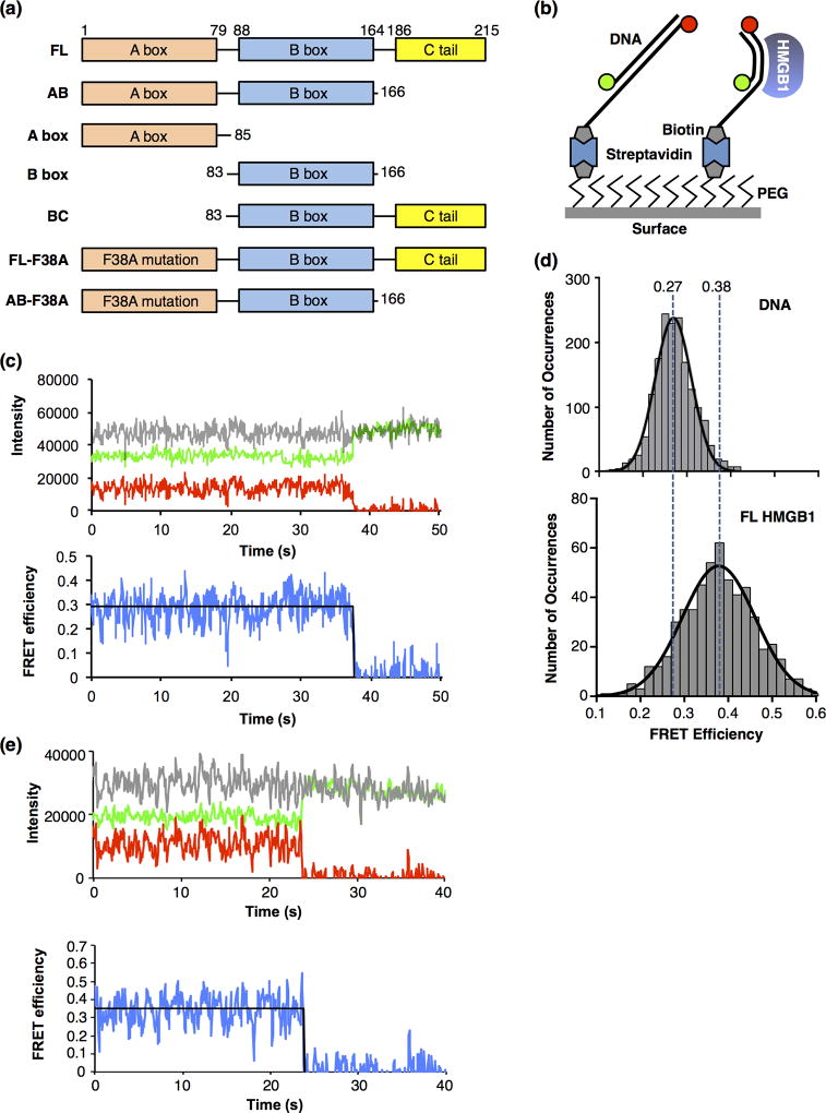
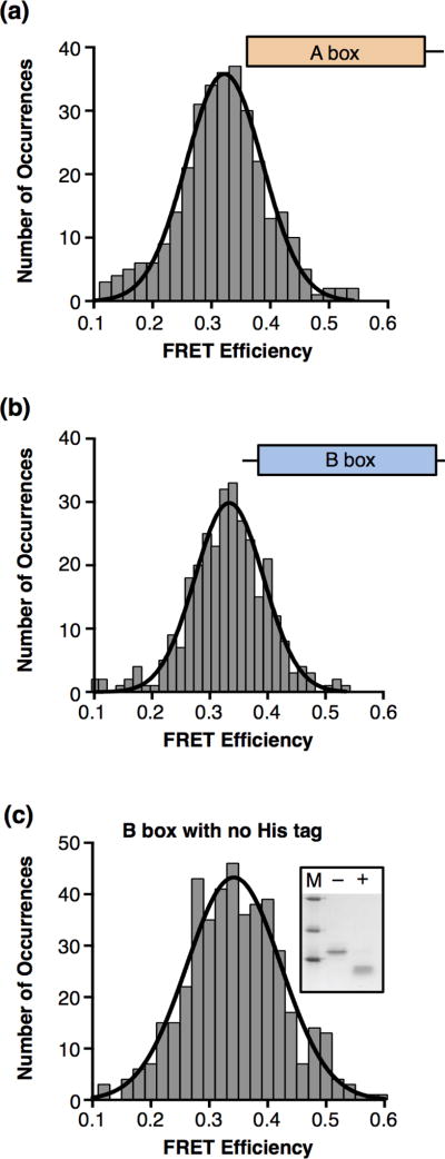
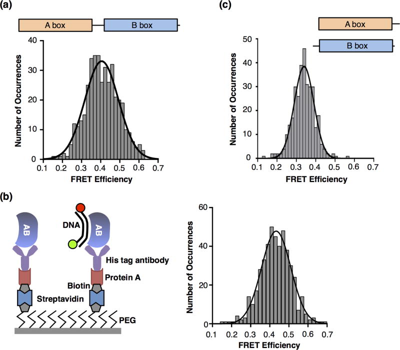
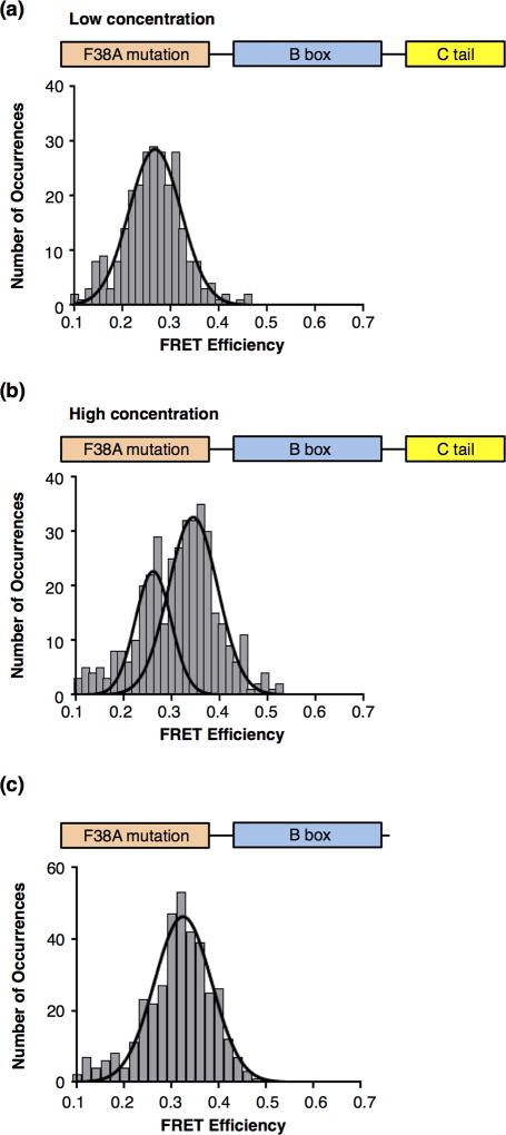
High mobility group box protein 1 (HMGB1) is an architectural protein that facilitates the formation of protein-DNA assemblies involved in transcription, recombination, DNA repair, and chromatin remodeling. Important to its function is the ability of HMGB1 to bend DNA non-sequence specifically. HMGB1 contains two HMG boxes that bind and bend DNA (the A box and the B box) and a C-terminal acidic tail. We investigated how these domains contribute to DNA bending by HMGB1 using single-molecule fluorescence resonance energy transfer (FRET), which enabled us to resolve heterogeneous populations of bent and unbent DNA. We found that full-length (FL) HMGB1 bent DNA more than the individual A and B boxes. Removing the C-terminal tail resulted in a protein that bent DNA to a greater extent than the FL protein. These data suggest that the A and B boxes simultaneously bind DNA in the absence of the C-terminal tail, but the tail modulates DNA binding and bending by one of the HMG boxes in the FL protein. Indeed, a construct composed of the B box and the C-terminal tail only bent DNA at higher protein concentrations. Moreover, in the context of the FL protein, mutating the A box such that it could not bend DNA resulted in a protein that bent DNA similar to a single HMG box and only at higher protein concentrations. We propose a model in which the HMGB1 C-terminal tail serves as an intramolecular damper that modulates the interaction of the B box with DNA.

Keywords: DNA bending; FRET; HMGB1; TIRF microscopy; single-molecule.


Figures

Figure 1
Figure 1
Full length HMGB1 bends DNA molecules, as detected using smFRET. (a) Domain structures of full length human HMGB1 (FL) and the truncations and mutations used in this study. (b) Depiction of the surface used for the smFRET experiments. The glass coverslip was coated with polyethylene glycol (PEG) to prevent nonspecific interactions of the proteins and DNA with the surface. Some PEG molecules were functionalized with biotin, which allowed immobilization of streptavidin and biotinylated DNA labeled with donor and acceptor fluorophores (green and red circles). When HMGB1 bends the DNA (right side), the donor and acceptor fluorophores move closer together, resulting in a higher FRET efficiency than the unbent DNA (left side). (c) A representative time trace of the fluorescent signals from a single molecule of linear DNA. The upper plot shows emission from the donor fluorophore (green), acceptor fluorophore (red), and the sum of the donor and acceptor (grey) over time. The lower plot shows the FRET efficiency (blue) calculated from the donor and acceptor emissions, and the fit of the FRET efficiency (black line) over time. The acceptor dye photobleached at 38 s, resulting in a loss of FRET and an anticorrelated change in the donor fluorophore. For purposes of display, the signals were smoothed by averaging 3 adjacent time frames. (d) HMGB1 (FL) bends DNA to a single state. The upper plot shows 1701 FRET occurrences for DNA in the absence of protein. The data were fit to a Gaussian and the mean FRET efficiency is 0.27. The lower plot shows 553 FRET occurrences for DNA in the presence of 50 nM FL HMGB1. The data were fit to a Gaussian and the mean FRET efficiency is 0.38. The vertical dashed lines are centered over the mean FRET efficiencies and illustrate the HMGB1-induced increase in FRET due to DNA bending. (e) A representative time trace of the fluorescent signals from a single molecule of DNA bent by HMGB1. The upper plot shows emission from the donor fluorophore (green), acceptor fluorophore (red), and the sum of the donor and acceptor (grey) over time. The lower plot shows the FRET efficiency (blue) calculated from the donor and acceptor emissions, and the fit of the FRET efficiency (black line) over time. The acceptor dye photobleached at ~24 s, resulting in a loss of FRET and an anticorrelated change in the donor fluorophore. For purposes of display, the signals were smoothed by averaging 3 adjacent time frames.
Figure 2
Figure 2
The HMGB1 A box and B box individually bend DNA to the same extent as the full length protein. (a) The HMGB1 A box bends DNA. Shown is a histogram of 307 occurrences obtained from single DNA molecules in the presence of 100 nM A box. The data were fit to a single Gaussian with a mean FRET efficiency of 0.32. (b) The HMGB1 B box bends DNA. The histogram shows 306 occurrences for DNA in the presence of 100 nM B box. The mean FRET efficiency obtained from the Gaussian fit is 0.33. (c) The B box with the His tag removed bends DNA to the same extent as the His-tagged B box protein. The inset gel shows the B box before (−) and after (+) incubation with enterokinase protease to cleave off the His tag; M designates markers of molecular weight 31, 21.5, and 14.4 kD. The histogram shows 440 occurrences for DNA in the presence of 100 nM cleaved B box. The mean FRET efficiency obtained from the Gaussian fit is 0.34.
Figure 3
Figure 3
The AB construct, which lacks the C-terminal tail, bends DNA to a higher FRET state, suggesting that both the A and the B boxes together bind and bend the DNA. (a) AB protein bends DNA to a higher FRET. 403 occurrences for DNA in the presence of 20 nM AB protein were histogrammed and fit to a Gaussian with mean FRET efficiency of 0.41. (b) AB protein immobilized on the slide surface bent DNA to a high FRET state. The schematic on the left depicts the surface used to immobilize the AB protein and observe bound and bent DNA via smFRET. The histogram on the right shows 466 occurrences after flowing in 0.5 nM DNA. The data were fit to a single Gaussian with a mean FRET efficiency of 0.43. (c) Observing the higher FRET state requires that the A box and B box be covalently linked. The histogram shows 333 occurrences after 75 nM A box plus 50 nM B box were flowed into the chamber. The data were fit to a single Gaussian with a mean FRET efficiency of 0.34.
Figure 4
Figure 4
The C-terminal tail reduces the apparent affinity with which the B box binds and bends DNA. (a) The BC protein does not bend DNA when used at the same concentration at which the B box alone bent DNA. BC protein (the B box plus the C-terminal tail) was flowed into the chamber at 50 nM; 292 occurrences were histogrammed. The Gaussian fit revealed a mean FRET efficiency of 0.27, consistent with unbent DNA. (b) A population of bent DNA is observed occurs at a higher concentration of the BC protein. In the presence of 200 nM BC protein, 190 occurrences were histogrammed. The histogram revealed two FRET populations that were fit with two Gaussians to yield mean FRET efficiencies of 0.28 and 0.33.
Figure 5
Figure 5
The C-terminal tail impedes DNA bending by the B box in the context of full length HMGB1 containing a mutant A box. (a) Removing the ability of the A box to bend DNA in the context of the full length protein resulted in no DNA bending at low protein concentrations. The plot shows 269 occurrences for DNA in the presence of 32 nM FL-F38A protein. The data were fit to a Gaussian with a mean FRET efficiency of 0.27. (b) At a higher concentration of FLF38A, a population of bent DNA is observed. 402 occurrences observed in the presence of 5.5 µM FL-F38A were histogrammed to reveal two populations with mean FRET efficiencies of 0.26 and 0.34, corresponding to unbent and bent DNA, respectively. (c) Removing the C-terminal tail from the protein with the mutant A box enabled DNA binding and bending at low concentrations of protein. AB-F38A (56 nM) was flowed into the chamber, 369 occurrences were histogrammed. The data revealed a single FRET state with a mean efficiency of 0.32, indicative of bent DNA.

References

    1. Stros M. HMGB proteins: interactions with DNA and chromatin. Biochim. Biophys. Acta. 2010;1799:101–113. - PubMed
    1. Agresti A, Bianchi ME. HMGB proteins and gene expression. Curr. Opin. Genet. Dev. 2003;13:170–178. - PubMed
    1. Thomas JO, Travers AA. HMG1 and 2, and related ‘architectural’ DNA-binding proteins. Trends Biochem. Sci. 2001;26:167–174. - PubMed
    1. Lange SS, Vasquez KM. HMGB1: the jack-of-all-trades protein is a master DNA repair mechanic. Mol. Carcinog. 2009;48:571–580. - PMC - PubMed
    1. Malarkey CS, Churchill ME. The high mobility group box: the ultimate utility player of a cell. Trends Biochem. Sci. 2012;37:553–562. - PMC - PubMed

Publication types