Review
. 2016 Aug 25:2:16061.
doi: 10.1038/nrdp.2016.61.

Ovarian cancer

Affiliations
Review

Ovarian cancer

Ursula A Matulonis et al. Nat Rev Dis Primers. .

Abstract

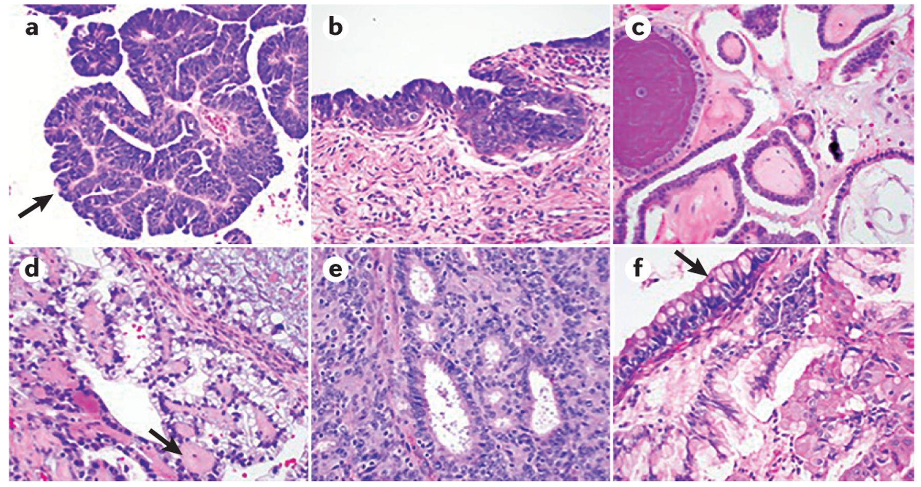
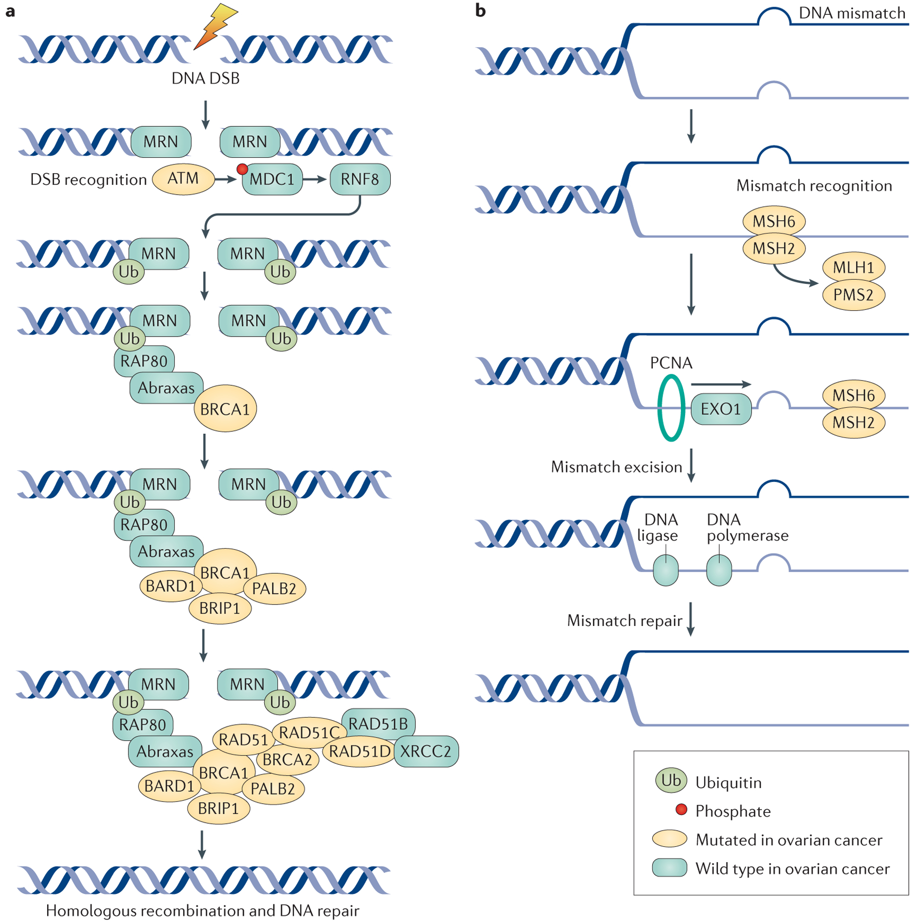
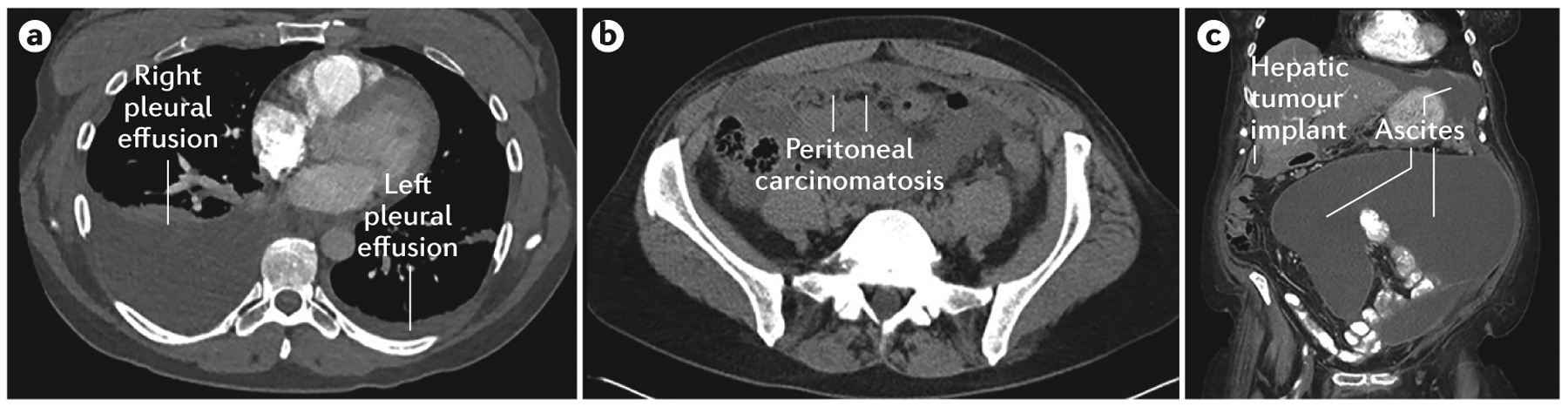
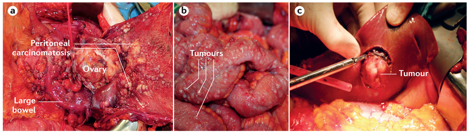
Ovarian cancer is not a single disease and can be subdivided into at least five different histological subtypes that have different identifiable risk factors, cells of origin, molecular compositions, clinical features and treatments. Ovarian cancer is a global problem, is typically diagnosed at a late stage and has no effective screening strategy. Standard treatments for newly diagnosed cancer consist of cytoreductive surgery and platinum-based chemotherapy. In recurrent cancer, chemotherapy, anti-angiogenic agents and poly(ADP-ribose) polymerase inhibitors are used, and immunological therapies are currently being tested. High-grade serous carcinoma (HGSC) is the most commonly diagnosed form of ovarian cancer and at diagnosis is typically very responsive to platinum-based chemotherapy. However, in addition to the other histologies, HGSCs frequently relapse and become increasingly resistant to chemotherapy. Consequently, understanding the mechanisms underlying platinum resistance and finding ways to overcome them are active areas of study in ovarian cancer. Substantial progress has been made in identifying genes that are associated with a high risk of ovarian cancer (such as BRCA1 and BRCA2), as well as a precursor lesion of HGSC called serous tubal intraepithelial carcinoma, which holds promise for identifying individuals at high risk of developing the disease and for developing prevention strategies.


Conflict of interest statement

Competing interests

U.A.M. has served as a consultant for AstraZeneca, ImmunoGen, Pfizer, Genentech and Merck. All other authors declare no competing interests.

Figures

Figure 1 |
Figure 1 |. Histological subtypes of ovarian cancer.
a | High-grade serous carcinoma (HGSC) is characterized by severe nuclear atypia, high nuclear-to-cytoplasmic ratio and abundant mitoses. Papillary architecture (arrow) is also often present. b | Serous tubal intraepithelial carcinoma (STIC) lesions share the same morphological features as HGSC, with severe atypia, mitoses and lack of polarity. STIC lesions are thought to be precursors for HGSC. c | Low-grade serous carcinoma (LGSC) shows papillary architecture, but only mild nuclear atypia and a lower nuclear-to-cytoplasmic ratio. d | Clear-cell carcinoma is characterized by large atypical tumour cells with frequent clearing of the cytoplasm and stromal hyalinization (arrow). e | Endometrioid adenocarcinoma is characterized by gland formation that recapitulates endometrial glands and is graded based on cellular architecture and nuclear atypia. f | Mucinous adenocarcinoma shows mucin-filled tumour cells, with frequent goblet cell forms present (arrow).
Figure 2 |
Figure 2 |. DNA repair mechanisms and ovarian cancer.
a | The double-stranded DNA break and homologous repair process begins with recognition and sensing of double-strand breaks (DSBs) by the meiotic recombination 11 homologue 1 (MRE11)–RAD50–Nijmegen breakage syndrome protein 1 (NBS1) (MRN) complex, which acts as an activation site for the serine-protein kinase ATM. ATM has a crucial role in DNA repair by coordinating homologous recombination. ATM phosphorylates the histone H2AX, which directly binds to mediator of DNA damage checkpoint protein 1 (MDC1) and NBS1 of the MRN complex, to enhance ATM binding. MDC1 phosphorylation results in a binding site for the E3 ubiquitin-protein ligase RING finger protein 8 (RNF8), which allows ubiquitin-mediated recruitment of downstream DNA damage response proteins, such as receptor-associated protein 80 (RAP80; encoded by UIMC1). RAP80 is an ubiquitin-interaction motif-containing protein that associates with the breast cancer type 1 susceptibility protein (BRCA1) complex through its interaction with Abraxas (encoded by FAM175A); Abraxas is thought to function as a central adaptor protein and contains domains required for BRCA1 interactions. The RAP80–Abraxas complex is crucial for recruiting BRCA1 to the site of DNA repair. BRCA1 and BRCA2 function as scaffolds for other proteins involved in DNA repair. BRCA1-associated RING domain protein 1 (BARD1) and BRCA1-interacting protein 1 (BRIP1; also known as Fanconi anaemia group J protein) bind directly to BRCA1; BARD1 forms a heterodimer with BRCA1, which is essential for mutual stability. BRIP1 also binds to BRCA1 and is required for S phase checkpoint activation. Partner and localizer of BRCA2 (PALB2) helps BRCA1 and BRCA2 bind at sites of DNA damage and helps load RAD51 proteins on to the BRCA proteins; the DNA repair protein XRCC2 is one of the five paralogues of RAD51. Mutations in genes involved in homologous repair lead to defective DNA repair mechanisms, the accumulation of DSBs and an increase in the risk of developing ovarian tumours. b | DNA mismatch repair is mediated by the MutS protein homologue 2 (MSH) proteins, as well as the endonuclease PMS2 and proliferating cell nuclear antigen (PCNA). DNA mismatch repair processes are aberrant in ovarian cancer due to mutations in the genes encoding MutL protein homologue 1 (MLH1), MSH2, MSH6 and PMS2. MSH2 and MSH6 form a heterodimeric complex, which initially identifies mismatched bases and initiates DNA repair. Binding of this complex to the mismatched bases enables the recruitment of MLH1 and PMS2. PCNA attaches to the sites of base mismatch and helps to recruit and tether exonuclease 1 (EXO1; a member of the RAD2 exonuclease family) to the sites of DNA damage. EXO1 excises the mismatched bases, which are then repaired by DNA polymerase and DNA ligase.
Figure 3 |
Figure 3 |. CT scans from a patient with stage IV ovarian cancer.
a | Right and left pleural effusions. b | Peritoneal carcinomatosis. c | Large volume ascites and a peritoneal hepatic implant.
Figure 4 |
Figure 4 |. Tumour burden in ovarian cancer.
a | Surgical removal of ovarian tumours in a patient 63 years of age with bilateral advanced-stage high-grade serous carcinoma (HGSC). b | A patient with a bilateral HGSC and peritoneal carcinomatosis with involvement of intestinal surfaces. c | Surgical removal of a serosal tumour implant located on the surface of the liver.

References

    1. Oswald AJ & Gourley C Low-grade epithelial ovarian cancer: a number of distinct clinical entities? Curr. Opin. Oncol 27, 412–419 (2015). - PubMed
    1. Groen RS, Gershenson DM & Fader AN Updates and emerging therapies for rare epithelial ovarian cancers: one size no longer fits all. Gynecol. Oncol 136, 373–383 (2015). - PubMed
    1. Mangili G et al. Unraveling the two entities of endometrioid ovarian cancer: a single center clinical experience. Gynecol. Oncol 126, 403–407 (2012). - PubMed
    1. Callegaro-Filho D et al. Small cell carcinoma of the ovary-hypercalcemic type (SCCOHT): a review of 47 cases. Gynecol. Oncol 140, 53–57 (2016). - PMC - PubMed
    1. Witkowski L et al. The influence of clinical and genetic factors on patient outcome in small cell carcinoma of the ovary, hypercalcemic type. Gynecol. Oncol 141, 454–460 (2016). - PMC - PubMed

MeSH terms