. 2016 Aug 25:6:32243.
doi: 10.1038/srep32243.

Autophagy-associated dengue vesicles promote viral transmission avoiding antibody neutralization

Affiliations

Autophagy-associated dengue vesicles promote viral transmission avoiding antibody neutralization

Yan-Wei Wu et al. Sci Rep. .

Abstract

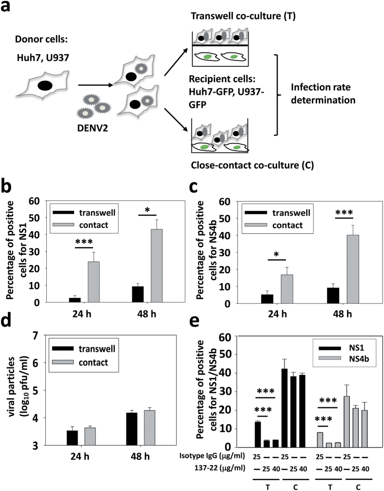
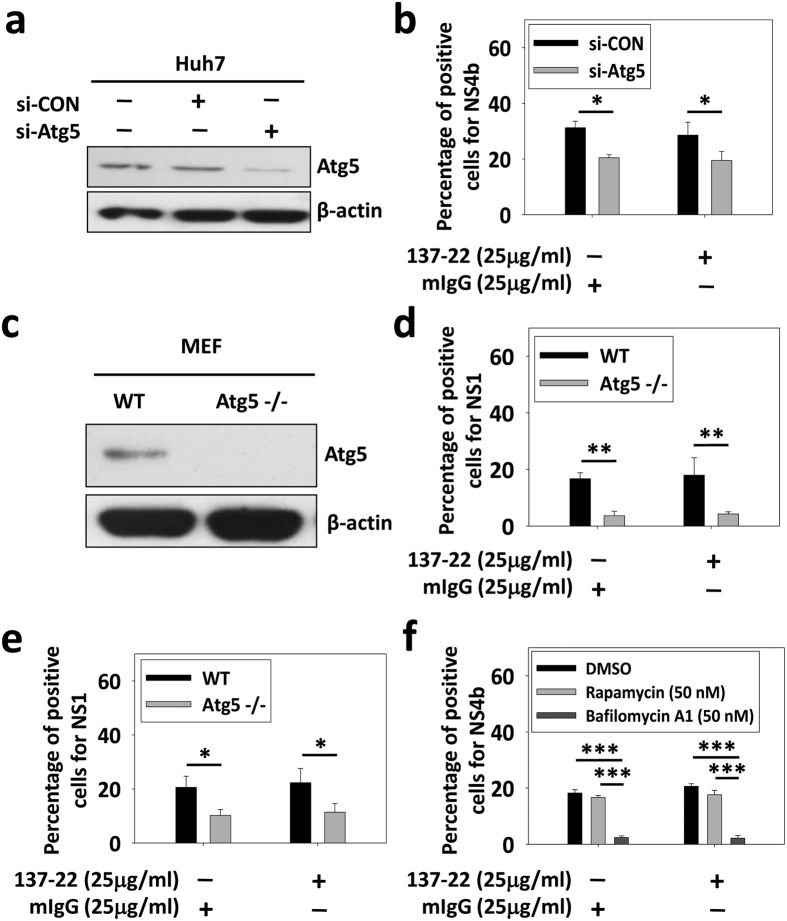
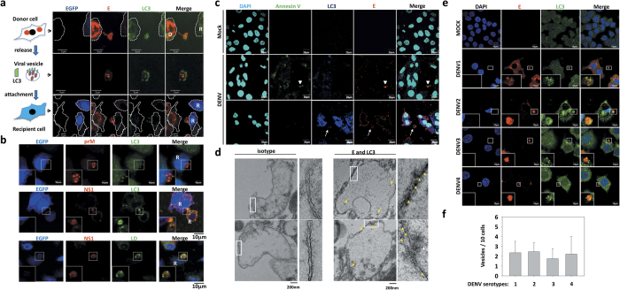
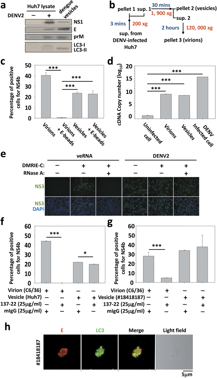
One of the major defense mechanisms against virus spread in vivo is the blocking of viral infectibility by neutralizing antibodies. We describe here the identification of infectious autophagy-associated dengue vesicles released from infected cells. These vesicles contain viral proteins E, NS1, prM/M, and viral RNA, as well as host lipid droplets and LC3-II, an autophagy marker. The viral RNA can be protected within the autophagic organelles since anti-dengue neutralizing antibodies do not have an effect on the vesicle-mediated transmission that is able to initiate a new round of infection in target cells. Importantly, such infectious vesicles were also detected in a patient serum. Our study suggests that autophagy machinery plays a new role in dengue virus transmission. This discovery explains the inefficiency of neutralizing antibody upon dengue infection as a potential immune evasion mechanism in vivo.


Figures

Figure 1
Figure 1. Efficient infection of DENV avoids neutralizing antibody in a direct-contact co-culture.
(a) Schematic illustration of the experimental procedure. (b,c) DENV2 (PL046)-infected Huh7 cells were used as donors to further infect Huh7-GFP cells harboring an integrated GFP gene. Infected Huh7 cells were either cultured in transwells on top of the recipient cells or directly with Huh7-GFP cells for 24 h. The infection rate was quantified using a specific antibody against dengue NS1 (b) or NS4b (c) and flow cytometry analysis. (d) Comparable virion production in transwell and close-contact co-culture conditions. DENV2-infected Huh7 cells were either cultured in transwells on top of the recipient cells or directly with Huh7-GFP cells for 24 h. The virion production in the culture media was quantified using plaque assay. (e) Two different concentrations of neutralizing antibody were added in transwell and close-contact co-culture during the infection. The infection rate of the recipient was detected by flow cytometry using NS1 or NS4b antibodies. Data represent the mean ± SD of three independent experiments, *P < 0.05; ***P < 0.001; paired t-test.
Figure 2
Figure 2. Autophagy-deficient donor cells decrease the infection rate of recipients in close-contact co-culture condition.
(a) Huh7 cells were transfected with control or specific siRNA against Atg5. The expression level of Atg5 was quantified by Western blotting. (b) Control (MOI = 1) and knockdown cells (MOI = 5) were infected with DENV2 and close-contact co-cultured with Huh7-GFP cells with the isotype control or neutralizing antibody 137-22 for 48 h. The infection rate of the recipient was detected by flow cytometry with an NS4b monoclonal antibody. (c) The expression levels of Atg5 in wild-type and Atg5−/− MEF (mouse embryonic fibroblast) were quantified by Western blotting. (d) DENV2-infected wild-type or Atg5−/− MEF with MOI = 5 were close-contact co-cultured with Huh7-GFP cells with the isotype control or neutralizing antibody 137-22 for 48 h. Dengue infection rate of the recipient was detected by flow cytometry with an NS1 monoclonal antibody. (e) DENV2-infected wild-type (MOI = 5) or Atg5−/− MEF (MOI = 10) were close-contact co-cultured with Huh7-GFP cells with the isotype control or neutralizing antibody 137-22 for 48 h. Infection rate of the recipient was detected by flow cytometry with an NS1 monoclonal antibody. (f) DENV2-infected Huh7 cells were co-cultured with Huh7-GFP cells for 24 h with rapamycin or bafilomycin A1. The infection rate was quantified using a specific antibody against dengue NS4b and flow cytometry analysis. Data represent the mean ± SD of three independent experiments, *P < 0.05; **P < 0.01; ***P < 0.001; paired t-test.
Figure 3
Figure 3. Dengue viral vesicles contain autophagy machinery.
(a) After DENV2 infection for 24 h (MOI = 5), Huh7 cells were close-contact co-cultured 12 h with Huh7-GFP cells. Cells were fixed, stained with anti-E and -LC3 antibodies and analyzed with confocal microscopy. Upper line emphasizes a donor cell field, line 2 a vesicle field and bottom line a recipient cell field. Donor cells (D); Recipient cells (R); Cell edge (dashed line). (b) The presence of other viral proteins, prM, NS1 and lipid droplet (LD) was also detected using specific antibodies and lipid droplet dye. R: recipient cells. (c) After DENV2 infection for 48 h (MOI = 5), Huh7 cells were fixed and stained with anti-dengue E, LC3, and annexin V antibodies and analyzed with confocal microscopy. Apoptotic body (arrow head); Vesicle (arrow bar). (d) The vesicles were detected by TEM, and LC3 and E were detected by 10 nm (arrow head) and 20 nm (arrow bar) immunogold, respectively. (e) Four serotypes of DENV produced by C6/36 cells were used to infect Huh7 cells. The presence of E and LC3 proteins in secreted viral vesicles was detected using confocal microscopy. (f) The number of vesicles was quantified from (e). Data represent the mean ± SD of three independent experiments; paired t-test.
Figure 4
Figure 4. The number of dengue vesicles is reduced in the autophagy-deficient cells.
(a) DENV2-infected Huh7 cells at an MOI of 1 were treated with rapamycin or bafilomycin A1. After 24 and 48 h of incubation, cells were fixed and stained with anti-E and -LC3 antibodies and analyzed with confocal microscopy. (b) DENV2-infected wild-type (MOI = 5) or Atg5−/− MEF (MOI = 10) were close-contact co-cultured with Huh7-GFP cells for 24 or 48 h. Cells were fixed, stained with anti-dengue E and LC3 antibodies and analyzed with confocal microscopy. Vesicle (arrow bar). (c,d) The numbers of vesicles from (a,b) were quantified and represent the mean ± SD of ten figures in each groups, *P < 0.05; **P < 0.01; ***P < 0.001; paired t-test.
Figure 5
Figure 5. Dengue vesicles containing viral RNA are infectious.
(a) Huh7 cells were infected by DENV2 at an MOI of 1, and the supernatant was harvested 48 h post-infection to obtain the vesicles. The E, prM, NS1 and LC3 expression levels were detected by Western blotting. (b) Schematic illustration of vesicle isolation. (c) Virions-depleted vesicles by anti-E-coated magnetic beads. Isolated virions and vesicles were incubated with anti-E-coated magnetic beads overnight and retreated to naïve Huh7 cells. After 48 h incubation, infection rates were detected by NS4b. (d) Total RNA was extracted from virions of C6/36, and vesicles from infected or non-infected Huh7. The presence of viral RNA was detected by qRT-PCR using specific primers pairing NS1 sequence. (e) BHK cells were infected or transfected (DMRIE-C reagent) by DENV2 or veRNA (RNA from purified vesicles) with or without RNase A treatment. After 24 or 72 h, the expression of viral NS3 was revealed by IFA. (f) Huh7 cells were infected with vesicles or virions isolated from the culture medium of infected cells. The infection rate in the presence or absence of neutralizing antibody 137-22 was quantified using an antibody against NS4b and flow cytometry analysis. (g) Huh7 cells were incubated with vesicles separated from a dengue patient (#18418187) or virions isolated from C6/36 cells in the absence or presence of the neutralizing antibody (137-22). The infection rate was quantified using an antibody against NS4b and flow cytometry analysis. (h) Dengue vesicles were isolated from serum of a dengue patient (#18418187) by centrifugation as described above. The sample was analyzed using anti-dengue E and LC3 antibodies and confocal microscopy. Data show one representative experiment out of 3, the mean ± SD of triplicates. *P < 0.05, ***P < 0.001; paired t-test.

References

    1. Bhatt S. et al.. The global distribution and burden of dengue. Nature 496, 504–507 (2013). - PMC - PubMed
    1. Guzman M. G. & Harris E. Dengue. Lancet 385, 453–465 (2015). - PubMed
    1. Screaton G., Mongkolsapaya J., Yacoub S. & Roberts C. New insights into the immunopathology and control of dengue virus infection. Nat. Rev. Immunol. 15, 745–759 (2015). - PubMed
    1. Diamond M. S. & Pierson T. C. Molecular insight into dengue virus pathogenesis and its implications for disease control. Cell 162, 488–492 (2015). - PMC - PubMed
    1. Simmons C. P. et al.. Recent advances in dengue pathogenesis and clinical management. Vaccine 33, 7061–7068 (2015). - PubMed

Publication types