. 2016 Aug 25:7:12593.
doi: 10.1038/ncomms12593.

Metal-responsive promoter DNA compaction by the ferric uptake regulator

Affiliations

Metal-responsive promoter DNA compaction by the ferric uptake regulator

Davide Roncarati et al. Nat Commun. .

Abstract

Short-range DNA looping has been proposed to affect promoter activity in many bacterial species and operator configurations, but only few examples have been experimentally investigated in molecular detail. Here we present evidence for a metal-responsive DNA condensation mechanism controlled by the Helicobacter pylori ferric uptake regulator (Fur), an orthologue of the widespread Fur family of prokaryotic metal-dependent regulators. H. pylori Fur represses the transcription of the essential arsRS acid acclimation operon through iron-responsive oligomerization and DNA compaction, encasing the arsR transcriptional start site in a repressive macromolecular complex. A second metal-dependent regulator NikR functions as nickel-dependent anti-repressor at this promoter, antagonizing the binding of Fur to the operator elements responsible for the DNA condensation. The results allow unifying H. pylori metal ion homeostasis and acid acclimation in a mechanistically coherent model, and demonstrate, for the first time, the existence of a selective metal-responsive DNA compaction mechanism controlling bacterial transcriptional regulation.


Figures
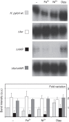
Figure 1
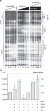
Figure 1. Transcriptional analysis of the arsR gene in response to metal ion treatment.
Primer extensions performed on total RNA extracted from wild-type, Δfur, ΔnikR and ΔfurΔnikR double mutants H. pylori strains grown to exponential phase and treated for 15 min with 1 mM (NH4)2Fe(SO4)2 (Fe2+), 1 mM NiSO4 (Ni2+) or 100 μM 2-2 dipyridyl (Dipy); untreated control RNA (−). Full gel is shown in Supplementary Fig. 6. Fold variation of the band intensities is reported in the graph. Error bars represent the standard deviation recorded in four independent experiments.
Figure 2
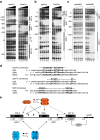
Figure 2. DNA footprinting of apo/holo-Fur and apo/holo-NikR at ParsR.
(a) apo-Fur (left gel) and holo-Fur (right gel) on the coding strand; (b) apo-Fur (left gel) and holo-Fur (right gel) on noncoding strand; (c) apo-NikR (left gel) and holo-NikR (right gel) on noncoding strand. Lanes 1–5: 0, 35, 70, 140 and 280 nM monomeric Fur or NikR, respectively. The scale bar on the left of each gel shows the distance from the TSS. A schematic representation of the promoter is presented on the right side of each gel with Fur and NikR footprints outlined as black and white boxes, respectively. Black arrowheads indicate persistent or hypersensitive bands. (d) Comparison of the Fur and NikR operator sequences on ParsR with the previously defined consensus motifs of the regulators. (e) Inferred schematic representation of the operator layout in the ParsR promoter. Fur operators named fOPI, fOPII and fOPIII are depicted as black boxes, while NikR operators named nOPI and nOPII are depicted as white boxes. The position of the ArsR operator aOP is mapped as reported in ref. . Positions are indicated with respect to the TSS.
Figure 3
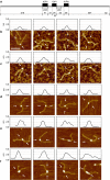
Figure 3. AFM images of Fur–DNA complexes.
(a) Scaled representation of the 818 bp DNA template used in the AFM experiments. Fur operator sites (black boxes) and NikR operator sites (white boxes) are indicated. Distances are in base pairs and the arrow indicates the midpoint of the template. (b) One apo-Fur dimer bound to the central fOPI site. (c) Two apo-Fur dimers bound to fOPI and fOPII sites. (d) One holo-Fur tetramer bound to the central fOPI site. (e) Two holo-Fur tetramers bound to fOPI and fOPII sites. (f) Three or more tetramers bound to fOPI, fOPII and fOPIII sites with consequent large DNA compaction. The image profile along the direction indicated by white arrows is shown on top of each panel. Scale bar, 100 nm. The profile plots have a width of 80 nm.
Figure 4
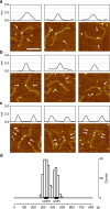
Figure 4. AFM images of holo-NikR–DNA complexes.
The image profile along the direction indicated by white arrows is shown on top of each panel. (a) One holo-NikR tetramer bound to the central nOPI site. (b) One holo-NikR tetramer bound to the slightly off-centre nOPII site. (c) Two holo-NikR tetramers bound to both nOPI and nOPII sites. (d) Bar chart representing the position of NikR bound along the DNA template (black line) with the nOPI and nOPII sites represented, in scale, as black boxes. Graph scale in base pairs. Scale bar, 100 nm. Width of the profile plot, 80 nm.
Figure 5
Figure 5. Functional analysis of Fur and NikR operators on ParsR.
Expression and transcription levels monitored through a ParsRlux reporter fusion. Histograms are shown for in vivo luminescence and in vitro RT–qPCR. (a) Wild-type ParsR promoter; (b) ParsR lacking the central holo-Fur operator (ΔfOPI); (c) ParsR lacking the distal Fur and NikR operators (ΔfOPII/nOPII); (d) ParsR mutated in the NikR proximal operator nOPI* (ATA→GGG). Fur and NikR operators are depicted as black and white boxes, respectively. From left to right, bars represent the untreated control (−), a 15 min treatment with iron (Fe2+), nickel (Ni2+) and iron chelator (Dipy). Error marks indicate the standard deviation of eight (luminescence) and four (RT–qPCR) independent experiments.
Figure 6
Figure 6. NikR impairs Fur binding to fOPII and fOPIII.
(a) Competitive footprinting experiment. Lanes 1–5: protection pattern with increasing holo-NikR concentration (0, 4, 8, 16 and 32 nM NikR tetramer, respectively). Lanes 6–10: protection pattern with increasing holo-Fur concentration (0, 4, 8, 17 and 35 nM Fur tetramer, respectively). Lanes 11–15: protection pattern with increasing holo-NikR concentration (as in lanes 1–5) in the presence of 70 nM Fur tetramer. A schematic map of the promoter region is depicted on the right side of the gels. Dashed boxes outline the competitive binding to the distal operator fOPII and impairment of Fur binding to fOPIII upon NikR binding to nOPI. (b) Functional analysis of the low-affinity holo-Fur operator (fOPIII) and of the NikR anti-repression element (nOPI). Bars represent the mRNA levels of the ParsRlux reporter fusion assayed by RT–qPCR in different genetic backgrounds: wild-type, ΔfOPI, ΔfOPIII, nOPI*, nOPI*-ΔfOPIII, nOPI*-Δfur and Δfur mutants. Error marks indicate the standard deviation of two biological replicates, each analysed twice in independent RT–qPCR runs.
Figure 7
Figure 7. ParsR regulation model.
Scheme of ParsR regulation. (a) AFM images of different holo-Fur/ParsR pseudo-knotting intermediates. The evident shortening of the DNA fragment and the concomitant enlargement of the nucleo-protein complex are indicative of a significant DNA condensation of the arsR promoter region and Fur oligomerization. (b) Proposed FeOFF-NikRON model for ParsR transcription regulation. At low iron concentration, apo-Fur forms dimers that bind to the upstream operators fOPI and fOPII, looping the intervening DNA but leaving the promoter elements open for RNAP binding and transcription. At high iron concentration, holo-Fur forms tetramers and higher order oligomers that condense the promoter DNA by binding to the three operator sites fOPI, fOPII and fOPIII, thus occluding RNAP binding (FeOFF). In the presence of nickel, holo-NikR binds to the operators nOPI and nOPII preventing Fur binding and promoter condensation, a condition that favours RNAP binding and arsR transcription (NikRON). An additional layer of control is introduced by the previously reported feedback regulation mechanism of arsR transcription. (c) Coherence of metal-dependent arsRS regulation with H. pylori acid acclimation needs: NikR-dependent antirepression of ParsR is possible only when the intracellular nickel levels are sufficiently high to cofactor the urease nickel-enzyme.

References

    1. Dorman C. J. Genome architecture and global gene regulation in bacteria: making progress towards a unified model? Nat. Rev. Microbiol. 11, 349–355 (2013). - PubMed
    1. Matthews K. S. DNA looping. Microbiol. Rev. 56, 123–136 (1992). - PMC - PubMed
    1. Schleif R. DNA looping. Annu. Rev. Biochem. 61, 199–223 (1992). - PubMed
    1. Hochschild A., Douhan J. & Ptashne M. How lambda repressor and lambda Cro distinguish between OR1 and OR3. Cell 47, 807–816 (1986). - PubMed
    1. Becker N. A., Greiner A. M., Peters J. P. & Maher L. J. Bacterial promoter repression by DNA looping without protein-protein binding competition. Nucleic Acids Res. 42, 5495–5504 (2014). - PMC - PubMed

Publication types

MeSH terms