. 2016 Dec;71(12):1119-1129.
doi: 10.1136/thoraxjnl-2015-208039. Epub 2016 Aug 24.

Chronic electronic cigarette exposure in mice induces features of COPD in a nicotine-dependent manner

Affiliations

Chronic electronic cigarette exposure in mice induces features of COPD in a nicotine-dependent manner

Itsaso Garcia-Arcos et al. Thorax. 2016 Dec.

Abstract

Background: The use of electronic (e)-cigarettes is increasing rapidly, but their lung health effects are not established. Clinical studies examining the potential long-term impact of e-cigarette use on lung health will take decades. To address this gap in knowledge, this study investigated the effects of exposure to aerosolised nicotine-free and nicotine-containing e-cigarette fluid on mouse lungs and normal human airway epithelial cells.

Methods: Mice were exposed to aerosolised phosphate-buffered saline, nicotine-free or nicotine-containing e-cigarette solution, 1-hour daily for 4 months. Normal human bronchial epithelial (NHBE) cells cultured at an air-liquid interface were exposed to e-cigarette vapours or nicotine solutions using a Vitrocell smoke exposure robot.

Results: Inhalation of nicotine-containing e-cigarettes increased airway hyper-reactivity, distal airspace enlargement, mucin production, cytokine and protease expression. Exposure to nicotine-free e-cigarettes did not affect these lung parameters. NHBE cells exposed to nicotine-containing e-cigarette vapour showed impaired ciliary beat frequency, airway surface liquid volume, cystic fibrosis transmembrane regulator and ATP-stimulated K+ ion conductance and decreased expression of FOXJ1 and KCNMA1. Exposure of NHBE cells to nicotine for 5 days increased interleukin (IL)-6 and IL-8 secretion.

Conclusions: Exposure to inhaled nicotine-containing e-cigarette fluids triggered effects normally associated with the development of COPD including cytokine expression, airway hyper-reactivity and lung tissue destruction. These effects were nicotine-dependent both in the mouse lung and in human airway cells, suggesting that inhaled nicotine contributes to airway and lung disease in addition to its addictive properties. Thus, these findings highlight the potential dangers of nicotine inhalation during e-cigarette use.

Keywords: COPD Pathology; COPD ÀÜ Mechanisms; Emphysema.


Conflict of interest statement

Competing interests: None declared.

Figures

Figure 1
Figure 1
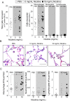
Lung function in mice exposed to inhaled phosphate-buffered saline (PBS), nicotine-free and nicotine-containing e-cigarette fluids. Mice were exposed daily to inhaled PBS, nicotine-free (EC vehicle, 0 mg/mL) or nicotine-containing (EC vehicle, 18 mg/mL) e-cigarette fluid for 4 months and nicotine absorption was confirmed by (A) plasma cotinine levels. (B) Airway hyperresponsiveness to increasing doses of methacholine was assessed in each animal group. (C) Negative pressure-driven forced expiratory and forced oscillation technique manoeuvres were performed to determine changes in pulmonary mechanics in all animal groups. Forced expiratory flow at 50% of FVC (FEF50) were determined in each animal. (D) Representative H&E-stained images of fixed lung parenchyma from mice exposed daily for 4 months (scale bar=100 µm) and quantification of surface area/unit volume, fractional volume and mean linear intercept. Graphs are represented as mean±SEM of three measurements. p Values shown, comparing both treatments connected by a line. All n≥6 per group. The comparisons between two individual groups were determined by Student's t-test. EC, e-cigarette.
Figure 2
Figure 2
Lung inflammation in mice exposed to inhaled phosphate-buffered saline (PBS) and to nicotine-free e-cigarette fluids. Mice were exposed daily to inhaled PBS, nicotine-free (0 mg/mL) or nicotine-containing (18 mg/mL) e-cigarette fluids for 4 months. (A) Total bronchoalveolar lavage fluid cells, macrophages, neutrophils and lymphocytes were quantified in mice exposed daily for 4 months. (B) Representative H&E-stained images of fixed lung parenchyma and macrophages from mice exposed daily for 4 months (scale bar=20 µm). (C) Tissue cellularity was quantified by measuring the average number of macrophages, neutrophils or lymphocytes per high powered field (×400). Ten randomly selected high powered images were used for quantification per mouse. Graphs are represented as mean±SEM of three measurements. p Values shown, comparing both treatments connected by a line. All n≥7 per group. The comparisons between two individual groups were determined by Student's t-test. BALF, bronchoalveolar lavage fluid. EC, e-cigarette.
Figure 3
Figure 3
Histological analysis of lungs from mice exposed to inhaled nicotine-free and nicotine-containing e-cigarette aerosol. Mice were exposed daily to inhaled nicotine-free (0 mg/mL) or nicotine-containing (18 mg/mL) e-cigarette aerosol for 4 months. Then, lungs were fixed and sectioned and stained for histological analysis. (A) Representative pictures of sections after Alcian blue stain and MUC5AC immunofluorescence (scale bar=20 µm). Graphs representing (A) qPCR analysis for Muc5ac are shown. (B) Representative images of sections after transferase dUTP nick end-labelling (TUNEL) (green) and DAPI (blue) staining from airway and alveolar regions of the lung (scale bar=20 µm). Percentage of TUNEL-positive cells quantified in each group and caspase 3/7 activity in lung tissue lysate are also shown. LU, luminescence units. Graphs are represented as mean±SEM of three measurements. p Values shown, comparing both treatments connected by a line. All n≥5 per group. The comparisons between two individual groups were determined by Student's t-test. DAPI, 4′,6-diamidino-2-phenylindole.
Figure 4
Figure 4
Cytokine and protease expression and activity in lungs from mice exposed to nicotine-free or nicotine-containing e-cigarette fluids. Mice were exposed to inhaled nicotine-free (0 mg/mL) or to nicotine-containing (18 mg/mL) e-cigarette fluid for 4 months. Gene expression analysis by qPCR for (A) the cytokines IL-1β, MCP-1, CXCL10, IL-6, CXCL2 and CXCL5 and (B) proteases MMP-3, MMP-9, MMP-12, CatK, CatL1 and CatS. (C) Collagenase and cathepsin activity were measured in lung bronchoalveolar lavage fluid. Graphs are represented as mean±SEM of three measurements. p Values shown, comparing both treatments connected by a line. All n≥5 per group. The comparisons between two individual groups were determined by Student's t-test.
Figure 5
Figure 5
PKCα and ERK activation in the lungs of mice exposed to inhaled vehicle or nicotine-containing e-cigarette fluid. Mice were exposed daily to inhaled nicotine-free (0 mg/mL) or to nicotine-containing (18 mg/mL) e-cigarette fluid for 4 months. (A) Representative western blots for total and phosphorylated PKCα (p-PKCα) and ERK in lung tissue homogenates. β-Actin was used as a loading control. (B) Optical intensity quantification and calculated ratios of phosphorylated/total protein for PKCα and ERK. Graphs are represented as mean±SEM of three measurements. p Values shown, comparing both treatments connected by a line. All n=9 per group for densitometry analysis. The comparisons between two individual groups were determined by Student's t-test.
Figure 6
Figure 6
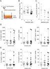
Effects of vehicle and nicotine-containing e-cigarette vapour on normal human bronchial epithelial (NHBE) cells. (A) Scheme showing how fully differentiated NHBE cells, grown at an air–liquid interface (ALI), were exposed to vapour from vehicle (0 mg/mL) or from nicotine-containing (36 mg/mL) e-cigarettes. (B) Ciliary beat frequency significantly decreased after exposure to nicotine-containing e-cigarette vapour. (C) Cells were stained with DAPI and antiacetylated tubulin to visualise cilia, 24 hours after exposures. The % ciliated area was determined using ImageJ software. (D) qPCR for gene expression of FOXJ1. (E) Cystic fibrosis transmembrane regulator (CFTR) ion conductance is significantly decreased after exposure to nicotine-containing e-cigarette vapour. (F) qPCR for gene expression of CFTR. (G) ATP-stimulated K+ (BK) ion conductance is significantly decreased after exposure to nicotine-containing e-cigarette vapour. (H) qPCR for gene expression of the α subunit of the BK channel (KCNMA1). (I) Measurement of airway surface liquid volume changes by refraction light microscopy. Graphs are represented as mean±SEM. p Values shown, comparing both treatments connected by a line. All n≥5 per group. The comparisons between two individual groups were determined by Student's t-test.
Figure 7
Figure 7
Effects of aerosolised nicotine on normal human bronchial epithelial (NHBE) cells. (A) Scheme showing how fully differentiated NHBE cells, grown at an air–liquid interface (ALI), were exposed to aerosolised saline containing 0 or 100 μM nicotine. Final concentration of nicotine in the airway surface liquid was estimated between 60 and 120 nM by microbalance deposition measurements. (B) Cystic fibrosis transmembrane regulator (CFTR) and (C) ATP-stimulated K+ (BK) ion conductances are significantly decreased in nicotine-exposed cell cultures compared with nicotine-free saline. Graphs are represented as mean±SEM. p Values shown, comparing both treatments connected by a line. All n≥4 per group. The comparisons between two individual groups were determined by Student's t-test.
Figure 8
Figure 8
Cystic fibrosis transmembrane regulator (CFTR) conductance, ciliary beat frequency (CBF) and cytokine expression in normal human bronchial epithelial (NHBE) cells exposed to basolateral nicotine. (A) Differentiated NHBE cells, grown at an air–liquid interface (ALI), were exposed to nicotine (0, 1 and 10 μM) basolaterally. (B) CBF and (C) CFTR ion conductance measurements were performed 12 hours after exposure. Graphs are represented as mean±SEM. One-way analysis of variance was used to compare the time-course curves and multiple comparisons were determined by the Dunn's method. (D–E) NHBE cells were exposed to culture media with or without nicotine for 5 days as outlined in Methods section. The concentration of interleukin (IL)-6, IL-8 and MCP-1 was measured in the apical wash (D) and in the basolateral media (E) following 5 days of exposure to culture media with or without nicotine. Graphs are represented as median±range. p Values shown, comparing both treatments connected by a line; n≥3 per group. (D–E) Data sets were analysed by non-parametric Friedman tests.
Figure 9
Figure 9
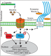
Possible signalling mechanism for nicotine-containing e-cigarette-induced lung damage. ERK, extracellular regulated kinase; IL, interleukin; nAChR, nicotinic acetylcholine receptor.

Comment in

References

    1. Murphy BS, Xu J, Kochanek KD. Deaths: preliminary data for 2010. In: Reports National Vital Statistics Report , ed. Hyattsville, MD: National Center for Health Statistics, 2012:61(4).
    1. Raherison C, Girodet PO. Epidemiology of COPD. Eur Respir Rev 2009;18:213–21. 10.1183/09059180.00003609 - DOI - PubMed
    1. Centers for Disease Control and Prevention (CDC). Cigarettes smoking among adults-United States, 2011. MMWR Morb Mortal Wkly Rep 2012;61:889–94. - PubMed
    1. US. Treating tobacco use and dependence: clinical practice guideline. In: Services DoHaH, ed. Department of Health and Human Services 2008 Report. 2008;1–32.
    1. Bullen C, Williman J, Howe C, et al. . Study protocol for a randomised controlled trial of electronic cigarettes versus nicotine patch for smoking cessation. BMC Public Health 2013;13:210 10.1186/1471-2458-13-210 - DOI - PMC - PubMed

Publication types

MeSH terms