Case Reports
. 2016 Dec;29(6):425-430.
doi: 10.1177/1971400916665384. Epub 2016 Aug 24.

Serial magnetic resonance imaging findings of intracerebral spread of listeria utilising subcortical U-fibres and the extreme capsule

Affiliations
Case Reports

Serial magnetic resonance imaging findings of intracerebral spread of listeria utilising subcortical U-fibres and the extreme capsule

Charlie Chia-Tsong Hsu et al. Neuroradiol J. 2016 Dec.

Abstract

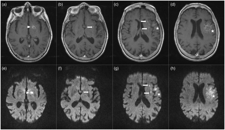
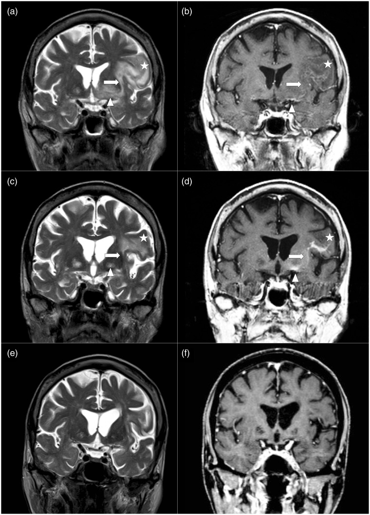
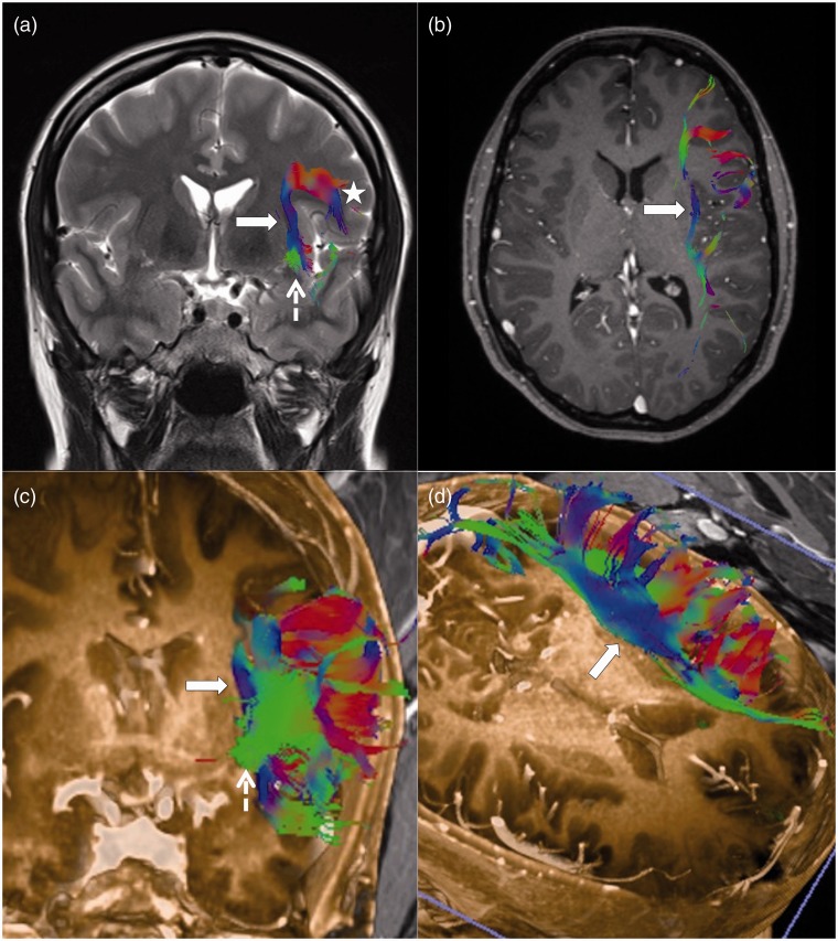
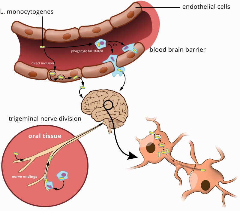
We present a case of Listeria monocytogenes cerebral abscess with axonal spread via the subcortical U-fibres and extreme capsule on magnetic resonance imaging, with follow-up studies demonstrating serial reduction in oedema and enhancement pattern of the white-matter fibre tracts following antimicrobial treatment. We discuss the microbiological mechanism of bacterial mobility to account for these unique imaging features. Recognition of this distinct pattern of spread of L. monocytogenes cerebral abscess may aid in diagnosis and enable early microbiological culture and treatment.

Keywords: Listeria monocytogenes; bacterial neural invasion; cerebral abscess; white-matter fibre tract.


Figures

Figure 1.
Figure 1.
MRI brain, gadolinium-enhanced T1-weighted images ((a)–(d)) showing clusters of rim-enhancing microabscesses in the left basal ganglia (arrow head) and pars opercularis (star) of the inferior left frontal lobe with continuity via the extreme capsule (arrow). These microabscesses showed restricted diffusion on the DWI images ((e)–(h)) with corresponding low ADC values (not shown). MRI: magnetic resonance imaging; DWI: diffusion-weighted imaging; ADC: apparent diffusion coefficient.
Figure 2.
Figure 2.
Serial brain MRI coronal T2 and post-gadolinium-enhanced T1 images at presentation ((a) and (b)), four months ((c) and (d)) and nine months ((e) and (f)), documenting response to antimicrobial therapy. Presentation MRI ((a) and (b)) demonstrates the spread of rim-enhancing cerebral microabscesses in the left basal ganglia (arrow head) and subcortical U-fibres of the pars opercularis of the inferior left frontal lobe (star) via the extreme capsule white-matter tract (arrow). MRI at four months ((c) and (d)) shows a favourable response to antimicrobial therapy with reduced perilesional oedema, but linear enhancement remains in the left basal ganglia, subcortical U-fibres of pars opercularis and extreme capsule. Final MRI ((e) and (f)) at nine months shows resolution, with no discernible enhancement or oedema, but minor gyral atrophy of the pars opercularis. MRI: magnetic resonance imaging.
Figure 3.
Figure 3.
MR tractography in a normal individual depicting the extreme capsule as a fan-shaped fibre tract which forms part of the ventral auditory-language pathway between Wernicke’s area and Broca’s area. (Subcortical U-fibres (star) in the pars opercularis of the inferior frontal lobe; extreme capsule (arrow) and inferior longidutinal fasciculus (dotted arrow).) Images obtained on a Siemens 3 T scanner. Diffusion tensor imaging sequence parameter: diffusion-weighted single-shot spin-echo echo-planar sequence along 30 different geometric directions. An effective b value of 1000 s/mm2 was used for each of the 30 diffusion-encoding directions. Tractography was performed on Siemens Neuro 3D syngo software with ROI placed in the pars opercularis and extreme capsule. Software setting: tract length 50–400 mm, two seed points per voxel length, angle threshold 30 degrees and FA threshold of 0.3. MR: magnetic resonance; ROI: region of interest; FA: fractional anisotropy.
Figure 4.
Figure 4.
Illustration showing possible routes of entry and spread of cerebral listeriosis. Three recognised routes of CNS entry are (1) direct invasion of endothelial cell membrane into the cytoplasm, where expression of F-actin protein by the bacteria enables intracellular or transcellular spread of the surrounding glial or neuronal cells or entry into the subarachnoid space; (2) phagocyte-facilitated route begins with bacterial ‘parasitisation’ of peripheral monocytes and subsequent monocytes adherence to CNS endothelial cells transmitting the bacteria from the monocyte to the endothelial cell. An infected monocyte also translocates into the CNS resulting in direct infection; and (3) the neural route of invasion can occur when oral mucosa is breached and recruited regional phagocyte become ‘parasitisation’ by the intracellular bacteria. Retrograde spread of infection occurs via invasion of cranial nerves, typically the divisions of the trigeminal nerve. The bacteria may migrate either transcellularly or intra-axonally ultimately reaching the CNS. Once Listeria have gained access to the CNS they are able to continue intercellular spread between neurons, glial cells or intra-axonally via white-matter fibre tracts utilising actin-based motility. (Adapted from Drevets et al.) CNS: central nervous system.

Similar articles

Cited by

References

    1. Clauss HE, Lorber B. Central nervous system infection with Listeria monocytogenes. Curr Infect Dis Rep 2008; 10: 300–306. - PubMed
    1. Crum NF. Update on Listeria monocytogenes infection. Curr Gastroenterol Rep 2002; 4: 287–296. - PubMed
    1. Drevets DA, Leenen PJ, Greenfield RA. Invasion of the central nervous system by intracellular bacteria. Clin Microbiol Rev 2004; 17: 323–347. - PMC - PubMed
    1. Bojanowski MW, Seizeur R, Effendi K, et al. Spreading of multiple Listeria monocytogenes abscesses via central nervous system fiber tracts: Case report. J Neurosurg 2015; 123: 1593–1599. - PubMed
    1. Perini G, Pravettoni R, Farina E, et al. Listeria brain abscesses during administration of mycophenolate mofetil for systemic lupus erythematosus: A case report. Neurol Sci 2015; 36: 1019–1020. - PubMed

Publication types

LinkOut - more resources