. 2016 Oct;162(10):1857-1869.
doi: 10.1099/mic.0.000358. Epub 2016 Aug 23.

Characterization of a protein-protein interaction within the SigO-RsoA two-subunit σ factor: the σ70 region 2.3-like segment of RsoA mediates interaction with SigO

Affiliations

Characterization of a protein-protein interaction within the SigO-RsoA two-subunit σ factor: the σ70 region 2.3-like segment of RsoA mediates interaction with SigO

Xiaowei Xue et al. Microbiology (Reading). 2016 Oct.

Abstract

σ factors are single subunit general transcription factors that reversibly bind core RNA polymerase and mediate gene-specific transcription in bacteria. Previously, an atypical two-subunit σ factor was identified that activates transcription from a group of related promoters in Bacillus subtilis. Both of the subunits, named SigO and RsoA, share primary sequence similarity with the canonical σ70 family of σ factors and interact with each other and with RNA polymerase subunits. Here we show that the σ70 region 2.3-like segment of RsoA is unexpectedly sufficient for interaction with the amino-terminus of SigO and the β' subunit. A mutational analysis of RsoA identified aromatic residues conserved amongst all RsoA homologues, and often amongst canonical σ factors, that are particularly important for the SigO-RsoA interaction. In a canonical σ factor, region 2.3 amino acids bind non-template strand DNA, trapping the promoter in a single-stranded state required for initiation of transcription. Accordingly, we speculate that RsoA region 2.3 protein-binding activity likely arose from a motif that, at least in its ancestral protein, participated in DNA-binding interactions.


Figures

Fig. 1.
Fig. 1.
Structural features of 79 amino acid B. subtilis RsoA. Alignment of partial sequences of group I (primary), III and IV (ECF) σ factors and full sequence of RsoA (bottom) aligned with full sequences of five closely related RsoA orthologues. Region 2.1, 2.2 and 2.3 delineations (top) are based on Lonetto et al. (1992). Key residues in regions 2.2 and 2.3 broadly conserved amongst most σ factors are shaded, including the seven universally conserved aromatic amino acids in type I σ region 2.3. Asterisks below alignment indicate conservation of residues amongst RsoA orthologues only. Arrows indicate RsoA residues mutated to alanine in this study. Lobed arrows indicate residues that were mutated to alanine and were also found mutated (i.e. F67S, E69V, I71N, M74T) in screen for random mutations that impair interaction with SigO. Numbered line indicates key amino acid positions between regions of RsoA. Secondary structure prediction (SS) for RsoA made using Jnet algorithm. H indicates high level of probability of α helix formation. RsoA orthologues are as follows: Bam, Bacillus amyloliquefaciens FZB42 (YP_001422598.1); Bmo, Bacillus mojavensis (WP_010331850.1); Bat, Bacillus atrophaeus 1942 (YP_003974761.1); Bpu, Bacillus pumilus SAFR-032 (YP_001486052.1); Bli, Bacillus licheniformis. Other protein abbreviations are as follows: Bsu, B. subtilis SigA; Eco, Escherichia coli σ70; Sco, Streptomyces coelicolor RpoD; Taq, Thermus aquaticus RpoD; SigH (B. subtilis); RpoH (E. coli); SigMXYW (B. subtilis); RpoE (E. coli σE); SigR (S. coelicolor σR).
Fig. 2.
Fig. 2.
Random mutations in RsoA that impair interaction with SigO. (a) Mutants isolated in the BACTH assay random mutagenesis screen (F67S, I71N, double mutant E69V/M74T). (b) Qualitative and quantitative BACTH analysis of the effects of the four mutations after regeneration in T25-RsoA fusion protein using site-directed mutagenesis. Quantitative assays conducted using β-galactosidase assays and activity reported as mean and sd (n=3). Qualitative activities shown as triplicate patches on selective medium containing X-Gal. Negative control is expression of T18-SigO protein in the absence of T25-RsoA expression. (c) Expression of wt and mutant T25-RsoA proteins tested in CyaA+ strain E. coli DH5α (see Methods). Upper panel is Coomassie blue-stained loading control. Lower panel is immunoblot detection of T25-RsoA fusion proteins tagged with HA epitope. For each allele, first lane is total protein and second lane is soluble (sedimentation-resistant) fraction. Control is pKT25 in E. coli DH5α.
Fig. 3.
Fig. 3.
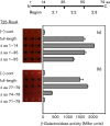
BACTH protein–protein interaction assay of T18-SigO fusion proteins co-expressed with (a) RsoA N-terminal and (b) RsoA C-terminal deletions derivatives. The full-length RsoA is 79 amino acids long and all derivatives are fused to the C-terminus of the T25 CyaA fragment. Quantitative assays conducted using β-galactosidase assays and activity reported as mean and sd (n=3). Negative control is expression of T18-SigO protein in the absence of T25-RsoA expression. Qualitative activities shown as triplicate spots on selective medium containing X-Gal. The line diagram indicates amino acid positions that delineate RsoA region 2.1, 2.2 and 2.3 sub-regions.
Fig. 4.
Fig. 4.
BACTH protein–protein interaction assay of T18-SigO fusion proteins co-expressed with wt and alanine substitution mutants of T25-RsoA fusion proteins. (a) Mutations in full-length RsoA (T25-RsoAFL) fused to T25 CyaA fragment and (b) mutations in the 24-amino acid RsoA region 2.3 (T25-RsoA2.3) fused to CyaA fragment. Quantitative assays conducted using β-galactosidase assays and activity reported as mean and sd (n=3). Qualitative activities shown as triplicate spots on selective medium containing X-Gal. Negative control is expression of T18-SigO protein in the absence of T25-RsoA expression. For the T25-RsoA2.3 alleles, total and soluble protein expression levels were tested in a CyaA+ background (Fig. S3).
Fig. 5.
Fig. 5.
BACTH assay testing interaction between N-terminus of β′ subunit (RpoC, amino acids 1–310) and deletion derivatives of RsoA. RpoC is expressed as a λcI fusion protein and RsoA derivatives are expressed as RpoA (α subunit) fusions in host strain E. coli FW Kan OL2-62 lac. Details of assay methodology are as described in Dove & Hochschild (2004) and MacLellan et al. (2009b).
Fig. 6.
Fig. 6.
Pull-down of RsoA-HA point mutants by SigONterm-FLAG. Proteins were co-expressed as CyaA T18 and T25 fusion proteins from separate plasmids in E. coli BL21. (a) Coomassie-stained loading control. (b) Western immunoblot using anti-FLAG antibodies. (c) Western immunoblot using anti-HA antibodies. For each strain, equal aliquots of total protein (first lane, crude) and proteins captured by anti-FLAG magnetic beads (second lane, bound) were separated using SDS-PAGE. Note that soluble RsoA accumulates poorly in the absence of SigO co-expression. (d) Bound protein band intensities from (b) and (c) were quantified, and the efficiency of RsoA co-sedimentation was reported as the ratio of RsoA to SigO in each treatment. This figure is a composite of several images from the same experiment.
Fig. 7.
Fig. 7.
Pull-down of RsoA-HA deletion mutants by SigONterm-FLAG. Proteins were co-expressed as CyaA T18 and T25 fusion proteins from separate plasmids in E. coli BL21. (a) Coomassie-stained loading control. (b) Western immunoblot using anti-FLAG antibodies. (c) Western immunoblot using anti-HA antibodies. For each strain, equal aliquots of total protein (first lane, crude) and proteins captured by anti-FLAG magnetic beads (second lane, bound) were separated using SDS-PAGE. ΔN, N-terminal deletions from RsoA (amino acids deleted in brackets). The full-length RsoA protein is 79 amino acids long. Note that soluble RsoA accumulates poorly in the absence of SigO co-expression. This figure is a composite of several images from the same experiment.
Fig. 8.
Fig. 8.
Transcription activation from PoxdC-lacZ fusion by wt and region 2.3 mutant alleles of RsoA co-expressed with SigO in B. subtilis. SigO and RsoA expression induced by 2 % xylose from separate ectopic (amyE and lacA, respectively) locations. Quantitative assays conducted using β-galactosidase assays and activity reported as mean and sd (n=3). Lower panel: immunoblot detection of RsoA-HA expression. A non-specific band (ns) acts as a loading control. This image is a composite of two immunoblots conducted in the same experiment. Negative control is expression of SigO protein in the absence of RsoA expression.

Similar articles

Cited by

References

    1. Arthur T. M., Burgess R. R.(1998). Localization of a sigma70 binding site on the N terminus of the Escherichia coli RNA polymerase beta' subunit. J Biol Chem 27331381–31387. 10.1074/jbc.273.47.31381 - DOI - PubMed
    1. Banta A. B., Chumanov R. S., Yuan A. H., Lin H., Campbell E. A., Burgess R. R., Gourse R. L.(2013). Key features of σS required for specific recognition by Crl, a transcription factor promoting assembly of RNA polymerase holoenzyme. Proc Natl Acad Sci U S A 11015955–15960. 10.1073/pnas.1311642110 - DOI - PMC - PubMed
    1. Bao X., Nickels B. E., Fan H.(2012). Chlamydia trachomatis protein GrgA activates transcription by contacting the nonconserved region of σ66. Proc Natl Acad Sci U S A 10916870–16875. 10.1073/pnas.1207300109 - DOI - PMC - PubMed
    1. Battesti A., Bouveret E.(2012). The bacterial two-hybrid system based on adenylate cyclase reconstitution in Escherichia coli. Methods 58325–334. 10.1016/j.ymeth.2012.07.018 - DOI - PubMed
    1. Bhandari V., Ahmod N. Z., Shah H. N., Gupta R. S.(2013). Molecular signatures for Bacillus species: demarcation of the Bacillus subtilis and Bacillus cereus clades in molecular terms and proposal to limit the placement of new species into the genus Bacillus. Int J Syst Evol Microbiol 632712–2726. 10.1099/ijs.0.048488-0 - DOI - PubMed

Publication types

MeSH terms

LinkOut - more resources