. 2016 Sep 13;134(11):817-32.
doi: 10.1161/CIRCULATIONAHA.115.016423. Epub 2016 Aug 24.

Glycoproteomics Reveals Decorin Peptides With Anti-Myostatin Activity in Human Atrial Fibrillation

Affiliations

Glycoproteomics Reveals Decorin Peptides With Anti-Myostatin Activity in Human Atrial Fibrillation

Javier Barallobre-Barreiro et al. Circulation. .

Abstract

Background: Myocardial fibrosis is a feature of many cardiac diseases. We used proteomics to profile glycoproteins in the human cardiac extracellular matrix (ECM).

Methods: Atrial specimens were analyzed by mass spectrometry after extraction of ECM proteins and enrichment for glycoproteins or glycopeptides.

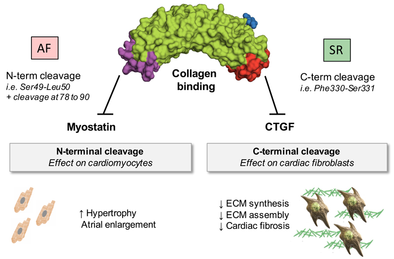
Results: ECM-related glycoproteins were identified in left and right atrial appendages from the same patients. Several known glycosylation sites were confirmed. In addition, putative and novel glycosylation sites were detected. On enrichment for glycoproteins, peptides of the small leucine-rich proteoglycan decorin were identified consistently in the flowthrough. Of all ECM proteins identified, decorin was found to be the most fragmented. Within its protein core, 18 different cleavage sites were identified. In contrast, less cleavage was observed for biglycan, the most closely related proteoglycan. Decorin processing differed between human ventricles and atria and was altered in disease. The C-terminus of decorin, important for the interaction with connective tissue growth factor, was detected predominantly in ventricles in comparison with atria. In contrast, atrial appendages from patients in persistent atrial fibrillation had greater levels of full-length decorin but also harbored a cleavage site that was not found in atrial appendages from patients in sinus rhythm. This cleavage site preceded the N-terminal domain of decorin that controls muscle growth by altering the binding capacity for myostatin. Myostatin expression was decreased in atrial appendages of patients with persistent atrial fibrillation and hearts of decorin null mice. A synthetic peptide corresponding to this decorin region dose-dependently inhibited the response to myostatin in cardiomyocytes and in perfused mouse hearts.

Conclusions: This proteomics study is the first to analyze the human cardiac ECM. Novel processed forms of decorin protein core, uncovered in human atrial appendages, can regulate the local bioavailability of antihypertrophic and profibrotic growth factors.

Keywords: atrial fibrillation; cardiovascular diseases; extracellular matrix; mass spectrometry; proteomics.


Figures

Figure 1
Figure 1. Glycoproteomics of the human cardiac ECM.
A) Atrial appendages were collected from patients in SR during cardiac surgery and subject to a decellularization-based three-step extraction for ECM proteins. First, GuHCl extracts were analyzed by LC-MS/MS. Then, ECM extracts were enriched for glycoproteins. The glycoprotein-enriched and unbound flow-through fractions were analysed by LC-MS/MS. Finally, enrichment was performed for glycoproteins as well as glycopeptides for direct glycopeptide identification by LC-MS/MS. B) Comparison of ECM-related glycoproteins in LAA and RAA (n=5, each). LTBP4 denotes latent transforming growth factor-beta binding protein 4; VTNC, vitronectin; FBLN2, fibulin 2; LUM, lumican; NID2, nidogen 2; CADH2, cadherin 2; BCAM, basal cell adhesion molecule. C) Validation by immunoblotting. Biglycan, mimecan (MIME) and Coomassie staining were included as controls. D) Quantification by densitometry. Bars represent mean ± SD. **p<0.01; ***p<0.001; A.U., arbitrary units.
Figure 2
Figure 2. Decorin after glycoprotein enrichment.
A) Proteomic comparison of the glycoprotein-enriched fraction and the flow-through. Relative abundances of glycoproteins (red dots) and non-glycosylated proteins (blue dots) are plotted for LAA and RAA (n=5, each). Decorin was the only glycoprotein consistently detected among the non-glycosylated proteins in the flow-through (yellow dots). B) The C-terminal half of decorin has three N-glycosylation sites (red triangles). C) According to the sequence coverage (yellow colour) in the three different fractions (input, glycoprotein-enriched fraction, non-glycosylated flow-through), only N-terminal (non-glycosylated) decorin peptides were identified in the flow-through.
Figure 3
Figure 3. Decorin processing in atrial tissue.
A) Decorin (DCN) showed the highest number of non-tryptic cleavage sites among all ECM proteins identified. Biglycan (BGN) is highlighted for comparison. B) Three different antibodies recognizing different epitopes were used to detect decorin. Numbers indicate amino acid positions. C) Comparison of LAA and LV. Note the C-terminal truncation of decorin in LAA compared to LV from non-failing hearts (Mw shift from 48kDa to 45kDa). The apparent reduction of decorin levels in LAA with the C-terminal antibody was not corroborated by antibodies to N-terminal and internal epitopes. Instead, they revealed a loss of ~3kDa in LAA compared to LV. Biglycan, a highly homologous SLRP, showed no such shift in Mw. The graphs show the densitometry results from immunoblots based on antibodies against three different decorin epitopes. Bars represent mean ± SD. A.U., arbitrary units.
Figure 4
Figure 4. Decorin processing in AF.
A) Representative pre-operative and postoperative echocardiography images from patients who stayed in SR throughout surgery and patients who developed postoperative AF. Rightmost panels depict left atrial structural remodeling observed in a patient with permanent, chronic AF after a 5-year follow-up. B) Immunoblotting for decorin in GuHCl extracts from LAA samples. Decorin was more abundant in AF than SR patients, and both bands (C-terminally truncated decorin at 45kDa and full-length at 48kDa) became detectable. The bottom chart shows the quantification (mean ± SD) in SR and AF patients (n=6, each) with the antibody against the internal region. **p<0.01 C) GuHCl extracts (n=6, each group) were analysed using LC-MS/MS. After a search using no enzyme, non-tryptic decorin peptides were identified and quantified. In addition to the four main cleavage sites of decorin in SR, a N-terminal cleavage site at position Ser49-Leu50 was detected in AF.
Figure 5
Figure 5. Decorin peptides bind myostatin.
A) qPCR analysis for myostatin expression in additional samples (SR-SR=14, SR-AF=10, AF=13). Bars represent mean ± SD. B) Schematic illustration of two downstream signaling pathways of myostatin (MSTN). “ActRIIB/ALK” denotes Activin receptor type-2B (ActRIIB)/ activin receptor-like kinase (ALK) receptor complex; “TAK1” transforming growth factor beta-activated kinase 1. C) Repression of MSTN-SMAD3 signaling. A synthetic N-terminal peptide of mouse decorin (amino acids 42-71 corresponding to the peptide generated by the Ser49-Leu50 cleavage site in humans) inhibited the CAGA-luciferase reporter activity in a dose-dependent manner. The trend for the effect was calculated based on Spearman’s correlation (rho, ρ). Bars are mean ± SD of triplicate experiments. D) Effect on rat cardiomyocytes. MSTN represses hypertrophy induced by pre-treatment with isoproterenol (Iso) and phenylephrine (PE). Cardiomyocyte size is given as mean ± standard error of the mean (SEM). E) Cardiomyocyte size was calculated after defining the cell perimeter to integrate the area (see bottom panels). F) The decorin N-terminal peptide attenuated the MSTN effect dose-dependently (0-20µM). Cardiomyocyte size is given as mean ± SEM based on a total of 300-400 cell measurements per experimental condition and compared to a control without decorin N-terminal peptide. *p<0.05; ***p<0.001.
Figure 6
Figure 6. Effect of decorin on cardiac metabolism.
A) Deficiency of decorin was associated with reduced expression of myostatin in murine hearts. Bars represent mean ± SD. B) In hearts of decorin null mice, the abundance of closely related SLRPs, such as biglycan and lumican (LUM) was not affected, but phosphorylation of SMAD2 was increased. C) Overlay of +H-NMR spectra from wild type and decorin null (DCN -/-) hearts. D) Metabolic profiles of cardiac tissue from DCN -/- mice revealed elevated levels of glutamine (Gln), succinate (Suc), aspartate (Asp) and nicotinamide adenine dinucleotides (NAD+, NADH). E) Myostatin represses AMPK activation in rat cardiomyocytes. Immunoblotting was performed with two different antibodies for AMPK phospho-Thr172. Levels of total AMPK (pan-AMPK) were unchanged. F) N-terminal decorin peptides from mouse or human origin (20µM) reversed the effect of myostatin on AMPK phosphorylation. Rat cardiomyocytes were stimulated with Iso and PE after incubation with myostatin for 2 hours. G) Schematic summary of the experiment in Langendorff perfused mouse hearts. H) Phosphorylation levels of AMPK and SMAD2 were used as readouts for inhibition of myostatin signaling by the N-terminal decorin peptide. **p<0.01; ***p<0.001.
Figure 7
Figure 7. Decorin in human atria.
Decorin cleavage constitutes a mechanism for local regulation of growth factor bioactivity in the human cardiac ECM. N-terminal cleavage of decorin was observed in patients with persistent AF and gives rise to peptides with anti-myostatin activity. C-terminal truncation is more common in atria than ventricles and releases the CTGF-binding domain of decorin.

References

    1. Pellman J, Lyon RC, Sheikh F. Extracellular matrix remodeling in atrial fibrosis: mechanisms and implications in atrial fibrillation. J Mol Cell Cardiol. 2010;48:461–467. - PMC - PubMed
    1. Dawson K, Wakili R, Ordög B, Clauss S, Chen Y, Iwasaki Y, Voigt N, Qi XY, Sinner MF, Dobrev D, Kääb S, et al. MicroRNA29: a mechanistic contributor and potential biomarker in atrial fibrillation. Circulation. 2013;127:1466–1475. - PubMed
    1. Chiang DY, Lebesgue N, Beavers DL, Alsina KM, Damen JM, Voigt N, Dobrev D, Wehrens XH, Scholten A. Alterations in the interactome of serine/threonine protein phosphatase type-1 in atrial fibrillation patients. J Am Coll Cardiol. 2015;65:163–173. - PMC - PubMed
    1. Mayr M, Yusuf S, Weir G, Chung YL, Mayr U, Yin X, Ladroue C, Madhu B, Roberts N, De Souza A, Fredericks S, et al. Combined metabolomic and proteomic analysis of human atrial fibrillation. J Am Coll Cardiol. 2008;51:585–594. - PubMed
    1. Barallobre-Barreiro J, Didangelos A, Schoendube FA, Drozdov I, Yin X, Fernández-Caggiano M, Willeit P, Puntmann VO, Aldama-López G, Shah AM, Doménech N, et al. Proteomics analysis of cardiac extracellular matrix remodeling in a porcine model of ischemia/reperfusion injury. Circulation. 2012;125:789–802. - PubMed

Publication types

MeSH terms