. 2016 Oct 1;197(7):2816-27.
doi: 10.4049/jimmunol.1600598. Epub 2016 Aug 24.

Mincle Signaling Promotes Con A Hepatitis

Affiliations

Mincle Signaling Promotes Con A Hepatitis

Stephanie H Greco et al. J Immunol. .

Abstract

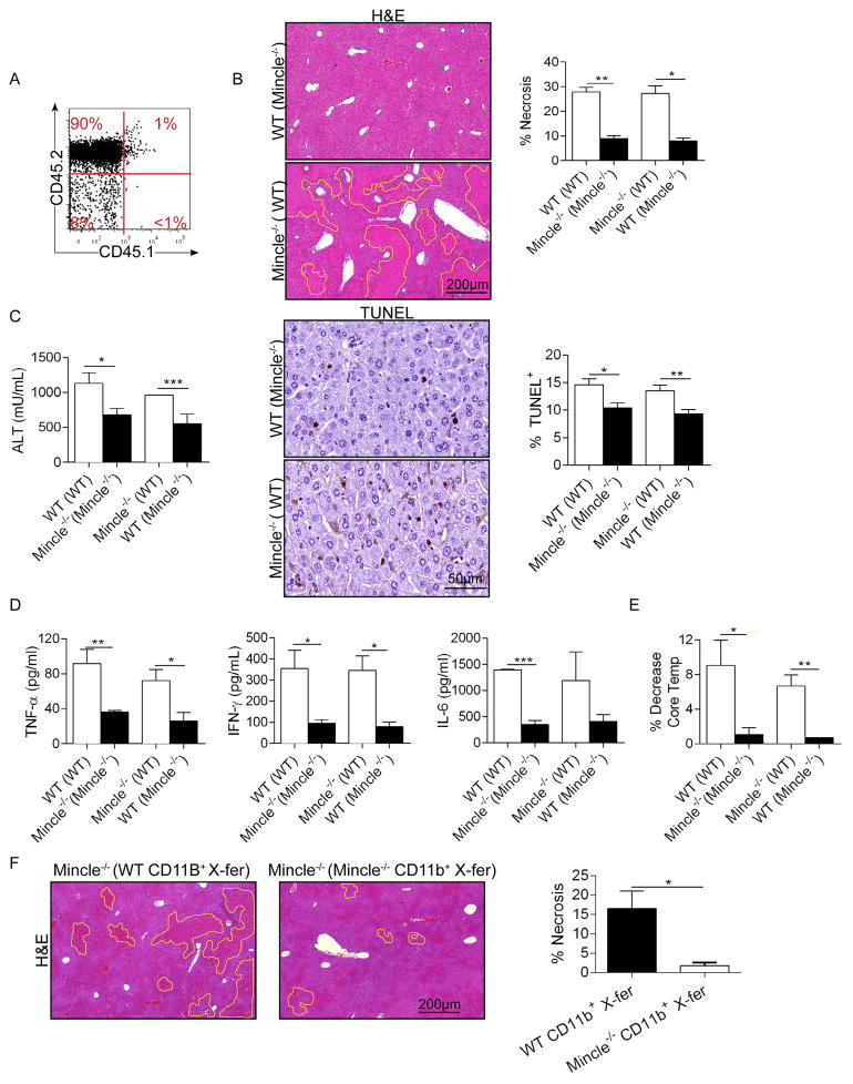
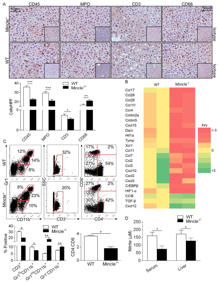
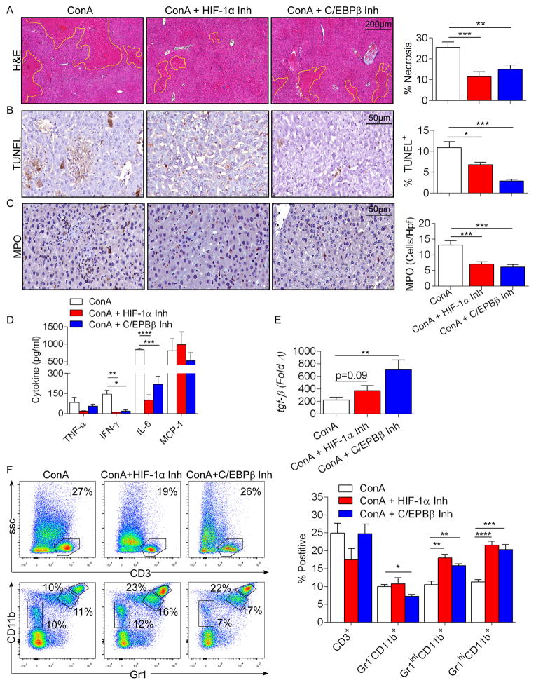
Con A hepatitis is regarded as a T cell-mediated model of acute liver injury. Mincle is a C-type lectin receptor that is critical in the immune response to mycobacteria and fungi but does not have a well-defined role in preclinical models of non-pathogen-mediated inflammation. Because Mincle can ligate the cell death ligand SAP130, we postulated that Mincle signaling drives intrahepatic inflammation and liver injury in Con A hepatitis. Acute liver injury was assessed in the murine Con A hepatitis model using C57BL/6, Mincle(-/-), and Dectin-1(-/-) mice. The role of C/EBPβ and hypoxia-inducible factor-1α (HIF-1α) signaling was assessed using selective inhibitors. We found that Mincle was highly expressed in hepatic innate inflammatory cells and endothelial cells in both mice and humans. Furthermore, sterile Mincle ligands and Mincle signaling intermediates were increased in the murine liver in Con A hepatitis. Most significantly, Mincle deletion or blockade protected against Con A hepatitis, whereas Mincle ligation exacerbated disease. Bone marrow chimeric and adoptive transfer experiments suggested that Mincle signaling in infiltrating myeloid cells dictates disease phenotype. Conversely, signaling via other C-type lectin receptors did not alter disease course. Mechanistically, we found that Mincle blockade decreased the NF-κβ-related signaling intermediates C/EBPβ and HIF-1α, both of which are necessary in macrophage-mediated inflammatory responses. Accordingly, Mincle deletion lowered production of nitrites in Con A hepatitis and inhibition of both C/EBPβ and HIF-1α reduced the severity of liver disease. Our work implicates a novel innate immune driver of Con A hepatitis and, more broadly, suggests a potential role for Mincle in diseases governed by sterile inflammation.


Conflict of interest statement

The authors have no conflicts of interest to declare.

Figures

Figure 1
Figure 1. High Mincle expression and signaling in Con-A hepatitis
(a) Mincle expression was assayed in mouse liver NPC by confocal microscopy compared with NPC from Mincle−/− liver. Mincle expression was also compared by flow cytometry in mouse liver and spleen CD45+ pan-liver leukocytes, Gr1CD11b+F480+ macrophages, and F480CD11c+MHCII+ dendritic cells. Representative histograms and quantitative data from 4 mice are shown. Grey histograms represent isotype controls. (b) Mincle expression was tested in murine CD45CD146+ liver sinusoidal endothelial cells and CD45CD146 hepatocytes by flow cytometry. Representative data indicating the percentage of positive cells relative to isotype control is shown. (c) Mincle expression was assayed in human liver NPC by confocal microscopy. Mincle expression was further tested by flow cytometry in human CD15+ liver monocytes and LinHLA-DR+ liver dendritic cells compared with their counterparts in PBMC. Representative histograms are shown. Similar results were obtained from 5 separate patients. (d) Mice were treated with Con-A (20μg/g) or PBS and livers were harvested and tested for SAP130 expression by ELISA at 12h (n=3). (e) Lysates from PBS- or Con-A-treated mice were immuno-precipitated using an α-SAP130 or control mAb and then tested for expression of SAP130 and Mincle. (f) Paraffin-embedded sections from livers of WT mice treated with PBS or Con-A were assayed for expression of p-Syk by immunohistochemistry at 12h. Isotype control is shown. Representative images and quantified data are shown (n=3/group). (g) The time course of Syk activation in Con-A hepatitis was tested by western blotting using liver lysate from 2 mice at each time point. β-actin was used as a loading control. (h) Inflammatory monocytes from the liver of PBS- and Con-A-treated mice were tested for expression of p-Syk at 12h after injury. Representative histograms and quantitative data from 3 mice based on median fluorescent indices (MFI) are shown (*p<0.05, ***p<0.001). All experiments were repeated at least twice with similar results.
Figure 2
Figure 2. Mincle deletion is protective against Con-A hepatitis
WT and Mincle−/− mice were treated with Con-A (20μg/g; n=5/group). (a) Livers were harvested at serial time intervals and examined by H&E staining. The fraction of non-viable liver area was bracketed and calculated by examining 10 HPFs per liver. Representative H&E-stained sections from the 12h time-point are shown. Paraffin-embedded sections from the 12h time-point were also examined by TUNEL staining. The fraction of TUNEL+ hepatocytes was calculated. (b) Serum levels of ALT and AST, (c) TNF-α, IL-10, IFN-γ, IL-6, and MCP-1 were calculated for each cohort at 12h. (d) Similarly, hepatic expression of FasL and Bcl2 were calculated by a Nanostring assay. (e) The decrease in mouse rectal temperature from baseline was calculated at 12h in the WT and Mincle−/− cohorts. Experiments were repeated more than 5 times with similar results (*p<0.05, **p<0.01, ***p<0.001). (f) WT and Mincle−/− mice were treated with a potentially lethal dose of Con-A (40μg/g) and survival was measured according to the Kaplan-Meier method (n=10–11/group; p=0.04).
Figure 3
Figure 3. Mincle or Syk blockade are protective in Con-A hepatitis but Mincle ligation exacerbates disease
(a, b) WT mice were treated with Con-A alone or in combination with a neutralizing α-Mincle mAb (n=5/group). (a) Livers were harvested at 12h and the fraction of necrotic liver area was calculated based on H&E staining. (b) Serum levels of pro-inflammatory cytokines were tested. (c, d) WT mice were treated with Con-A alone or in combination with the Syk inhibitor Piceatannol (n=5/group). (c) Livers were harvested at 12h and the fraction of necrotic liver area was calculated based on H&E staining. (d) Serum levels of pro-inflammatory cytokines were tested. (e, f) WT mice were treated with PBS, TDB alone, Con-A alone, or Con-A + TDB (n=5/group). (e) Livers were harvested at 12h and the fraction of necrotic liver area was calculated based on H&E staining. (f) Serum levels of pro-inflammatory cytokines were tested (*p<0.05, **p<0.01, ***p<0.001, ****p<0.0001).
Figure 4
Figure 4. Mincle deletion does not mitigate TAA-induced liver fibrosis and Dectin-1 deletion does not protect against ConA hepatitis
(a–c) WT and Mincle−/− mice (n=5/group) were serially treated with TAA for 12 weeks. Livers were examined by (a) H&E and (b) trichrome staining and the percent fibrotic area was calculated. (c) CD45+ pan-leukocyte infiltrate were determined by immunohistochemistry. (d–f) WT and Dectin-1−/− mice were treated with Con-A (20μg/g). (d) Livers were harvested at 12h and examined by H&E staining. Representative H&E-stained sections are shown and the fraction of non-viable liver was calculated. (e) Serum levels of AST, (f) TNF-α, IL-10, IL-6, and MCP-1 were calculated for each cohort (n=5 group; p=ns for all comparisons). In vivo experiemnts were repeated twice with similar results.
Figure 5
Figure 5. Mincle signaling in liver infiltrating inflammatory cells modulates Con-A hepatitis
(a) CD45.1 mice were irradiated and made chimeric with bone marrow derived from congenic CD45.2 mice. The extent of chimerism was tested at 7 weeks by flow cytometry and found to be near-complete. (b) WT mice were made chimeric with WT bone marrow ‘WT (WT)’ or Mincle−/− bone marrow ‘WT (Mincle−/−)’. Similarly, Mincle−/− mice were made chimeric with WT bone marrow ‘Mincle−/− (WT)’ or Mincle−/− bone marrow ‘Mincle−/− (Mincle−/−)’. Seven weeks later, all four cohorts were treated with Con-A (20μg/g; n=5/group). Livers were harvested at 12h and the percentage of non-viable liver was calculated based on H&E staining. Hepatocyte apoptosis was determined by TUNEL staining. (c) Serum ALT was calculated. (d) Serum TNF-α, IFN-γ, and IL-6 were measured. (e) The decrease in mouse rectal temperature from baseline was calculated for each cohort. (f) Mincle−/− mice were adoptively transferred one hour prior to Con-A administration with CD11b+ hepatic leukocytes (1×10⁶ cells, i.v.) harvested from either WT or Mincle−/− liver. Mice were sacrificed at 12h and the percentage of non-viable liver was calculated based on H&E staining (*p<0.05, **p<0.01, ***p<0.001).
Figure 6
Figure 6. Mincle deletion modulates intra-hepatic inflammation and nitrite production in Con-A hepatitis
(a–c) WT and Mincle−/− mice were treated with Con-A (20μg/g). (a) CD45+ pan-leukocyte, MPO+ neutrophil, CD3+ T cell, and CD68+ macrophage infiltrate were determined by immunohistochemistry at 12h. Results were quantified by examining 10 HPFs per slide (n=5/group). (b) Intra-hepatic inflammation was determined by mRNA levels of diverse inflammatory mediators in whole liver tisues. A heat map analysis showing fold change in Mincle−/− mRNA expression levels relative to WT controls is shown (n=2/group). (c) Cellular ractions of myeloid and T cell subsets in livers of Con-A-treated WT and Mincle−/− mice were determined by flow cytometry. Representative dot plots and quantifications based on replicates, including the CD4:CD8 ratio, are shown. Flow cytometry experiments were reproduced more than 3 times using 3–5 mice per cohort. (d) The Griess assay was used to determine nitrite levels in the liver and serum of WT and Mincle−/− mice at 12h after Con-A administration. (n=5/group; *p<0.05, **p<0.01, ***p<0.001).
Figure 7
Figure 7. Inhibition of HIF-1α or C/EBPβ is protective against Con-A hepatitis
(a–e) WT mice were treated with Con-A (20μg/g) alone or in combination with a HIF-1α or C/EBPβ inhibitor. Injury was determined by (a) the percent necrotic liver area based on H&E staining, (b) the percentage of TUNEL+ hepatocytes, (c) number of infiltrating MPO+ cells, and (d) serum levels of TNF-α, IFN-γ, IL-6, and MCP-1. (e) Hepatic expression of TGF-β was tested at 12h by qPCR. Data was normalized to PBS-treated controls. (f) Cellular fractions of T cells and myeloid cellular subsets in livers of mice in each cohort were determined by flow cytometry. Representative pseudocolor plots and quantifications based on replicates are shown. This experiment was repeated twice (n=5/group; *p<0.05, **p<0.01, ***p<0.001, ****p<0.0001).

References

    1. Murphy N. An update in acute liver failure: when to transplant and the role of liver support devices. Clin Med (Lond) 2006;6:40–6. - PMC - PubMed
    1. Chun LJ, Tong MJ, Busuttil RW, Hiatt JR. Acetaminophen hepatotoxicity and acute liver failure. J Clin Gastroenterol. 2009;43:342–9. - PubMed
    1. Singanayagam A, Bernal W. Update on acute liver failure. Curr Opin Crit Care. 2015;21:134–41. - PubMed
    1. Khashab M, Tector AJ, Kwo PY. Epidemiology of acute liver failure. Curr Gastroenterol Rep. 2007;9:66–73. - PubMed
    1. Wang HX, Liu M, Weng SY, Li JJ, Xie C, He HL, Guan W, Yuan YS, Gao J. Immune mechanisms of Concanavalin A model of autoimmune hepatitis. World J Gastroenterol. 2012;18:119–25. - PMC - PubMed

Publication types