. 2016 Sep 6;113(36):10127-32.
doi: 10.1073/pnas.1612594113. Epub 2016 Aug 24.

Biallelic hypomorphic mutations in a linear deubiquitinase define otulipenia, an early-onset autoinflammatory disease

Affiliations

Biallelic hypomorphic mutations in a linear deubiquitinase define otulipenia, an early-onset autoinflammatory disease

Qing Zhou et al. Proc Natl Acad Sci U S A. .

Abstract

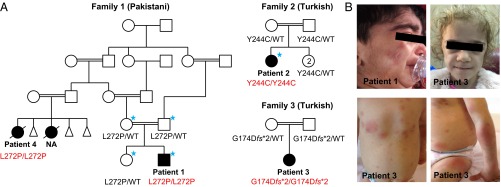
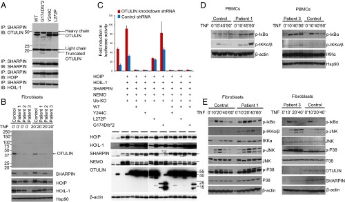
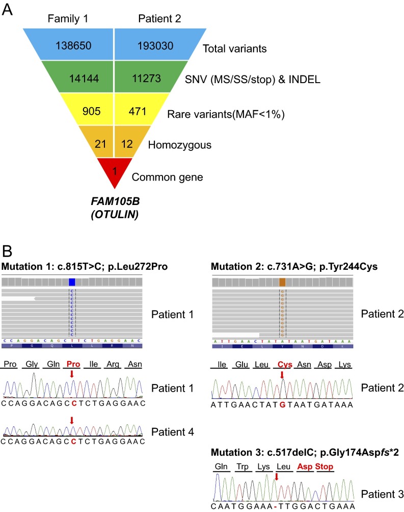
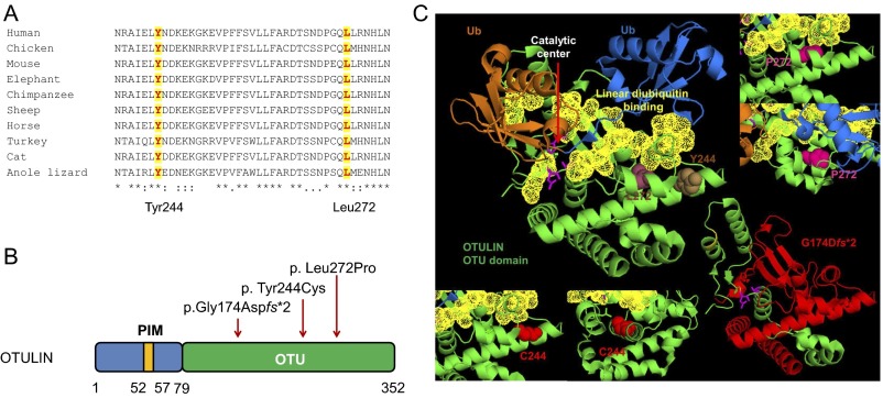
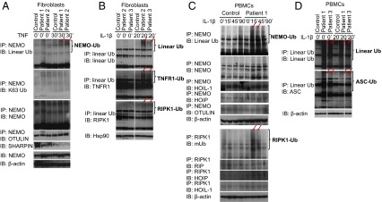
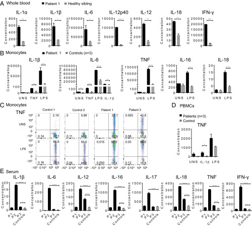
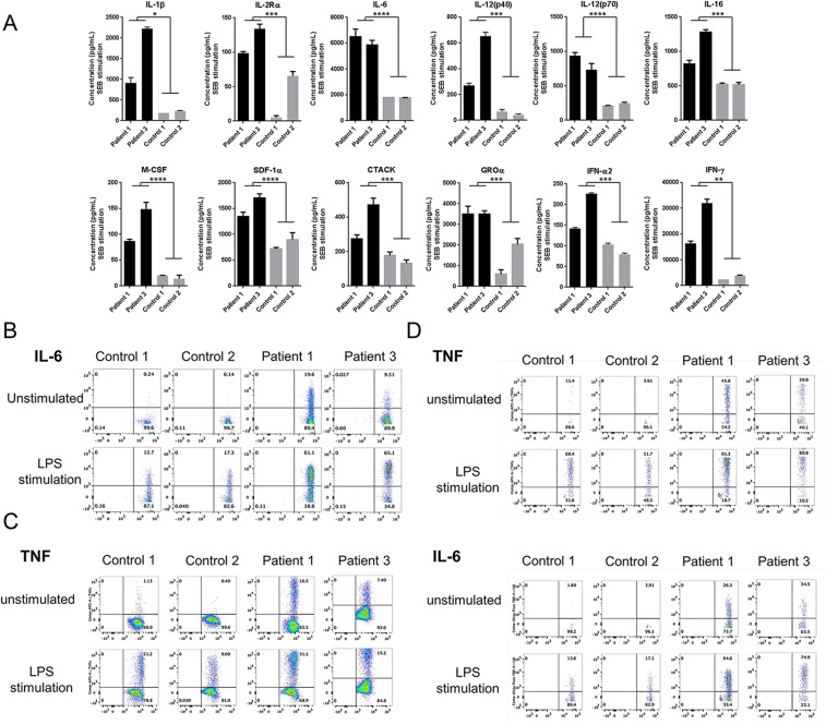
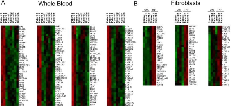
Systemic autoinflammatory diseases are caused by mutations in genes that function in innate immunity. Here, we report an autoinflammatory disease caused by loss-of-function mutations in OTULIN (FAM105B), encoding a deubiquitinase with linear linkage specificity. We identified two missense and one frameshift mutations in one Pakistani and two Turkish families with four affected patients. Patients presented with neonatal-onset fever, neutrophilic dermatitis/panniculitis, and failure to thrive, but without obvious primary immunodeficiency. HEK293 cells transfected with mutated OTULIN had decreased enzyme activity relative to cells transfected with WT OTULIN, and showed a substantial defect in the linear deubiquitination of target molecules. Stimulated patients' fibroblasts and peripheral blood mononuclear cells showed evidence for increased signaling in the canonical NF-κB pathway and accumulated linear ubiquitin aggregates. Levels of proinflammatory cytokines were significantly increased in the supernatants of stimulated primary cells and serum samples. This discovery adds to the emerging spectrum of human diseases caused by defects in the ubiquitin pathway and suggests a role for targeted cytokine therapies.

Keywords: NF-κB pathway; OTULIN; autoinflammatory disease; cytokines; linear deubiquitinase.


Conflict of interest statement

Conflict of interest statement: S.Ö. received royalties for consulting and speaking from Novartis and SOBI. All other authors declare no conflict of interest.

Figures

Fig. 1.
Fig. 1.
Mutations in OTULIN cause severe early-onset systemic autoinflammatory disease. (A) Pedigrees and the identified genotypes in three families with mutations in OTULIN. WT indicates wild-type OTULIN alleles. The individuals selected for exome sequencing are marked with blue asterisks. NA: an affected cousin of patient 1 had similar disease, but her DNA sample was not available for this study. (B) Clinical manifestations of three patients with otulipenia. Top two panels show facial features of the patients, including cushingoid appearance (Left) and prominent fat loss (lipodystrophy) (Right). Bottom two panels show erythematous skin lesions and subcutaneous nodules.
Fig. 2.
Fig. 2.
Induced NF-κB activity in cells with mutant OTULIN. (A) OTULIN mutants do not disrupt interaction with LUBAC. HEK293 cells were transiently transfected with plasmids encoding the LUBAC components (SHARPIN, HOIP, and HOIL-1), and WT or mutant OTULIN plasmids. Whole-cell lysates were collected 36 h later, and subjected to immunoprecipitation with antibodies against SHARPIN. The precipitates were then immunoblotted with OTULIN, SHARPIN, HOIP, and HOIL-1. (B) OTULIN and LUBAC complex expression in patients’ fibroblasts. Whole-cell lysates from the OTULIN-deficient patients and one healthy donor were immunoblotted with antibodies for OTULIN, SHARPIN, HOIP, HOIL-1, and Hsp90. (C) NF-κB luciferase assay in transiently transfected HEK293 cells with endogenous OTULIN down-regulated by shRNA. OTULIN mutants p.L272P and p.G174Dfs*2 do not inhibit LUBAC-induced NF-κB activation in HEK293 cells transfected with firefly NF-κB reporter plasmid, a Renilla luciferase control vector, and expression plasmids for WT or mutant OTULIN, together with LUBAC (SHARPIN, HOIL-1, HOIP), Ub-KO (ubiquitin mutant with all lysines mutated to arginines, which only forms linear polyubiquitin chains), and LUBAC linear ubiquitination substrate NEMO. Results are plotted as firefly normalized to Renilla luciferase activity to account for variance in transfection efficiency and cell number. One representative result of three independent experiments is shown. Values are reported as the means of technical triplicates ± SEM. (Lower) Whole-cell lysates from transfected cells were immunoblotted with antibodies for HOIP, HOIL-1, SHARPIN, NEMO, OTULIN, and β-actin. (D) PBMCs from OTULIN-deficient patients showed increased levels of phosphorylated IκBα and phosphorylated IKKα/IKKβ compared with a healthy control. Whole-cell lysates from TNF-stimulated PBMCs were immunoblotted for respective target proteins. (E) Stimulated fibroblasts from OTULIN-deficient patients sustained increased levels of phosphorylated IκBα, increased phosphorylated IKKα/IKKβ, and increased phosphorylated JNK and P38. Fibroblasts from patients 1 and 3 were stimulated with TNF for the time periods indicated. Whole-cell lysates were immunoblotted for respective target proteins. Two healthy individuals’ fibroblasts served as controls.
Fig. S1.
Fig. S1.
Identification of FAM105B/OTULIN mutations using exome sequencing and Sanger sequencing. (A) Whole-exome sequencing leads to identification of a common gene FAM105B. Schematic representation of the exome data-filtering approach used to select for novel and homozygous inherited variants segregating with disease in family 1 and patient 2. OTULIN is the only gene in common between the family 1 and patient 2. INDEL, frameshift and nonframeshift insertions and deletions; SNV, single-nucleotide variants including missense variants, splice site variants, and stop codon variants. (B) Electropherograms for the three OTULIN mutations identified in four patients from three families.
Fig. S2.
Fig. S2.
Schematic of OTULIN protein domains, and positions and conservation of the three disease-associated mutations. (A) Amino acid sequence alignment of the OTU domain of OTULIN orthologs. The two missense mutations, p.Tyr244Cys and p.Leu272Pro, affect amino acid residues that are highly conserved. The protein sequences of different species were aligned by using CLUSTALW. The two conserved amino acids Tyr244 and Leu272 are highlighted. (B) OTULIN is a 352-residue protein that consists of N-terminal LUBAC-binding PUB-interacting motif (PIM) and C-terminal ovarian tumor (OTU) domain that mediates deubiquitinase activity of OTULIN (79–352 aa). All three mutations are located in the OTU domain, and red arrows indicate their positions. (C) In silico modeling of OTULIN mutations based on the crystal structure of human OTULIN (3ZNZ). The distal Ub is marked in blue, the proximal Ub is in brown, and the OTU domain is in green. The linear ubiquitin binding regions (95–96 aa, 124–126 aa, 255–259 aa, 283–289 aa, and 336–338 aa) are shown in yellow, and catalytic sites (residues Asp126, Cys129, and His339) are shown as pink sticks. The left top panel shows the native residue of Leu272 (in raspberry) and Tyr244 (in brown). The left bottom two panels show the mutant Cys244 from front and side views. The right top two panels show the mutant Pro272 from two different angles. The mutations Tyr244Cys and Leu272Pro are located near or on the S1 distal Ub binding site. These two missense mutations likely affect enzymatic activity or linear ubiquitin binding. The right bottom panel shows location of the p.G174Dfs*2 mutation, with the truncated part of the OTU domain shown in red.
Fig. S3.
Fig. S3.
Patient 1 responded to infliximab treatment. (A) Image of patient 1 at the age of 11 y, 4 mo (Left); his height is at the 10th to 25th percentile for his age (Right). (B) Patient 1’s CRP and white blood cell (WBC) level before and after infliximab treatment from March 2006 to September 2008. Patient 1 was treated with prednisone but continued to have fevers and skin lesions before the start of infliximab treatment.
Fig. S4.
Fig. S4.
Normal T- and B-cell proliferation and normal NF-κB activation in response to TCR and B-cell receptor (BCR) stimulation in patient 3 compared with controls. (A) Normal T- and B-cell proliferation in patient 3 compared with a healthy control. Total peripheral blood mononuclear cells (PBMCs) were stained with CellTrace Violet and cultured with (dark gray) or without (light gray) anti-CD3 and anti-CD28 antibodies (1 μg/mL) for 4 d. For B-cell proliferation, PBMCs were stimulated with a-IgM (10 μg/mL), CD40L (1 μg/mL), and IL-4 (50 ng/mL) for 4 d. Numbers indicate percentage of cells having undergone at least one cellular division, assessed by dye dilution. (B) Normal NF-κB activation in response to TCR stimulation in patient 3 compared with controls. (C) Normal NF-κB activation in response to BCR stimulation in patient 3 compared with controls.
Fig. S5.
Fig. S5.
Increased NF-κB in OTULIN-deficient cells. (A) NF-κB luciferase assay in transiently transfected HEK293T cells. OTULIN mutants p.L272P and p.G174Dfs*2 do not inhibit LUBAC-induced NF-κB activation in HEK293T cells transfected with firefly NF-κB reporter plasmid, a Renilla luciferase control vector, and expression plasmids for WT or mutant OTULIN, together with LUBAC (SHARPIN, HOIL-1, HOIP), Ub-WT. Results are plotted as firefly luciferase activity normalized to Renilla luciferase activity to account for variance in transfection efficiency differences and cell number differences. One representative result of three independent experiments is shown. Values are shown reported as the means of technical triplicates ± SEM. (B) Increased NF-κB signaling in fibroblasts from patient 2 compared with a healthy control. Stimulated fibroblasts from patient 2 showed increased levels of phosphorylated IκBα, increased phosphorylated IKKα/IKKβ, and increased phosphorylation of JNK and P38. Fibroblasts derived from patient 2 and a healthy control were stimulated with TNF for the time periods indicated. Whole-cell lysates were immunoblotted for respective target proteins. (C) Ubiquitination defect of RIPK1 was less noticeable in cells transfected with the mutant monoubiquitin plasmid (Ub-KO), which can only form linear ubiquitin chains.
Fig. 3.
Fig. 3.
Mutant OTULIN plasmids show impaired deubiquitinase function in transfected HEK293 cells. (A–D) OTULIN mutants failed to deubiquitinate Met1-linked linear ubiquitin chains from molecules targeted by LUBAC. HEK293 cells were transiently transfected with expression plasmids for one of the OTULIN target proteins, including NEMO (A), RIPK1 (B), ASC (C), or TNFR1 (D) together with plasmids for the LUBAC (SHARPIN, HOIL-1, and HOIP), Ub plasmids (Ub-KO: ubiquitin mutant with all lysines mutated to arginines, which only forms linear polyubiquitin chains; Ub-WT: WT ubiquitin, which forms linear polyubiquitin chains and other polyubiquitin chains, such as K48-Ub, K63-Ub chains), and WT or mutant OTULIN plasmids. Whole-cell lysates were subjected to immunoprecipitation with antibodies against NEMO (A), RIPK1 (B), ASC (C), and linear ubiquitination chain (C and D). High–molecular-weight (HMW) ubiquitin aggregates (Top) were detected by immunoblotting the precipitates with linear Ub antibody (A–D). Cell lysates were also blotted with antibody against NEMO, RIPK1, TNFR1, ASC, HOIP, SHARPIN, HOIL-1, OTULIN, and Hsp90 or β-actin.
Fig. 4.
Fig. 4.
Patient-derived PBMCs and fibroblasts lose their ability to deubiquitinate Met1-linked linear ubiquitin chains. Whole-cell lysates were immunoprecipitated with antibodies for NEMO (A and C), linear Ub (B and D), and RIPK1 (C), and the precipitates were blotted with antibodies against linear ubiquitin. The precipitates were also blotted with antibodies against NEMO, TNFR1, RIPK1, K63-linked ubiquitin, HOIP, HOIL-1, or OTULIN, and the cell lysates were blotted with antibodies against Hsp90 or β-actin. Red arrows point to the differences for comparison. (A) Patients’ TNF-stimulated fibroblasts showed increased abundance and molecular weight of linear-ubiquitinated NEMO as a result of the impaired OTULIN deubiquitinase activity. K63-ubiquitinated NEMO is mainly unaffected (second panel). Fibroblasts from patient 1, patient 2, and a healthy control were stimulated with TNF for 30 min. (B) Patients’ IL-1β–stimulated fibroblasts showed increased abundance of linear ubiquitinated TNFR1 and RIPK1 and accumulation of high-molecular linear-ubiquitin chains. Fibroblasts from patient 2, patient 3, and a healthy control were stimulated with IL-1β for 20 min. (C and D) Patients’ IL-1β–stimulated PBMCs showed increased linear ubiquitination of NEMO (C, Upper), RIPK1 (C, Lower), and ASC (D, Lower), and accumulation of high-molecular linear-ubiquitin chains (D, Upper). PBMCs from patients 1 and 3, and a healthy control were stimulated with IL-1β for the indicated time. mUB is a mouse monoclonal antibody that is not linear Ub specific (C).
Fig. 5.
Fig. 5.
Patient-derived immune cells display a strong inflammatory signature. Cytokine profiles are compared for OTULIN-deficient patients and healthy controls. Cytokine concentration shown in y axis is in picograms per milliliter. Values are represented as means ± SEM. *P ≤ 0.05; **P ≤ 0.01; ***P ≤ 0.001; ****P ≤ 0.0001. (A) Whole-blood samples from patient 1 and his unaffected sibling were stimulated with bacterial LPS (1 μg/mL) for 24 h. A total of 48 cytokines or growth factors listed in SI Materials and Methods were assayed in duplicates. (B) Cytokine levels from the supernatant of stimulated purified monocytes from patient 1 compared with three healthy controls. Cells were unstimulated, TNF stimulated (20 ng/mL), LPS stimulated (1 μg/mL), or IL-1β stimulated (10 ng/mL) for 48 h. A total of 48 cytokines or growth factors were assayed; however, only the most significant results are shown. Samples were assayed in duplicates. (C) Intracellular staining of TNF in monocytes from patient 1 and patient 3 compared with two healthy controls before stimulation and following LPS stimulation (1 μg/mL). PBMCs from patient 2 were not available due to sample limitation. (D) TNF levels in the supernatants of PBMCs derived from three patients and one healthy control, at the basal level and after stimulation with IL-1β (10 ng/mL), and LPS (1 μg/mL). Samples were assayed in triplicates. (E) Serum cytokine levels from 3 patients and 12 healthy controls. Patient 1 (P1) has been treated with the TNF inhibitor, infliximab, and had no evidence of active disease at the time of sampling. Patient 2 (P2) has been treated with the IL-1 inhibitor, anakinra, and had active disease at the time of sampling. Patient 3 (P3) has been treated with the TNF inhibitor, etanercept, and still had some symptoms at the time of sampling. Patients’ samples were assayed in triplicates.
Fig. S6.
Fig. S6.
Increased cytokine production in patients. (A) SEB stimulation in whole blood. Cytokine profiles are compared for OTULIN-deficient patients and healthy controls. Cytokine concentration shown in y axis is in picograms per milliliter. Values are represented as means ± SD. *P ≤ 0.05; **P ≤ 0.01; ***P ≤ 0.001; ****P ≤ 0.0001. Whole-blood samples from patient 1 and patient 3 compared with age-matched healthy controls were stimulated with staphylococcal enterotoxin B (SEB) at 1 μg/mL for 24 h. A total of 48 cytokines or growth factors listed in SI Materials and Methods were assayed in triplicates. (B) Intracellular cytokine staining for IL-6 in PBMCs gated on CD14+/CD11+ for monocytes. (C) Intracellular cytokine staining TNF in PBMCs gated on CD3+/CD45RO+ for T cells. Flow cytometry showed higher TNF levels at baseline in T cells of patients compared with two healthy controls. (D) Intracellular cytokine staining for TNF and IL-6 in PBMCs gated on CD14+/CD11+/CD123 for dendritic cells. Flow cytometry showed increased TNF and IL-6 staining in dendritic cells, at basal level and after LPS stimulation, from two patients compared with two healthy controls.
Fig. S7.
Fig. S7.
TNF-induced inflammatory signature in whole blood and fibroblasts. (A) Gene expression of immunologically related genes in unstimulated whole-blood samples from three patients and five healthy controls by Nanostring assay. Transcriptional profiles reflect the disease activity of each patient at the time of sampling. Patient 1 had inactive disease, and his profile is similar to control samples. Patient 2 and patient 3 had more active disease as shown by red color in the heat map. (B) TNF-induced inflammatory signature in fibroblasts. Gene expression profiles in stimulated fibroblasts from two patients and a healthy control. Fibroblasts from patients 2 and 3 showed increased inflammatory signatures enriched for NF-κB, Jak-STAT, and TNF signaling.

References

    1. Gerlach B, et al. Linear ubiquitination prevents inflammation and regulates immune signalling. Nature. 2011;471(7340):591–596. - PubMed
    1. Tokunaga F, et al. SHARPIN is a component of the NF-κB-activating linear ubiquitin chain assembly complex. Nature. 2011;471(7340):633–636. - PubMed
    1. Boisson B, et al. Immunodeficiency, autoinflammation and amylopectinosis in humans with inherited HOIL-1 and LUBAC deficiency. Nat Immunol. 2012;13(12):1178–1186. - PMC - PubMed
    1. Boisson B, et al. Human HOIP and LUBAC deficiency underlies autoinflammation, immunodeficiency, amylopectinosis, and lymphangiectasia. J Exp Med. 2015;212(6):939–951. - PMC - PubMed
    1. Ombrello MJ, Kastner DL, Milner JD. HOIL and water: The two faces of HOIL-1 deficiency. Nat Immunol. 2012;13(12):1133–1135. - PubMed

Publication types

MeSH terms