. 2016 Oct 15;27(20):3040-3051.
doi: 10.1091/mbc.E16-01-0003. Epub 2016 Aug 24.

C9orf72 binds SMCR8, localizes to lysosomes, and regulates mTORC1 signaling

Affiliations

C9orf72 binds SMCR8, localizes to lysosomes, and regulates mTORC1 signaling

Joseph Amick et al. Mol Biol Cell. .

Abstract

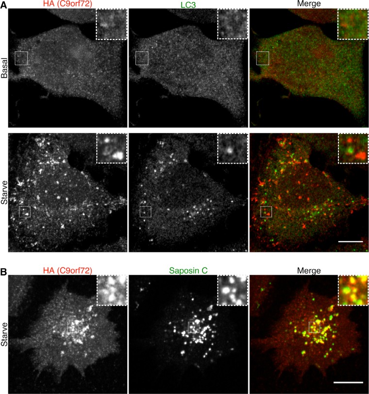
Hexanucleotide expansion in an intron of the C9orf72 gene causes amyotrophic lateral sclerosis and frontotemporal dementia. However, beyond bioinformatics predictions that suggested structural similarity to folliculin, the Birt-Hogg-Dubé syndrome tumor suppressor, little is known about the normal functions of the C9orf72 protein. To address this problem, we used genome-editing strategies to investigate C9orf72 interactions, subcellular localization, and knockout (KO) phenotypes. We found that C9orf72 robustly interacts with SMCR8 (a protein of previously unknown function). We also observed that C9orf72 localizes to lysosomes and that such localization is negatively regulated by amino acid availability. Analysis of C9orf72 KO, SMCR8 KO, and double-KO cell lines revealed phenotypes that are consistent with a function for C9orf72 at lysosomes. These include abnormally swollen lysosomes in the absence of C9orf72 and impaired responses of mTORC1 signaling to changes in amino acid availability (a lysosome-dependent process) after depletion of either C9orf72 or SMCR8. Collectively these results identify strong physical and functional interactions between C9orf72 and SMCR8 and support a lysosomal site of action for this protein complex.


Figures

FIGURE 1:
FIGURE 1:
C9orf72 interacts robustly and directly with SMCR8. (A) Immunoblotting reveals the successful detection of 2xHA-C9orf72 expressed from its endogenous locus in HEK293FT cells. Suppression of this signal by two distinct C9orf72 siRNAs confirms specificity. (B) Anti-HA immunoprecipitation of 2xHA-C9orf72 from genome-edited HEK293FT cells, followed by immunoblotting for the indicated endogenously expressed proteins to assess their interaction with C9orf72. Nonspecific band on SMCR8 blot is indicated by an asterisk. (C) After their cotransfection, anti-GFP immunoprecipitation and subsequent immunoblotting experiments reveal the interaction of SMCR8-tdTomato with GFP-C9orf72 but not with GFP alone. (D) Coomassie staining of an SDS–PAGE gel with resolved anti-GFP immunoprecipitates from cells transfected with SMCR8-tdTomato and GFP or GFP-C9orf72. (E) Anti-HA immunoprecipitates from 2xHA-C9orf72 cells were washed with the indicated salt and detergent concentrations, followed by immunoblotting detection of 2xHA-C9orf72 and SMCR8. (F) Immunoblot analysis of siRNA-mediated depletion of SMCR8 and C9orf72 depletion in HEK293FT cells. Nonspecific band on SMCR8 blot is indicated by an asterisk.
FIGURE 2:
FIGURE 2:
Amino acid availability regulates C9orf72 recruitment to lysosomes. Immunofluorescence images showing 2xHA-tagged endogenous C9ORF72’s localization in (A) basal, (B) starved (1-h serum and amino acid–free RPMI), and (C) starvation followed by amino acid refeeding (15 min). C9ORF72’s recruitment to lysosomes is enhanced by amino acid starvation, as revealed by its colocalization with LAMP1 (a lysosomal integral membrane protein) in starved conditions. Insets, higher magnification of boxed regions of interest. Experiments were performed in HEK293FT cells, and images were obtained by spinning-disk confocal microscopy. Scale bar, 10 μm. (D) Anti-HA immunoprecipitations from 2xHA-C9orf72 cells harvested under starvation and amino acid–fed conditions revealed similar levels of SMCR8 coimmunoprecipitation. Analysis of S6 phosphorylation served as a positive control for assessing cellular responses to changes in amino acid availability via the mTORC1 signaling pathway.
FIGURE 3:
FIGURE 3:
C9orf72 localizes more robustly to lysosomes than autophagosomes under starvation conditions. (A) Immunofluorescence images of endogenously expressed 2xHA-C9orf72 and LC-3 (autophagosome marker) in basal cell growth conditions and starved (1-h serum and amino acid–free RPMI) conditions. (B) Immunofluorescence images of 2xHA-C9orf72 and Saposin C (protein of the lysosome lumen) in starved conditions. Scale bar, 10 μm.
FIGURE 4:
FIGURE 4:
Analysis of C9orf72 and SMCR8 KO cell lines. (A) Immunoblot analysis of SMCR8 and C9orf72 abundance in KO cell lines. Asterisk indicates nonspecific band in SMCR8 immunoblots. (B) LAMP1 immunofluorescence in cells of the indicated genotypes (maximum intensity projections of confocal image z-stacks acquired through full cell thickness). Cell edges are indicated with a dotted white line. Boxes indicate regions of interest displayed in higher-magnification insets. Scale bar, 10 μm.
FIGURE 5:
FIGURE 5:
SMCR8 KO causes an increase in cell size. (A) F-actin (phalloidin) and nuclear (DAPI) labeling of cells of the indicated genotypes reveals the enlargement of SMCR8 KO cells. Scale bar, 10 μm. (B) Scatterplot of cell areas as determined by measuring the perimeter of phalloidin-stained cells (>30 cells measured/experiment, three experiments, mean ± SEM summarized by lines and whiskers; ****p < 0.0001, analysis of variance (ANOVA) with Tukey’s multiple comparisons posttest). (C) Scatterplot of nuclear areas as determined by measuring the outline of DAPI-stained nuclei (>30 cells measured/experiment, three experiments, mean ± SEM summarized by lines and whiskers; ****p < 0.0001, ANOVA with Tukey’s multiple comparisons posttest). (D) Cell diameter distributions of WT, C9orf72 KO, SMCR8 KO, and C9orf72/SMCR8 double-KO cell lines. Cell diameters were measured in suspension by flow cytometry (data represent the average of results from two independent experiments with >1800 cells measured for each genotype per experiment).
FIGURE 6:
FIGURE 6:
Impaired regulation of mTORC1 in C9orf72 and SMCR8 KO cells. (A) Immunoblot analysis of ribosomal protein S6 phosphorylation (S235/S236) under normal growth conditions. (B) Summary of S6 phosphorylation levels. Mean ± SEM; ****p < 0.0001 (ANOVA with Dunnett’s posttest); three to seven experiments per genotype (three for the double-KO line). (C) Increased cell size after SMCR8 depletion is mTOR dependent. Flow cytometry analysis of HeLa cell diameter after treatment with the indicated siRNAs ± 200 nM torin 1 (1300 cells measured/condition). (D) Immunoblot analysis of HeLa cells treated with indicated siRNAs and/or 200 nM torin 1 confirms the effectiveness of SMCR8 depletion and mTORC1 inhibition. (E) Immunoblot analysis of S6 phosphorylation after starvation (1.5 h) and subsequent amino acid refeeding (15 min). (F) Summary of S6 phosphorylation levels after starvation and amino acid refeeding (WT refed condition was normalized to 1, mean ± SEM; **p < 0.01, ****p < 0.0001, ANOVA with Dunnett’s posttest; four to eight experiments, four for double-KO cell line).
FIGURE 7:
FIGURE 7:
Intact regulation of mTOR localization in C9orf72 KO, SMCR8 KO, and double-KO cells. mTOR localization was observed under basal growth conditions and starvation (1.5 h) and amino acid refed (15 min) conditions in WT (A), C9orf72 (B), SMCR8 (C), and double-KO (D) cells. The lysosomal colocalization of this punctate mTOR staining has been thoroughly established in previous studies (Petit et al., 2013; Ferguson, 2015). Scale bar, 10 μm.

References

    1. Atanasio A, Decman V, White D, Ramos M, Ikiz B, Lee HC, Siao CJ, Brydges S, LaRosa E, Bai Y, et al. C9orf72 ablation causes immune dysregulation characterized by leukocyte expansion, autoantibody production, and glomerulonephropathy in mice. Sci Rep. 2016;6:23204. - PMC - PubMed
    1. Atkinson RA, Fernandez-Martos CM, Atkin JD, Vickers JC, King AE. C9ORF72 expression and cellular localization over mouse development. Acta Neuropathol Commun. 2015;3:59. - PMC - PubMed
    1. Baba M, Hong SB, Sharma N, Warren MB, Nickerson ML, Iwamatsu A, Esposito D, Gillette WK, Hopkins RF, 3rd, Hartley JL, et al. Folliculin encoded by the BHD gene interacts with a binding protein, FNIP1, and AMPK, and is involved in AMPK and mTOR signaling. Proc Natl Acad Sci USA. 2006;103:15552–15557. - PMC - PubMed
    1. Baker M, Mackenzie IR, Pickering-Brown SM, Gass J, Rademakers R, Lindholm C, Snowden J, Adamson J, Sadovnick AD, Rollinson S, et al. Mutations in progranulin cause tau-negative frontotemporal dementia linked to chromosome 17. Nature. 2006;442:916–919. - PubMed
    1. Bar-Peled L, Sabatini DM. Regulation of mTORC1 by amino acids. Trends Cell Biol. 2014;24:400–406. - PMC - PubMed