. 2016 Aug 24;36(34):8882-94.
doi: 10.1523/JNEUROSCI.1470-16.2016.

Phosphorylation of Synaptojanin Differentially Regulates Endocytosis of Functionally Distinct Synaptic Vesicle Pools

Affiliations

Phosphorylation of Synaptojanin Differentially Regulates Endocytosis of Functionally Distinct Synaptic Vesicle Pools

Junhua Geng et al. J Neurosci. .

Abstract

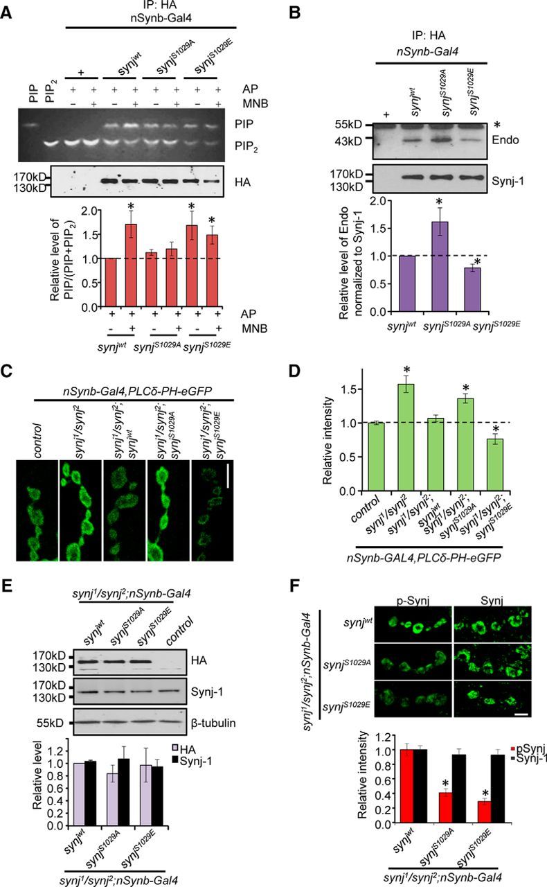
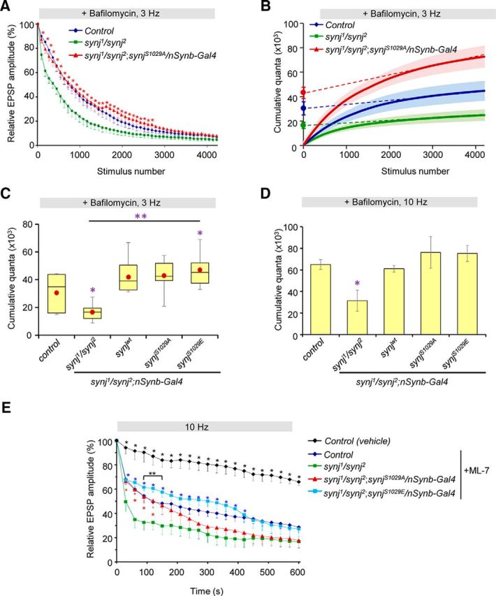
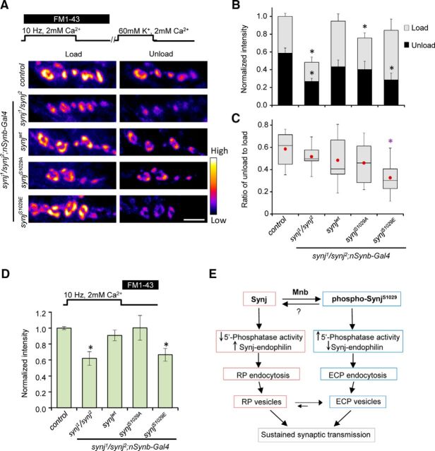
The rapid replenishment of synaptic vesicles through endocytosis is crucial for sustaining synaptic transmission during intense neuronal activity. Synaptojanin (Synj), a phosphoinositide phosphatase, is known to play an important role in vesicle recycling by promoting the uncoating of clathrin following synaptic vesicle uptake. Synj has been shown to be a substrate of the minibrain (Mnb) kinase, a fly homolog of the dual-specificity tyrosine phosphorylation-regulated kinase 1A (DYRK1A); however, the functional impacts of Synj phosphorylation by Mnb are not well understood. Here we identify that Mnb phosphorylates Synj at S1029 in Drosophila We find that phosphorylation of Synj at S1029 enhances Synj phosphatase activity, alters interaction between Synj and endophilin, and promotes efficient endocytosis of the active cycling vesicle pool (also referred to as exo-endo cycling pool) at the expense of reserve pool vesicle endocytosis. Dephosphorylated Synj, on the other hand, is deficient in the endocytosis of the active recycling pool vesicles but maintains reserve pool vesicle endocytosis to restore total vesicle pool size and sustain synaptic transmission. Together, our findings reveal a novel role for Synj in modulating reserve pool vesicle endocytosis and further indicate that dynamic phosphorylation and dephosphorylation of Synj differentially maintain endocytosis of distinct functional synaptic vesicle pools.

Significance statement: Synaptic vesicle endocytosis sustains communication between neurons during a wide range of neuronal activities by recycling used vesicle membrane and protein components. Here we identify that Synaptojanin, a protein with a known role in synaptic vesicle endocytosis, is phosphorylated at S1029 in vivo by the Minibrain kinase. We further demonstrate that the phosphorylation status of Synaptojanin at S1029 differentially regulates its participation in the recycling of distinct synaptic vesicle pools. Our results reveal a new role for Synaptojanin in maintaining synaptic vesicle pool size and in reserve vesicle endocytosis. As Synaptojanin and Minibrain perturbations are associated with various neurological disorders, such as Parkinson's, autism, and Down syndrome, understanding mechanisms modulating Synaptojanin function provides valuable insights into processes affecting neuronal communication.

Keywords: endocytosis; minibrain; phosphorylation; synaptojanin.


Figures

Figure 1.
Figure 1.
Mnb phosphorylates Synaptojanin at S1029 in vitro. A, Protein sequence alignment of partial Drosophila Synj-1 PRD domain with mouse and human Synj-1 PRD domain (Clustal Omega). Phosphorylation of Ser1029 by Mnb precedes a Proline residue, consistent with Mnb being a proline-directed kinase (highlighted in yellow). Gray shaded region represents the peptide fragment identified from MS. B, C, Validation that Mnb phosphorylates Synj at Ser1029 using the indicated antibodies. HA-tagged Synjwt, SynjS1029A, and SynjS1029E were expressed in flies, immunoprecipitated using HA-agarose beads, and incubated with or without purified Mnb kinase following alkaline phosphatase (AP) treatment. Representative blots are shown, and results were confirmed in at least 3 independent experiments. Mutation at S1029 abolished the increased phosphorylation by Mnb.
Figure 2.
Figure 2.
Phosphorylation of Synaptojanin at S1029 in vivo. A, Western blot of wild-type head protein extracts treated with and without alkaline phosphatase (AP) and detected using p-SynjS1029 antibody and total Synj-1 antibody. B, Staining of Synj in the third intar NMJ using p-Synj and Synj-1 antibodies for the indicated genotypes in wild-type background. Scale bar, 5 μm. C, Quantification of relative staining intensity for p-Synj and Synj signals normalized to control (top), and the relative p-Synj/Synj signals. Error propagation was used to calculate fold change and SE. n > 12 NMJ per genotype from at least 4 larvae per genotype. D, Different Synj PRD constructs are indicated by different color. Lower gel represents Coomassie staining of His-PRD constructs purified from bacteria. Lane labeled “Beads” indicates negative control done in parallel using bacteria transformed with empty PET-15b vector backbone. Arrows indicate purified PRD proteins. Full-PRD indicates full-length Synj PRD. E, Top blots, Western blots detected with pSynj antibody. Bottom, Direct blue 71(DB71) staining of the nitrocellulose membrane, showing the amount of protein loaded in each lane. Right, p-Synj does not detect signals well following alkaline phosphatase (AP) treatment, confirming the antibody is sensitive to phosphorylation status of Synj. F, Staining of p-Synj and Synj-1 antibodies of the third instar NMJ for the indicated genotypes in mnb1 mutant background. Scale bar, 5 μm. G, Quantification of relative staining intensity for p-Synj/Synj signals. n = 11 NMJ from ≥ 4 animals per genotype. Data are mean ± SEM. *p < 0.05 compared with control.
Figure 3.
Figure 3.
Phosphorylation of Synaptojanin at S1029 enhances Synaptojanin phosphoinositide phosphatase activity and alters its interaction with endophilin. A, TLC showing conversion of BODIPY-PIP2 to BODIPY-PIP by Synj constructs. Western blot represents levels of total Synj. Graph represents quantification of relative PIP to total phospholipid (PIP+PIP2) level. n = 8 independent experiments per genotype per condition. B, Immunoprecipitation (IP) experiment using flies overexpressing Synj tagged with HA reveals that SynjS1029A has strong interaction with Endophilin. *Background band due to IgG. Graph represents quantification of relative levels of endophilin-Synj interaction. n = 7 independent experiments. Values have been normalized to total Synj and then fold change calculated relative to control. C, PIP2 levels in the NMJ measured by PLCδ-PH-GFP for the indicated genotypes. D, Quantification of PIP2 levels in the synapse. n > 12 NMJ from ≥4 larvae per genotype. Data are mean ± SEM. *p ≤ 0.05. E, Representative Western blots showing similar levels of transgene expression in synj mutant background detected with the indicated antibodies. Graph represents quantification of relative protein levels for the indicated lines normalized to β-tubulin as loading control. For HA staining, the level was normalized to synjwt in synj mutant background because control flies do not express HA. For total Synj signal, the level was normalized to control flies (data not shown because the level was set to 1). n = 4 independent experiments. F, Staining of the third intar NMJ using p-Synj and Synj-1 antibodies for the indicated genotypes in synj mutant background. Scale bar, 5 μm. Quantification of the levels of Synj and p-Synj at the NMJ is graphed. n ≥12 NMJ from at least 4 larvae per genotype.
Figure 4.
Figure 4.
Phosphorylation of synaptojanin at S1029 is required for normal synaptic vesicle endocytosis. A, Representative images of the NMJs after FM1-43 loading and unloading for the indicated genotypes in synj1/synj2 background. The 60 mm K+ stimulation for 5 min was used to load the dye, and 1 min stimulation with 60 mm K+ was used to unload FM1-43. Scale bar, 5 μm. B, Quantification of relative load and unload intensity for FM1-43 dye with normalization to loading intensity of the control. C, Quantification of FM1-43 signal removed during unloading normalized to amount of FM1-43 loading. n ≥ 7 NMJ from n ≥ 7 larvae per genotype. Data are mean ± SEM. *p < 0.05 compared with control of the same condition.
Figure 5.
Figure 5.
Expression of either phospho-null or phospho-mimetic Synaptojanin can rescue neurotransmission defect of synj mutant at high stimulation frequency. A, Average evoked EPSP recorded using HL-3 containing 0.4 mm Ca2+. n = 6 NMJ from 6 larvae per genotype. B, Representative EPSP recordings during 10 Hz stimulation for 10 min in HL-3 containing 2 mm Ca2+. C, Relative EPSP amplitude plotted over time for the indicated genotypes. n > 6 NMJ from > 6 larvae for each genotype. *p < 0.05 compared with control. Data are mean ± SEM.
Figure 6.
Figure 6.
Phosphorylation of Synaptojanin differentially affects the size of the ECP and RP. A, Relative EPSP amplitude plotted over time for indicated genotypes in the presence of 2 μm bafilomycin A1 with 3 Hz stimulation. Expression of the phospho-null SynjS1029A restores the size of the ECP. Data are mean ± SEM. *p < 0.05, comparing synj1/synj2 with phospho-null SynjS1029A. B, Cumulative quantal content plot. Linear regression analysis was used to back extrapolate from points between stimulus pulses 2500 and 4200 for indicated genotypes. ECP estimates were observed from the y-intercepts. C, Box plot showing ECP estimates obtained from linear regression analysis of cumulative quantal content plot for the indicated genotypes. Red dot indicates the mean value. *p < 0.05, compared with control. **p < 0.05, indicated genotypes compared with synj1/synj2. A–C, n = 6 NMJ from 6 larvae for each genotypes. D, Total synaptic vesicle pool size estimates for the indicated genotypes. synj1/synj2 has a reduced total pool size. n = 4 per genotype. Data are mean ± SEM. *p < 0.05 compared with control. E, Relative EPSP amplitude in the presence of ML-7 treatment (to inhibit mobilization of the RP vesicles) or vehicle control. n > 6 NMJ from >6 larvae per genotype. Data are mean ± SEM. *(black), p < 0.05, comparing control treated with vehicle (DMSO) to synj mutant. *(red), p < 0.05, comparing synjS1029A expression in synj mutant background to synj mutant. *(blue), p < 0.05, comparing control or synjS1029E expression in synj mutant background to synj mutant. **(and bracket), p < 0.05, comparing control with SynjS1029E expression in synj mutant background. Nonlinear summation correction was used to determine quantal content.
Figure 7.
Figure 7.
Phosphorylation of Synaptojanin affects endocytosis of the RP. A, Representative images of the NMJ after FM1-43 loading and unloading. The 10 Hz electrical stimulation for 10 min plus an additional 5 min waiting was used to load both the ECP and RP. A subsequent 5 min stimulation using 60 mm K+ was used to unload the ECP, revealing the amount of RP endocytosis. Scale bar, 5 μm. B, Relative FM1-43 loading and unloading intensities for indicated genotypes with normalization to the loading intensity of control. Data are mean ± SEM. *p < 0.05 compared with control of the same loading/unloading condition. C, Box plot showing the relative amount of RP endocytosis by normalizing FM1-43 dye remaining after ECP unloading to the total FM1-43 dye loading. Red dot indicates the mean. n > 6 NMJ from >6 larvae per genotype. *p < 0.05 compared with control. D, Relative FM1-43 loading of the RP pool for the indicated genotypes. RP was loaded by adding in FM1-43 dye immediately following cessation of electrical stimulation. Data are mean ± SEM. *p < 0.05 compared with control. n > 5 NMJ from >5 larvae per genotype. E, Model depicting recycling of distinct functional vesicle pools modulated by phosphorylated-Synj or dephosphorylated Synj.

Similar articles

Cited by

References

    1. Adayev T, Chen-Hwang MC, Murakami N, Wang R, Hwang YW. MNB/DYRK1A phosphorylation regulates the interactions of synaptojanin 1 with endocytic accessory proteins. Biochem Biophys Res Commun. 2006;351:1060–1065. doi: 10.1016/j.bbrc.2006.10.169. - DOI - PMC - PubMed
    1. Akbergenova Y, Bykhovskaia M. Stimulation-induced formation of the reserve pool of vesicles in Drosophila motor boutons. J Neurophysiol. 2009;101:2423–2433. doi: 10.1152/jn.91122.2008. - DOI - PMC - PubMed
    1. Arai Y, Ijuin T, Takenawa T, Becker LE, Takashima S. Excessive expression of synaptojanin in brains with Down syndrome. Brain Dev. 2002;24:67–72. doi: 10.1016/S0387-7604(01)00405-3. - DOI - PubMed
    1. Chen CK, Bregere C, Paluch J, Lu JF, Dickman DK, Chang KT. Activity-dependent facilitation of Synaptojanin and synaptic vesicle recycling by the Minibrain kinase. Nat Commun. 2014;5:4246. doi: 10.1038/ncomms5246. - DOI - PMC - PubMed
    1. Cheung G, Cousin MA. Synaptic vesicle generation from activity-dependent bulk endosomes requires calcium and calcineurin. J Neurosci. 2013;33:3370–3379. doi: 10.1523/JNEUROSCI.4697-12.2013. - DOI - PMC - PubMed

Publication types

MeSH terms

LinkOut - more resources