. 2016 Aug 24;36(34):8902-20.
doi: 10.1523/JNEUROSCI.1496-16.2016.

P2X4 Receptor Reporter Mice: Sparse Brain Expression and Feeding-Related Presynaptic Facilitation in the Arcuate Nucleus

Affiliations

P2X4 Receptor Reporter Mice: Sparse Brain Expression and Feeding-Related Presynaptic Facilitation in the Arcuate Nucleus

Ji Xu et al. J Neurosci. .

Abstract

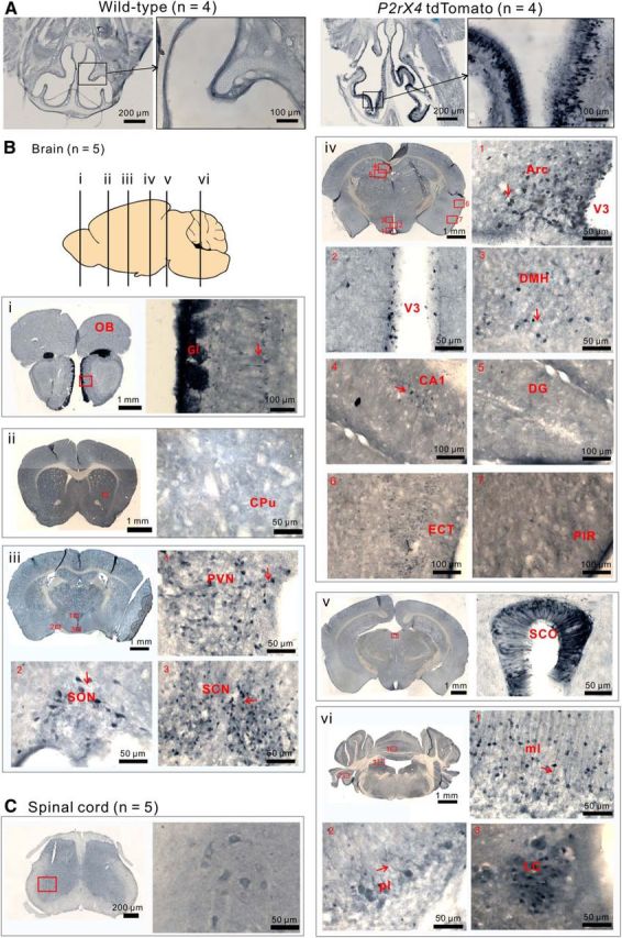
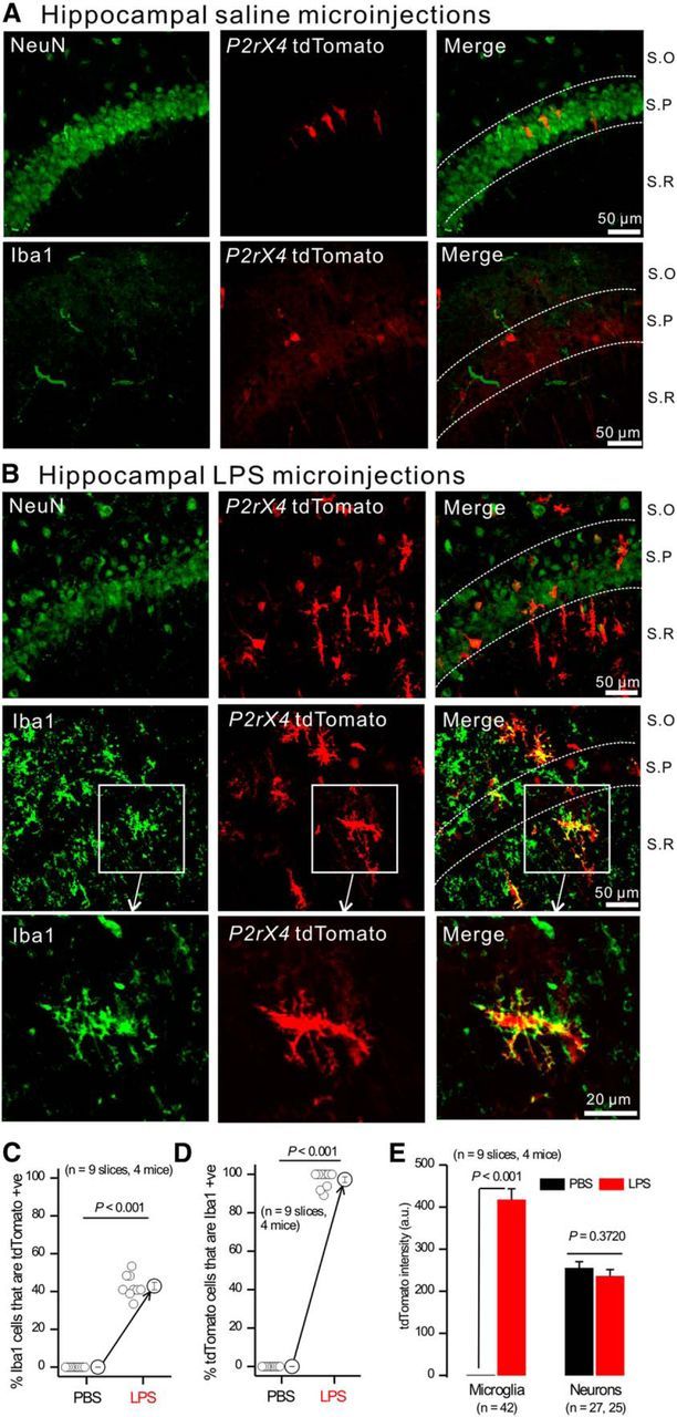
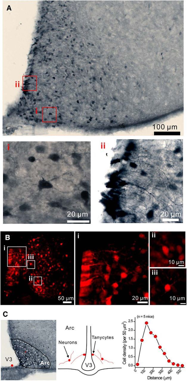
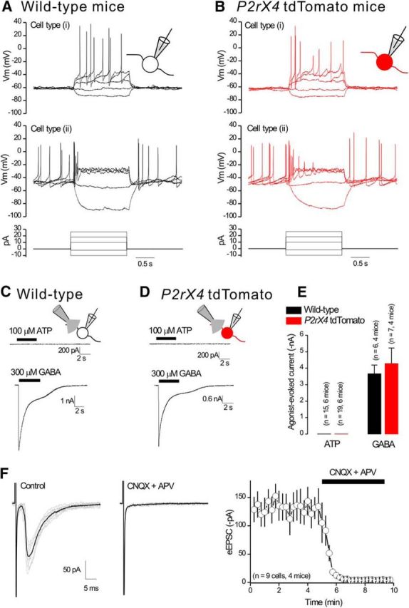
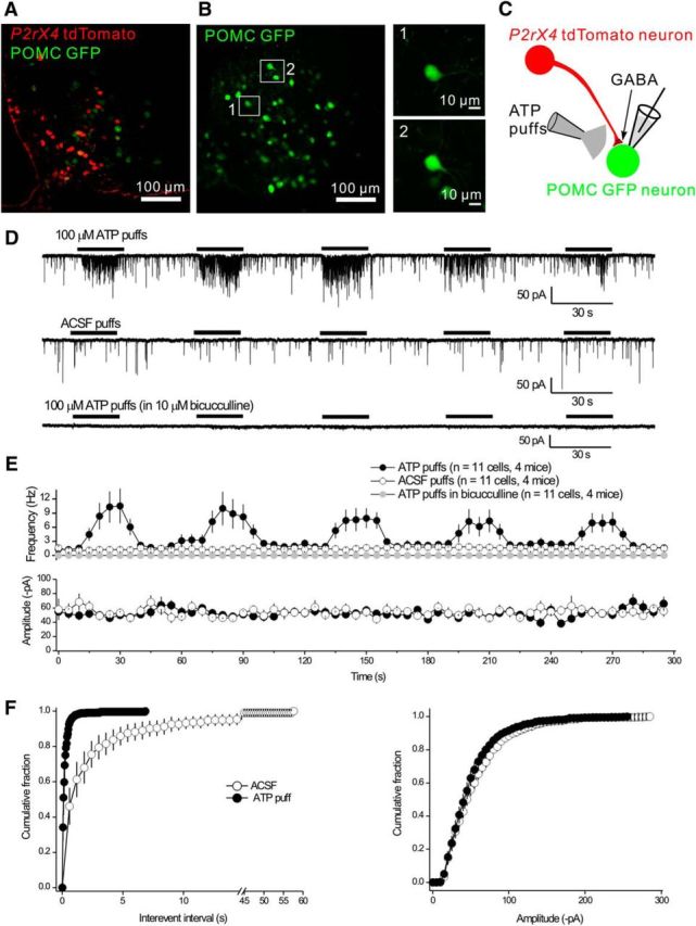
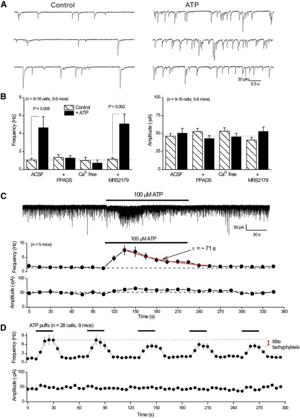
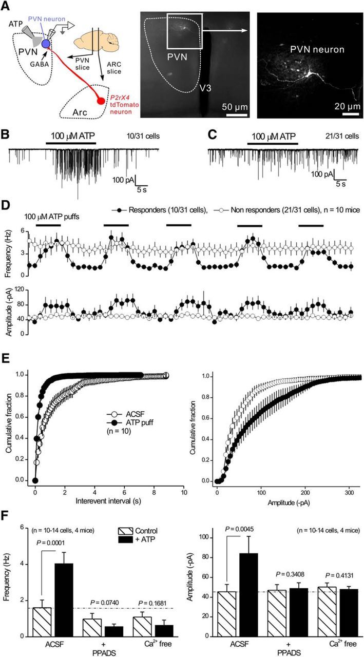
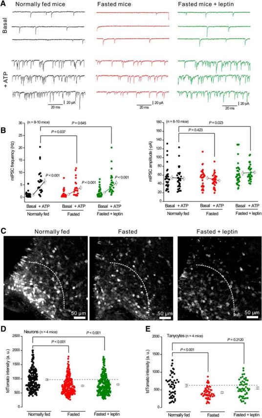
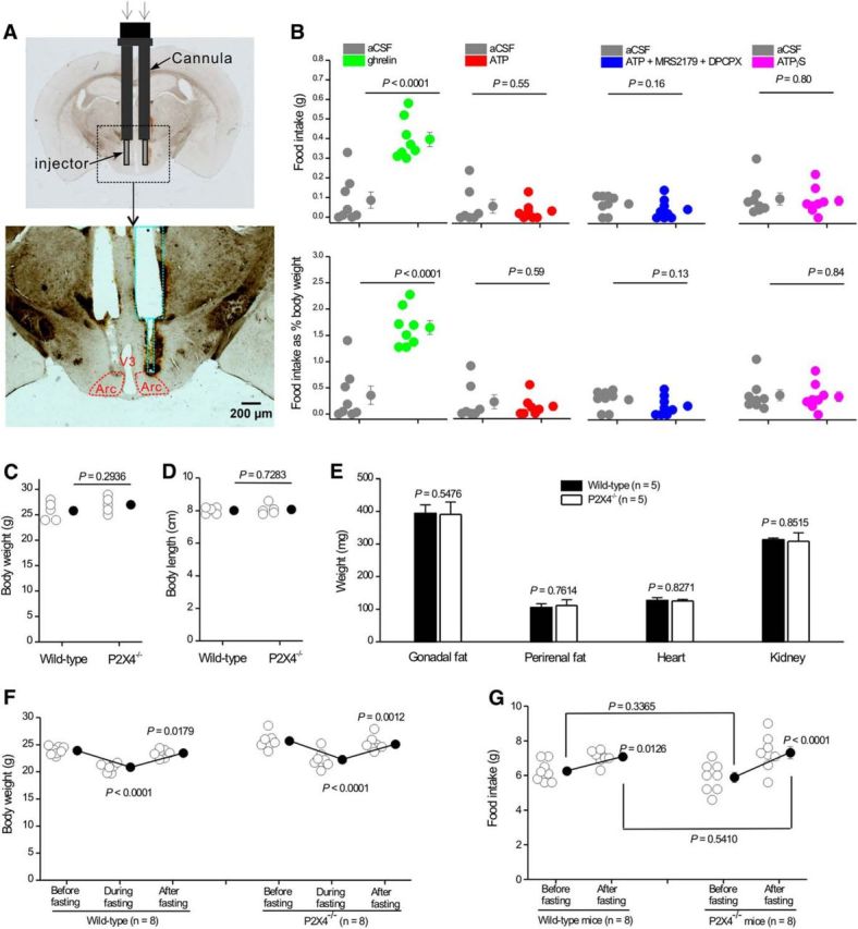
P2X4 receptors are ATP-gated cation channels that are widely expressed in the nervous system. To identify P2X4 receptor-expressing cells, we generated BAC transgenic mice expressing tdTomato under the control of the P2X4 receptor gene (P2rx4). We found sparse populations of tdTomato-positive neurons in most brain areas with patterns that matched P2X4 mRNA distribution. tdTomato expression within microglia was low but was increased by an experimental manipulation that triggered microglial activation. We found surprisingly high tdTomato expression in the hypothalamic arcuate nucleus (Arc) (i.e., within parts of the neural circuitry controlling feeding). Immunohistochemistry and genetic crosses of P2rx4 tdTomato mice with cell-specific GFP reporter lines showed that the tdTomato-expressing cells were mainly AgRP-NPY neurons and tanycytes. There was no electrophysiological evidence for functional expression of P2X4 receptors on AgRP-NPY neuron somata, but instead, we found clear evidence for functional presynaptic P2X4 receptor-mediated responses in terminals of AgRP-NPY neurons onto two of their postsynaptic targets (Arc POMC and paraventricular nucleus neurons), where ATP dramatically facilitated GABA release. The presynaptic responses onto POMC neurons, and the expression of tdTomato in AgRP-NPY neurons and tanycytes, were significantly decreased by food deprivation in male mice in a manner that was partially reversed by the satiety-related peptide leptin. Overall, we provide well-characterized tdTomato reporter mice to study P2X4-expressing cells in the brain, new insights on feeding-related regulation of presynaptic P2X4 receptor responses, and the rationale to explore extracellular ATP signaling in the control of feeding behaviors.

Significance statement: Cells expressing ATP-gated P2X4 receptors have proven problematic to identify and study in brain slice preparations because P2X4 expression is sparse. To address this limitation, we generated and characterized BAC transgenic P2rx4 tdTomato reporter mice. We report the distribution of tdTomato-expressing cells throughout the brain and particularly strong expression in the hypothalamic arcuate nucleus. Together, our studies provide a new, well-characterized tool with which to study P2X4 receptor-expressing cells. The electrophysiological studies enabled by this mouse suggest previously unanticipated roles for ATP and P2X4 receptors in the neural circuitry controlling feeding.

Keywords: ATP; P2X; arcuate; ion channel; mouse model; receptor.


Figures

Figure 1.
Figure 1.
tdTomato expression in P2rx4 tdTomato mice. A, Representative images of tdTomato IHC in olfactory tissue from wild-type and P2rx4 tdTomato reporter mice. B, Representative images of tdTomato-positive cells in the brain. Diagram represents the positions of coronal sections shown in Bi–Bvi. OB, Olfactory bulb; Gl, glomerular layer; CPu, corpus putamen; SON, supraoptic nucleus; SCN, suprachiasmatic nucleus; Arc, arcuate nucleus; V3, third ventricle; DMH, dorsal medial hypothalamus; DG, dentate gyrus; ECT, entorhinal cortex; PIR, piriform cortex; SCO, subcomissural organ; ml, molecular layer; pl, Purkinje layer; LC, locus ceruleus. C, IHC images of tdTomato-positive cells in spinal cord. The neurons stained in the ventral horn were motor neurons.
Figure 2.
Figure 2.
tdTomato expression was upregulated in microglia from P2rx4 tdTomato mice by LPS. A, B, Representative IHC images of NeuN and tdTomato (top) as well as Iba1 and tdTomato (bottom) from CA1 regions from P2rx4 tdTomato mice injected with PBS (A) or 4 μg LPS (B). S.O, Stratum oriens; S.P, stratum pyramidale; S.R, stratum radiatum. C, Percentage of Iba1-positive cells that were tdTomato-positive as measured from panels, such as in A, B. D, Percentage of tdTomato-positive cells that were Iba1-positive as measured from panels, such as in A, B. E, tdTomato fluorescence intensity within microglia and neurons from experiments, such as in A, B.
Figure 3.
Figure 3.
tdTomato-positive cells in the arcuate nucleus. A, Representative IHC images of tdTomato-positive cells in the arcuate nucleus. Ai, Representative image of neurons. Aii, Representative image of tanycytes. B, Confocal images of tdTomato-positive cells from live brain slices. Bi, Tanycytes. Bii, Biii, Neurons. C, Distance of Arc tdTomato-positive neurons from the bottom of the third ventricle (medium eminence). Diagram represents the positions of tanycytes and neurons. Right panels, The cell density of tdTomato-positive neurons per 50 μm2 was plotted against the distance to the medium eminence.
Figure 4.
Figure 4.
Cellular identity of tdTomato-positive cells in the arcuate nucleus. A–E, Representative images for colocalization of tdTomato-positive cells with markers for tanycytes (vimentin), neurons (HuC/HuD), astrocytes (GFAP), as well as GFP from POMC GFP and NPY GFP reporter mice. Average data for the percentage of P2X4-tdTomato cells that colocalized with these cell-specific markers are reported in the text.
Figure 5.
Figure 5.
Electrophysiological properties of neurons from the arcuate nucleus in wild-type and P2rx4 tdTomato mice. A, B, Representative waveforms of membrane potentials recorded in current clamp from arcuate nucleus neurons in wild-type mice (A) and tdTomato-positive neurons from P2rx4 tdTomato mice (B). Bottom panels, Current injection protocol. C, D, Representative traces for ATP-evoked and GABA-evoked currents recorded from arcuate nucleus neurons from wild-type mice (C) and tdTomato-positive neurons in P2rx4 tdTomato mice (D) in response to 5 s puff applications of 100 μm ATP or 300 μm GABA. No ATP-evoked currents were detected. E, Average data for experiments, such as those in C, D. F, Representative traces of eEPSCs recorded from tdTomato-positive neurons in the presence of 10 μm bicuculline. Right, The eEPSCs were completely blocked by 10 μm CNQX and 30 μm APV.
Figure 6.
Figure 6.
Presynaptic facilitation of mIPSCs onto Arc POMC neurons by ATP. A, There was no colocalization between tdTomato and GFP in the Arc from double-transgenic P2rx4 tdTomato and POMC GFP reporter mice. B, Images of fluorescent GFP-positive neurons in live slices from POMC GFP mice that were targeted for mIPSC recording in whole-cell mode. C, Schematic representation of the experimental strategy: tdTomato-positive neurons send GABAergic projections onto POMC neurons. We recorded mIPSCs from POMC GFP neurons and applied ATP locally via a puffer. D, Representative traces of mIPSCs recorded from POMC GFP neurons in response to five consecutive 20 s applications of 100 μm ATP or aCSF. The mIPSCs were abolished by 10 μm bicucculline. E, Plots of the average frequency and amplitude of mIPSCs over time from experiments, such as those in D. F, Cumulative probability plots of mIPSC interevent interval and amplitude during applications of aCSF and ATP from the experiments shown in E.
Figure 7.
Figure 7.
Properties of ATP-evoked presynaptic mIPSC facilitation onto POMC neurons. A, Representative traces of mIPSCs before and during ATP applications. There is a clear increase in frequency with no change in amplitude. B, Histograms summarize experiments where the ATP effect was evaluated under the indicated conditions. For these experiments, we used 10 μm PPADS, nominally Ca2+ free solutions, and 30 μm MRS-2179. C, Representative trace of mIPSCs during a prolonged 2 min 100 μm ATP application. Bottom, Average mIPSC frequency and amplitude during the recording before, during, and after ATP. D, Average frequency and amplitude of mIPSCs in response to five consecutive 20 s applications of 100 μm ATP or aCSF. The fifth application of ATP showed little tachyphylaxis compared with the first application.
Figure 8.
Figure 8.
Properties of ATP-evoked presynaptic mIPSC facilitation onto PVN neurons. A, Schematic of the GABAergic projection of tdTomato-positive neurons from the Arc to the PVN. The PVN neurons were targeted for whole-cell recording of mIPSCs. Right panels, Confocal images of a PVN neuron filled with biocytin via the patch pipette. B, Representative trace of mIPSCs recorded from a PVN neuron responding to 100 μm ATP. C, Representative trace of mIPSCs recorded from a PVN neuron that did not respond to 100 μm ATP. D, Average frequency and amplitudes of mIPSCs recorded from responding and nonresponding PVN neurons as shown in B, C. E, Cumulative probability plots of mIPSC interevent intervals and amplitudes recorded from PVN neurons that responded during applications of ATP. F, Histograms represent mIPSC frequency and amplitude from PVN neurons responding to ATP under various conditions, including control, 10 μm PPADS, and nominally Ca2+ free solutions.
Figure 9.
Figure 9.
Regulation of ATP-evoked mIPSC facilitation onto POMC neurons and tdTomato expression in AgRP-NPY neurons by metabolic state. A, Representative traces of mIPSCs before and during ATP applications recorded from POMC neurons in normally fed mice, fasted mice, and fasted mice injected with leptin (1 μg/g) 3 h before the experiments. B, Summary of mIPSC frequency and amplitude from experiments, such as those in A. C, Representative tdTomato IHC images for Arc from normally fed mice, fasted mice, and fasted mice injected with leptin. Dotted lines indicate the area in which tdTomato intensity was measured. D, E, tdTomato intensity in neurons (D) and tanycytes (C) from experiments, such as those shown in C.
Figure 10.
Figure 10.
Assessment of feeding in response to ATP and in P2X4 knock-out mice. A, Schematic illustrating the location of the guide cannula and injector for intra-Arc ATP and ghrelin injections. Top, Bilateral guide cannula and injectors projecting out of the cannula implanted in the mouse brain. Bottom, Track of the bilateral cannula with injectors, as marked by green dotted lines in relation to Arc (red). B, Food intake (top graphs) or food intake as a percentage of body weight (bottom graphs) during a 1 h period after bilateral delivery of 500 nl ghrelin (0.7 mm), ATP (3 mm), ATP (3 mm) plus MRS2179 (1 mm), and DPCPX (1 mm) or ATPγS (3 mm). C, D, Body weight (C) and length (D) of 2-month-old wild-type and P2X4 knock-out mice. E, Plot of gonadal fat, perirenal fat, heart, and kidney weights from 2-month-old wild-type and P2X4 knock-out mice. F, Body weights of wild-type and P2X4 knock-out mice before, during, and after fasting. G, Plot of food intake for wild-type and P2X4 knock-out mice before and after fasting.

Similar articles

Cited by

References

    1. Ase AR, Honson NS, Zaghdane H, Pfeifer TA, Séguéla P. Identification and characterization of a selective allosteric antagonist of human P2X4 receptor channels. Mol Pharmacol. 2015;87:606–616. doi: 10.1124/mol.114.096222. - DOI - PubMed
    1. Atasoy D, Betley JN, Su HH, Sternson SM. Deconstruction of a neural circuit for hunger. Nature. 2012;488:172–177. doi: 10.1038/nature11270. - DOI - PMC - PubMed
    1. Baxter AW, Choi SJ, Sim JA, North RA. Role of P2X4 receptors in synaptic strengthening in mouse CA1 hippocampal neurons. Eur J Neurosci. 2011;34:213–220. doi: 10.1111/j.1460-9568.2011.07763.x. - DOI - PMC - PubMed
    1. Besser S, Sicker M, Marx G, Winkler U, Eulenburg V, Hülsmann S, Hirrlinger J. A transgenic mouse line expressing the red fluorescent protein tdTomato in GABAergic neurons. PLoS One. 2015;10:e0129934. doi: 10.1371/journal.pone.0129934. - DOI - PMC - PubMed
    1. Bo X, Zhang Y, Nassar M, Burnstock G, Schoepfer R. A P2X purinoceptor cDNA conferring a novel pharmacological profile. FEBS Lett. 1995;375:129–133. doi: 10.1016/0014-5793(95)01203-Q. - DOI - PubMed

Publication types

MeSH terms

LinkOut - more resources