Multicenter Study
. 2016 Aug 25;11(8):e0161626.
doi: 10.1371/journal.pone.0161626. eCollection 2016.

Plasma Level of Placenta-Derived Macrophage-Stimulating Protein -Chain in Preeclampsia before 20 Weeks of Pregnancy

Affiliations
Multicenter Study

Plasma Level of Placenta-Derived Macrophage-Stimulating Protein -Chain in Preeclampsia before 20 Weeks of Pregnancy

Yonggang Zhang et al. PLoS One. .

Abstract

Object: This study aimed to investigate the diagnostic value of placenta-derived macrophage-stimulating protein α-chain (MSP-α) before the 20th week of gestation for the early diagnosis of preeclampsia (PE).

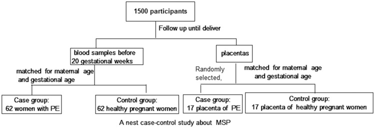
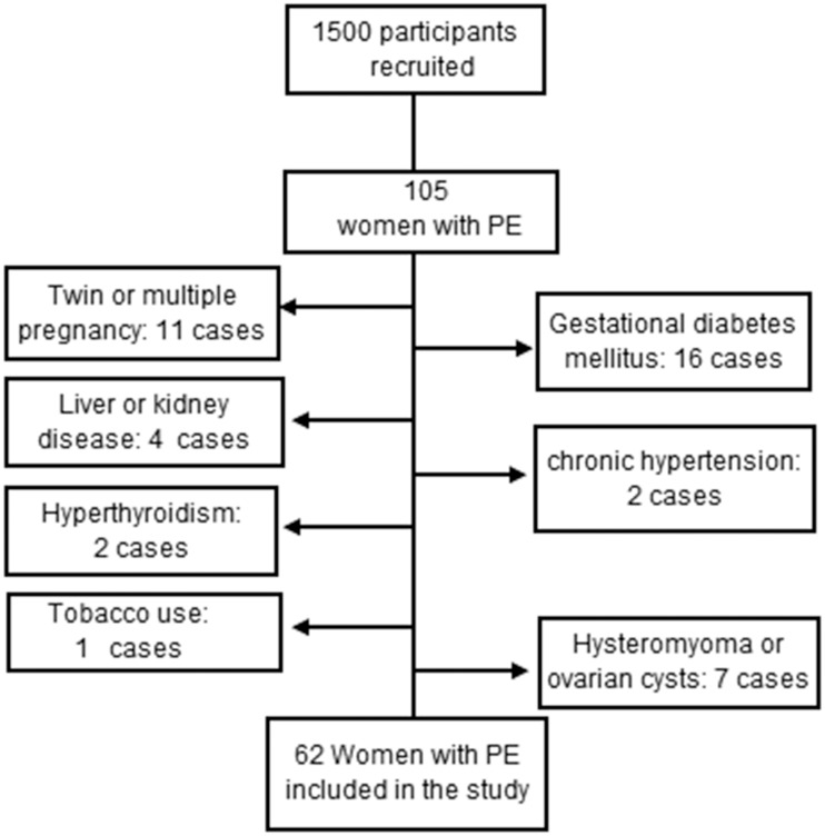
Methods and materials: Two parts of this nested case-control study were simultaneously executed, and 1500 pregnant women were recruited. A total of 124 pregnant women were included in the plasma analysis part of this study. The MSP-α plasma level was measured before the 20th week of gestation, and the participants were followed until delivery. A case group of 62 women with PE and a control group of 62 women matched by gestational age, maternal age, and pre-pregnancy BMI (with normotensive pregnancies) were evaluated. In the placenta analysis part of this nested case-control study, the placentas of 34 pregnant women were randomly obtained. The placental levels of MSP were measured in 17 individuals with PE (case group) and in 17 women with a normotensive pregnancy matched by gestational age and maternal age (control group).

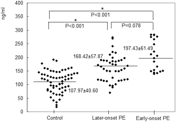
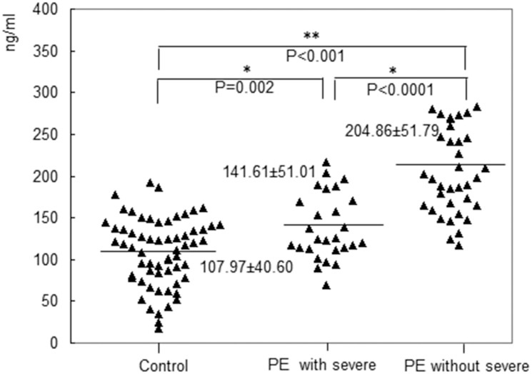
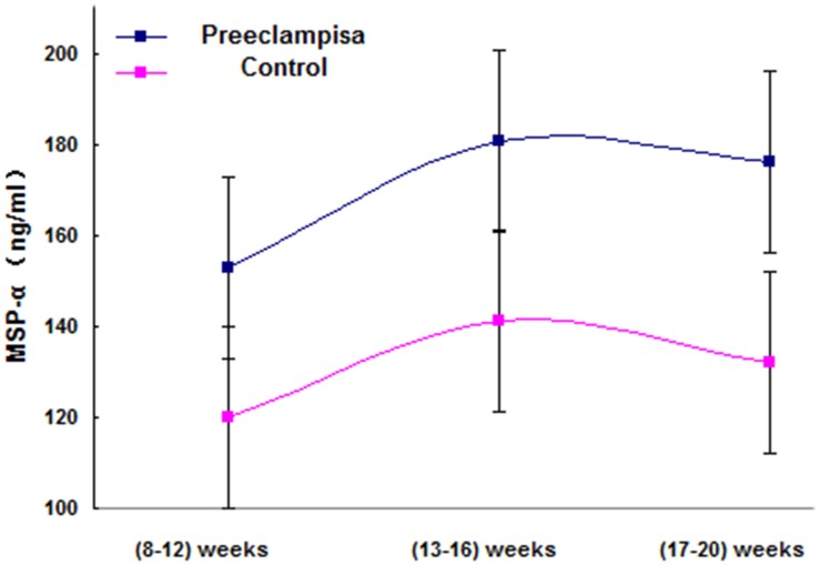
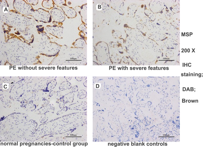
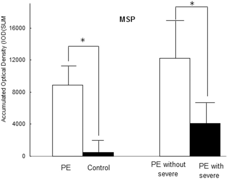
Results: The plasma level of MSP-α was higher in the PE group than in the control group before the 20th week of gestation (p < 0.001). In addition, compared to the women with severe features in the PE group, those without severe features had a significantly higher plasma MSP-α level before the 20th week of gestation (p < 0.001). The area under the receiver operating characteristic curve (AUC) of MSP-α before the 20th week of gestation was 0.905 (95% CI, 0.811-0.962) for the women with early-onset PE without severe features. With regard to the placenta, the PE group (accumulated optical density, IOD [SUM] = 8862.37 ± 2064.42) exhibited increased MSP staining (more intense MSP staining or more extensive staining) compared with the control group (normal pregnancies (IOD [SUM] = 447.92 ± 114.72, P < 0.001). Furthermore, increased MSP staining was detected among the women without severe features compared with those with severe features in the PE group (IOD [SUM]: 12192.65 ± 5325.56 vs. 4104.83 ± 2383.06, P = 0.021).

Conclusion: According to the findings of this study, the plasma level of MSP-α may be associated with PE, and MSP-α may be considered a candidate protein for further analysis in studies of PE. Multicenter studies with larger sample sizes must be performed in the future to obtain accurate results regarding the predictive value of MSP-α in combination with other protein factors for the early diagnosis of PE.


Conflict of interest statement

The authors have declared that no competing interests exist.

Figures

Fig 1
Fig 1. The study design.
Fig 2
Fig 2. A flow diagram of the participants in the plasma MSP study.
Fig 3
Fig 3. The plasma level of MSP-α was significantly higher in the patients with early-onset preeclampsia (197.43± 61.49 ng/mL) compared to the controls (107.97± 40.60 ng/mL) before the 20th week of gestation (p<0.001).
Before 20 weeks of pregnancy, significant differences in the plasma levels of MSP-α between the patients with late-onset preeclampsia and the controls were observed (168.42± 57.87 ng/mL vs. 107.97± 40.60 ng/mL, p < 0.001).
Fig 4
Fig 4. The plasma level of MSP-α in the patients with severe features was significantly lower than that in the patients without severe features before the 20th week of gestation (141.61 ± 51.01 ng/mL vs. 204.86 ± 51.79 ng/mL, p < 0.001).
The levels in the case groups were significantly higher compared with that in the control group (204.86 ± 51.79 ng/mL vs. 141.61 ± 51.01 ng/mL vs. 107.97± 40.60 ng/mL, p < 0.001).
Fig 5
Fig 5. In women with preeclampsia and in those with normal pregnancy, before the 20th week of gestation, fluctuations in the MSP-α level were examined at three time points (8–12, 13–16, and 17–20 gestational weeks), revealing that the mean (±SE) levels of MSP-α (in the weeks prior to the 20th week of gestation) in each group did not significantly differ.
Fig 6
Fig 6. MSP expression in trophoblast cells in placental sections from pregnant women, as determined by immunohistochemical staining.
The PE group exhibited strong MSP staining in the placenta compared with the control group. Furthermore, increased MSP staining was detected in the sections from the patients without severe features vs. those with severe features in the PE group (DAB, brown: images A-D, 200X magnification). (images A: PE without severe features; images B: PE with severe features; images C: normal pregnancies in the control group; images D: negative blank controls).
Fig 7
Fig 7. Immunohistochemical analysis of the placentas revealed that the patients with PE exhibited significantly increased plasma MSP staining (IOD [SUM] = 8862.37±2064.42) compared with the healthy pregnant controls (IOD [SUM] = 447.92±114.72, P <0.001).
A significant difference in MSP staining was observed between the PE group without severe features (IOD [SUM]: 12192.65±5325.56) and the PE group with severe features (4104.83±2383.06; P = 0.021).
Fig 8
Fig 8. Immunohistochemical analysis of the placentas revealed that the patients with PE exhibited significantly increased plasma RON staining (IOD [SUM] = 2540.15±637.76) compared with the healthy pregnant controls (IOD [SUM] = 1375.87±365.03, P <0.001).
A significant difference in RON staining was detected between the PE group without severe features (IOD (SUM):3611.78±1020.86) and the PE group with severe features (1009.25±158.36; P = 0.017).

References

    1. Hypertension in pregnancy. Report of the American College of Obstetricians and Gynecologists' Task Force on Hypertension in Pregnancy. Obstet Gynecol 2013,122:1122–31. 10.1097/01.AOG.0000437382.03963.88 - DOI - PubMed
    1. Myatt L, Clifton RG, Roberts JM, Spong CY, Hauth JC, Varner MW et al. The utility of uterine artery Doppler velocimetry in prediction of preeclampsia in a low-risk population. Obstet Gynecol 2012, 120:815–22. - PMC - PubMed
    1. Baumann MU, Bersinger NA, Mohaupt MG, Raio L, Gerber S, Surbek DV. First-trimester serum levels of soluble endoglin and soluble fms-like tyrosine kinase-1 as first-trimester markers for late-onset preeclampsia. Am J Obstet Gynecol 2008, 199:261–6. - PubMed
    1. Levine RJ, Lam C, Qian C, Yu KF, Maynard SE, Sachs BP, et al. Soluble endoglin and other circulating antiangiogenic factors in preeclampsia. N Engl J Med 2006, 355:992–1005. - PubMed
    1. Yao HP, Zhou YQ, Zhang R, Wang MH. MSP-RON signalling in cancer: pathogenesis and therapeutic potential. Nat Rev Cancer 2013, 13:466–81. 10.1038/nrc3545 - DOI - PubMed

Publication types

Substances

LinkOut - more resources