. 2016 Oct 25;115(9):1058-1068.
doi: 10.1038/bjc.2016.259. Epub 2016 Aug 25.

Establishment and molecular characterisation of seven novel soft-tissue sarcoma cell lines

Affiliations

Establishment and molecular characterisation of seven novel soft-tissue sarcoma cell lines

Abdulazeez Salawu et al. Br J Cancer. .

Abstract

Background: Soft-tissue sarcomas (STS) are a diverse group of malignancies that remain a diagnostic and therapeutic challenge. Relatively few reliable cell lines currently exist. Rapidly developing technology for genomic profiling with emerging insights into candidate functional (driver) aberrations raises the need for more models for in vitro functional validation of molecular targets.

Methods: Primary cell culture was performed on STS tumours utilising a differential attachment approach. Cell lines were characterised by morphology, immunocytochemistry, proliferation assays, short tandem repeat (STR) and microarray-based genomic copy number profiling.

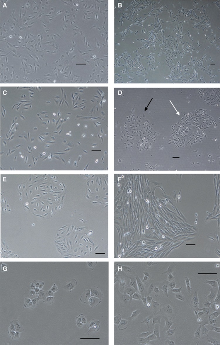
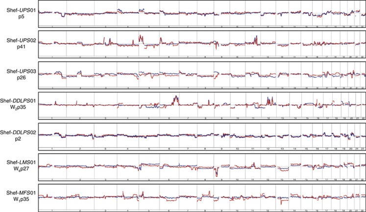
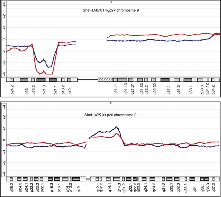
Results: Of 47 STS cases of various subtypes, half formed adherent monolayers. Seven formed self-immortalised cell lines, including three undifferentiated pleomorphic sarcomas, two dedifferentiated liposarcomas (one of which had received radiotherapy), a leiomyosarcoma and a myxofibrosarcoma. Two morphologically distinct yet genetically identical variants were established in separate cultures for the latter two tumours. All cell lines demonstrated genomic and phenotypic features that not only confirm their malignant characteristics but also confirm retention of DNA copy number aberrations present in their parent tumours that likely include drivers.

Conclusions: These primary cell lines are much-needed additions to the number of reliable cell lines of STS with complex genomics available for initial functional validation of candidate molecular targets.


Figures

Figure 1
Figure 1
Representative phase-contrast micrographs of STS primary cell lines.(A) Shef-UPS 01-spindle-shaped cells at passage 69 without distinct colony formation in culture. (B) Shef-UPS 03 cultures at passage 35 showing homogeneous cultures of long spindle-shaped cells growing in loose colonies. (C) Shef-DDLPS 01 cultures at passage 71 showing pleomorphic cells without distinct colony formation. (D) Shef-DDLPS 02 cultures at passage 22 composed of a combination of spindle-shaped cells (white arrows) and round, histiocyte-like cells (black arrows), both growing in distinct colonies. (E, F) Cells derived from Shef-LMS 01 growing in separate cultures designated as Ws (passage 69) and w1 (passage 56), respectively. (G, H) Morphologically distinct cells derived from Shef-MFS 01 growing in separate adherent cultures designated as w1 (passage 35) and w2 (passage 31), respectively. Scale bars=100 μm. DDLPS=dedifferentiated liposarcoma; LMS=leiomyosarcoma; MFS=myxofibrosarcoma.
Figure 2
Figure 2
Genomic copy number profile comparisons of seven soft-tissue sarcoma (STS) primary cell lines (shown on the left) with their parent tumours.Individual cell lines and passage number at which genomic DNA was extracted are shown to the left of the corresponding autosome ideograms. The overlaid red and blue lines represent the moving average of log2 ratios of the cultured cells and parent tumour tissue, respectively. Deviations above and below the horizontal baseline represent amplifications and deletions, respectively. Relative amplitude of deviation shows the log2 ratio and represents DNA copy number at the corresponding genomic locus. Note the close similarity and/or near-identical breakpoints in the moving average patterns in each case over the majority of the genome. Copy number analysis was performed on the Agilent 4 × 180K DNA microarray platform and data were analysed using Agilent Genomic Workbench Software v.6.0 (Agilent Technologies).
Figure 3
Figure 3
Selected chromosome copy number profile comparisons of soft-tissue sarcoma (STS) primary cell lines with their parent tumours.Ideograms of specific chromosomes are as shown at the top of each panel with the corresponding regions at the bottom. The overlaid red and blue lines represent the moving average of log2 ratios (vs normal genomic DNA) of the cultured cells and parent tumour tissue, respectively. Deviations above and below the horizontal baseline represent amplifications and deletions, respectively. Amplitude of deviation shows the relative log2 ratio and represents relative DNA copy number. Note the close similarity and/or near-identical breakpoints in the moving average patterns in each case. Copy number analysis was performed on the Agilent 4 × 180K DNA microarray platform and data analysed using Agilent Genomic Workbench Software v.6.0 (Agilent Technologies).

Similar articles

Cited by

  • Experimental Models for SARS-CoV-2 Infection.
    Kim T, Lee JS, Ju YS. Kim T, et al. Mol Cells. 2021 Jun 30;44(6):377-383. doi: 10.14348/molcells.2021.0094. Mol Cells. 2021. PMID: 34187969 Free PMC article. Review.
  • Establishment and characterization of the novel myxofibrosarcoma cell line, SMU-MFS.
    Nakahashi N, Emori M, Takada K, Murahashi Y, Shimizu J, Murase K, Tsukahara T, Sugita S, Takasawa A, Iba K, Teramoto A, Osanai M. Nakahashi N, et al. Hum Cell. 2024 Dec 3;38(1):25. doi: 10.1007/s13577-024-01157-9. Hum Cell. 2024. PMID: 39625530
  • Sarcoma treatment in the era of molecular medicine.
    Grünewald TG, Alonso M, Avnet S, Banito A, Burdach S, Cidre-Aranaz F, Di Pompo G, Distel M, Dorado-Garcia H, Garcia-Castro J, González-González L, Grigoriadis AE, Kasan M, Koelsche C, Krumbholz M, Lecanda F, Lemma S, Longo DL, Madrigal-Esquivel C, Morales-Molina Á, Musa J, Ohmura S, Ory B, Pereira-Silva M, Perut F, Rodriguez R, Seeling C, Al Shaaili N, Shaabani S, Shiavone K, Sinha S, Tomazou EM, Trautmann M, Vela M, Versleijen-Jonkers YM, Visgauss J, Zalacain M, Schober SJ, Lissat A, English WR, Baldini N, Heymann D. Grünewald TG, et al. EMBO Mol Med. 2020 Nov 6;12(11):e11131. doi: 10.15252/emmm.201911131. Epub 2020 Oct 13. EMBO Mol Med. 2020. PMID: 33047515 Free PMC article. Review.
  • Primary Culture of Undifferentiated Pleomorphic Sarcoma: Molecular Characterization and Response to Anticancer Agents.
    De Vita A, Recine F, Mercatali L, Miserocchi G, Spadazzi C, Liverani C, Bongiovanni A, Pieri F, Casadei R, Riva N, Fausti V, Amadori D, Ibrahim T. De Vita A, et al. Int J Mol Sci. 2017 Dec 8;18(12):2662. doi: 10.3390/ijms18122662. Int J Mol Sci. 2017. PMID: 29292724 Free PMC article.
  • Oncopig Soft-Tissue Sarcomas Recapitulate Key Transcriptional Features of Human Sarcomas.
    Schachtschneider KM, Liu Y, Mäkeläinen S, Madsen O, Rund LA, Groenen MAM, Schook LB. Schachtschneider KM, et al. Sci Rep. 2017 Jun 1;7(1):2624. doi: 10.1038/s41598-017-02912-9. Sci Rep. 2017. PMID: 28572589 Free PMC article.

References

    1. American Type Culture Collection Standards Development Organization Workgroup ASN (2010) Cell line misidentification: the beginning of the end. Nat Rev Cancer 10(6): 441–448. - PubMed
    1. Anderson K, Lutz C, van Delft FW, Bateman CM, Guo Y, Colman SM, Kempski H, Moorman AV, Titley I, Swansbury J, Kearney L, Enver T, Greaves M (2011) Genetic variegation of clonal architecture and propagating cells in leukaemia. Nature 469(7330): 356–361. - PubMed
    1. Barretina J, Caponigro G, Stransky N, Venkatesan K, Margolin AA, Kim S, Wilson CJ, Lehar J, Kryukov GV, Sonkin D, Reddy A, Liu M, Murray L, Berger MF, Monahan JE, Morais P, Meltzer J, Korejwa A, Jane-Valbuena J, Mapa FA, Thibault J, Bric-Furlong E, Raman P, Shipway A, Engels IH, Cheng J, Yu GK, Yu J, Aspesi P Jr, de Silva M, Jagtap K, Jones MD, Wang L, Hatton C, Palescandolo E, Gupta S, Mahan S, Sougnez C, Onofrio RC, Liefeld T, MacConaill L, Winckler W, Reich M, Li N, Mesirov JP, Gabriel SB, Getz G, Ardlie K, Chan V, Myer VE, Weber BL, Porter J, Warmuth M, Finan P, Harris JL, Meyerson M, Golub TR, Morrissey MP, Sellers WR, Schlegel R, Garraway LA (2012) The Cancer Cell Line Encyclopedia enables predictive modelling of anticancer drug sensitivity. Nature 483(7391): 603–607. - PMC - PubMed
    1. Barretina J, Taylor BS, Banerji S, Ramos AH, Lagos-Quintana M, Decarolis PL, Shah K, Socci ND, Weir BA, Ho A, Chiang DY, Reva B, Mermel CH, Getz G, Antipin Y, Beroukhim R, Major JE, Hatton C, Nicoletti R, Hanna M, Sharpe T, Fennell TJ, Cibulskis K, Onofrio RC, Saito T, Shukla N, Lau C, Nelander S, Silver SJ, Sougnez C, Viale A, Winckler W, Maki RG, Garraway LA, Lash A, Greulich H, Root DE, Sellers WR, Schwartz GK, Antonescu CR, Lander ES, Varmus HE, Ladanyi M, Sander C, Meyerson M, Singer S (2010) Subtype-specific genomic alterations define new targets for soft-tissue sarcoma therapy. Nat Genet 42(8): 715–721. - PMC - PubMed
    1. Beroukhim R, Mermel CH, Porter D, Wei G, Raychaudhuri S, Donovan J, Barretina J, Boehm JS, Dobson J, Urashima M, Mc Henry KT, Pinchback RM, Ligon AH, Cho YJ, Haery L, Greulich H, Reich M, Winckler W, Lawrence MS, Weir BA, Tanaka KE, Chiang DY, Bass AJ, Loo A, Hoffman C, Prensner J, Liefeld T, Gao Q, Yecies D, Signoretti S, Maher E, Kaye FJ, Sasaki H, Tepper JE, Fletcher JA, Tabernero J, Baselga J, Tsao MS, Demichelis F, Rubin MA, Janne PA, Daly MJ, Nucera C, Levine RL, Ebert BL, Gabriel S, Rustgi AK, Antonescu CR, Ladanyi M, Letai A, Garraway LA, Loda M, Beer DG, True LD, Okamoto A, Pomeroy SL, Singer S, Golub TR, Lander ES, Getz G, Sellers WR, Meyerson M (2010) The landscape of somatic copy-number alteration across human cancers. Nature 463(7283): 899–905. - PMC - PubMed