Review
. 2017 Mar 1;525(4):1034-1054.
doi: 10.1002/cne.24105. Epub 2016 Sep 27.

Stem and progenitor cells of the mammalian olfactory epithelium: Taking poietic license

Affiliations
Review

Stem and progenitor cells of the mammalian olfactory epithelium: Taking poietic license

James E Schwob et al. J Comp Neurol. .

Abstract

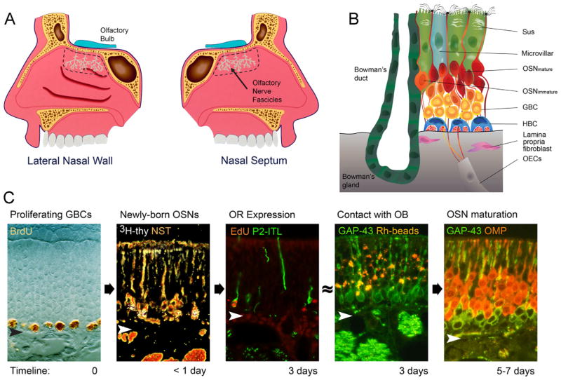
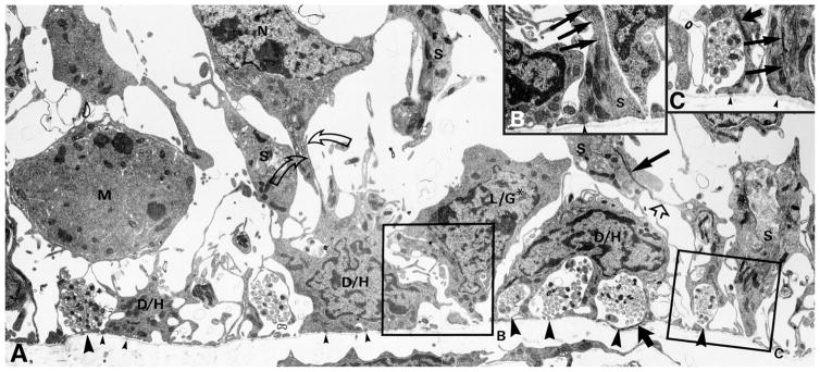
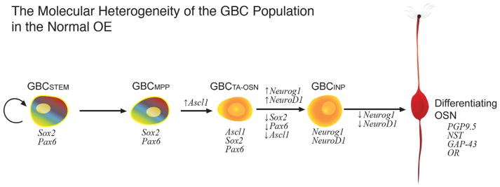
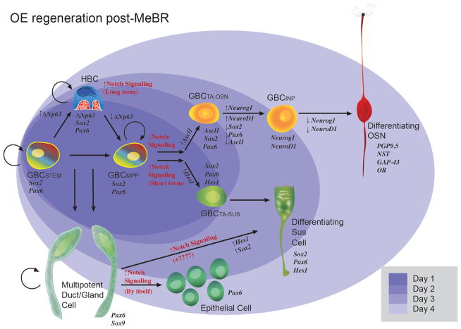
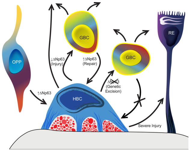
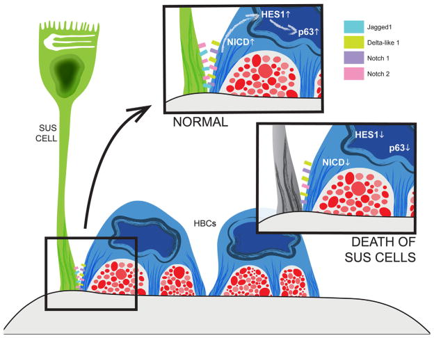
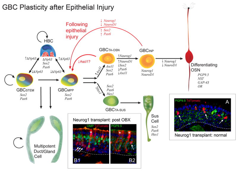
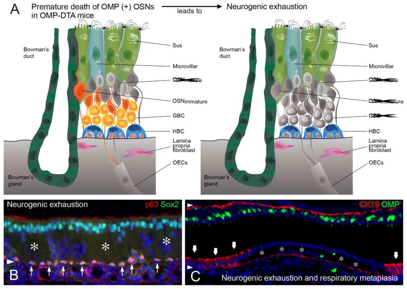
The capacity of the olfactory epithelium (OE) for lifelong neurogenesis and regeneration depends on the persistence of neurocompetent stem cells, which self-renew as well as generating all of the cell types found within the nasal epithelium. This Review focuses on the types of stem and progenitor cells in the epithelium and their regulation. Both horizontal basal cells (HBCs) and some among the population of globose basal cells (GBCs) are stem cells, but the two types plays vastly different roles. The GBC population includes the basal cells that proliferate in the uninjured OE and is heterogeneous with respect to transcription factor expression. From upstream in the hierarchy to downstream, GBCs encompass 1) Sox2+ /Pax6+ stem-like cells that are totipotent and self-renew over the long term, 2) Ascl1+ transit-amplifying progenitors with a limited capacity for expansive proliferation, and 3) Neurog1+ /NeuroD1+ immediate precursor cells that make neurons directly. In contrast, the normally quiescent HBCs are activated to multipotency and proliferate when sustentacular cells are killed, but not when only OSNs die, indicating that HBCs are reserve stem cells that respond to severe epithelial injury. The master regulator of HBC activation is the ΔN isoform of the transcription factor p63; eliminating ΔNp63 unleashes HBC multipotency. Notch signaling, via Jagged1 ligand on Sus cells and Notch1 and Notch2 receptors on HBCs, is likely to play a major role in setting the level of p63 expression. Thus, ΔNp63 becomes a potential therapeutic target for reversing the neurogenic exhaustion characteristic of the aged OE. J. Comp. Neurol. 525:1034-1054, 2017. © 2016 Wiley Periodicals, Inc.

Keywords: active stem cell; aging; dedifferentiation; globose basal cell; horizontal basal cell; neurogenesis; p63; reserve stem cell.


Conflict of interest statement

CONFLICT OF INTEREST STATEMENT

The authors have no conflicts of interest.

Figures

Figure 1
Figure 1
A: Schematic representation of the olfactory area (outlined by the dashed line) on the lateral nasal wall and nasal septum of the bisected nose along with the fibers of the olfactory nerve (teal). B: The major cellular constituents of the olfactory epithelium (OE). Note particularly the two populations of basal cells, globose basal cells (GBC) and horizontal basal cells (HBC). Sus, sustentacular cells; OSN, olfactory sensory neuron; OECs, olfactory ensheathing cells in fascicles of the olfactory nerve. C: Progression from proliferating GBC to mature OSN as a function of time after terminal S-phase (timeline). Proliferating GBCs are marked by the incorporation of BrdU or other thymidine analogues. Less than 24 hours after injection of 3H-thymidine (thy) or other thymidine analogue, the label is chased into differentiating OSNs marked by neuron-specific tubulin (NST). approximately 3 days after a neuron becomes postmitotic, olfactory receptors (OR) such as P2, which is marked by the labeling with tau-fused beta galactosidase (P2-ITL), are expressed. At about the same time, immature, GAP-43-expressing OSNs can be labeled by retrograde transport of rhodamine-labeled latex microspheres (Rh beads) following injections in the olfactory bulb (OB). Finally, morphological and functional maturation is evident by 5–7 days later, as marked by the transition from expression of GAP-43 to the olfactory marker protein (OMP; Verhaagen et al., 1989; Schwob, 1991; Schwob et al., 1992; Rodriguez-Gil et al., 2015). Arrowheads designate the basal lamina.
Figure 2
Figure 2
Electron microscopic investigation illustrates the characteristics of the basal region of the OE. A–C: The HBCs (D/H) tightly adhere to the basal lamina (small arrowheads indicate hemidesmosomes) forming cytoplasmic bridges that enfold clusters of olfactory axons (large arrowheads). The GBCs (L/G* and M) are found at a remove from the basal lamina, including those that are undergoing mitosis (M). The GBC marked by the asterisk (L/G*) has a process that touches the basal lamina (in contrast to the one marked M) and apparently corresponds to a “third type” of basal cell described previously (Graziadei and Monti Graziadei, 1979), the significance of which is unclear. Note the numerous adherens junctions between cells in the basal region: Sus cell (S) and HBCs (straight open arrow), HBCs and neurons (N; curved open arrows). Cellular identification is based on morphology, i.e., the foot process of Sus cells, and the presence of characteristic intermediate filaments: in Sus cells (thin straight solid arrows) and in HBCs (thick straight solid arrows). B,C: Higher magnification electron micrographs taken from the region of the boxed areas in A but from a nearby section. Modified from Holbrook et al. (1995).
Figure 3
Figure 3
Diagram illustrating the molecular hetereogeneity of the GBCs in the normal olfactory epithelium (OE) and the progression from stem cell through to differentiating olfactory sensory neuron (OSN). A relatively simple linear sequence proceeds from GBC stem cells through multiple molecularly discrete stages, with marker genes designated in italics and potential multipotency designated by the rainbow gradient; that multipotency is not evident in the normal epithelium in situ but is observed following transplantation from normal OE into the lesioned environment (Goldstein et al., 1998; Chen et al., 2004). HBCs are not shown and do not contribute to the OSN population in the normal OE (Caggiano et al., 1994). GBCSTEM, stem cell-like GBC expressing Sox2 and Pax6 that is mitotically quiescent (thymidine label-retaining); GBCMPP, multipotent GBC expressing Sox2 and Pax6 and first to appear during development and regeneration; GBCTA-OSN, transit-amplifying GBC restricted to a neuronal fate and expressing Sox2, Pax6, and Ascl1; GBCINP, GBC functioning as an immediate neuronal precursor, i.e., giving rise to a small number of OSNs, and expressing Neurog1 and NeuroD1. The evidence that some GBCs are stem cells is described in the text.
Figure 4
Figure 4
Diagram illustrating the process of epitheliopoiesis in the OE during recovery from methyl bromide (MeBr) injury, which is substantially more complex than the progression observed in the normal OE (cf. Fig. 3). The cellular lineages to which basal cells give rise and the timing of their re-emergence after damage (shaded ovals in the background) are indicated. The rainbow gradient filling the GBCSTEM and GBCMPP symbolizes their active multipotency, i.e., those GBCs that generate, in aggregate, all of the epithelial cell types following epithelial injury, including HBCs. The self-renewal capacity of the GBCSTEM is indicated by the circular arrow; self renewal is strongly suggested by the persistence of neurogenesis when the contribution of HBCs has been eliminated (see Fig. 5 and text for details). Thus, some GBCs satisfy both the multipotency and self-renewal criteria for being classified as stem cells. The retention of thymidine label by some GBCs also suggests stem-like quiescence. As an example of GBC multipotency, 1 day after MeBr (darkest purple oval), the epithelium consists of HBCs and GBCs, characterized by the indicated transcription factor profiles, including the selective HBC marker p63. Some among the GBCs express Hes1, which marks the GBCs that are transitioning directly into Sus cells. Notch signaling is key to the determination of neuronal vs. nonneuronal differentiation (Herrick and Schwob, 2015). Also indicated is the dual origin of the Sus cells from GBCs and from gland/duct cells, which has been demonstrated by retroviral lineage tracing and transplantation experiments, as summarized in the text (Huard et al., 1998). GBCSTEM, stem cell-like GBC expressing Sox2 and Pax6 that is mitotically quiescent (thymidine label-retaining); GBCMPP, multipotent GBC expressing Sox2 and Pax6 and appearing first during development and regeneration; GBCTA-OSN, transit-amplifying GBC restricted to the neuronal lineage and expressing Sox2, Pax6, and Ascl1; GBCINP, GBC functioning as an immediate neuronal precursor, i.e., giving rise to a small number of OSNs, and expressing Neurog1 and NeuroD1.
Figure 5
Figure 5
p63 Is the master regulator of the formation and activation–dormancy of the HBCs, the second category of OE stem cells, which are held in reserve normally. HBCs are formed from basally migrating olfactory placodal precursors (OPP) when ΔNp63 is upregulated during perinatal development (Packard et al., 2011b). In the absence of direct epithelial damage, HBCs are dormant (mitotically quiescent and nonparticipatory in the generation of replacement neurons; Huard and Schwob, 1995; Leung et al., 2007). With injury, ΔNp63 levels decline, and many of the HBCs differentiate into GBCs, which in turn give rise to neurons and other epithelial cell types (Packard et al., 2011b; Fletcher et al., 2011; Schnittke et al., 2015). The suggestion that HBCs may directly give rise to other cell types following injury is indicated by the upward-pointing arrow (Schwob et al., 1995). During epithelial repair, GBCs that initiate ΔNp63 expression in response to local cues or as a consequence of retroviral transduction give rise to HBCs, which return to quiescence (Schnittke et al., 2015). Genetic excision of ΔNp63 activates HBCs, generates GBCs that remain active for more than 6 months indicating stem cell capacity, and simultaneously prevents the regeneration of HBCs from GBCs bearing the mutated gene (Schnittke et al., 2015). Thus, expression of ΔNp63 is both necessary and sufficient for the formation of HBCs, and conversely downregulation of ΔNp63 is both necessary and sufficient for HBCs to activate. As a result of very severe injury, in which the population of GBCs and possibly gland/duct cells is completely depleted, the activated HBCs contribute to respiratory metaplasia, i.e., give rise to ciliated columnar cells like those found in respiratory epithelium (RE; red profiles between HBC and basal lamina represent olfactory axons; Xie et al., 2013).
Figure 6
Figure 6
TdTomato labeling of HBCs in normal OE (A1,2) or in OE harvested 24 hours after MeBr lesion (B) from mice in which the K5-CreERT2 transgene excises a floxed(stop) motif in the ROSA26 locus in response to tamoxifen, thereby driving expression of TdTomato exclusively in HBCs. The shape of the marked HBCs is illustrated in whole mounts of the OE subjected to processing using the CLARITY technique (for details see Schnittke et al., 2015). Note the retraction of processes following lesion. The arrows in A1 indicate two adjacent HBCs.
Figure 7
Figure 7
Death of Sus cells (as a result of the Cyp2G1-driven expression of diphtheria toxin A) causes downregulation of ΔNp63 in HBCs and, thereby, HBC proliferation and activation to multipotency (Herrick and Schwob, 2015). The effect on ΔNp63 is mediated, at least in part, by changes in Notch signaling. HBCs express both Notch1 and Notch2 receptors as well as the Delta-like1 ligand, whereas Sus cells express the Jagged1 ligand as well as the Notch2 receptor. Constitutive activation of Notch signaling by overexpression of the Notch1 intracellular domain selectively in HBCs elevates the downstream factor Hes1 as well as the levels of ΔNp63 expression. Conversely, excision of Notch1 from HBCs decreases their expression of ΔNp63 and enhances the rate of HBC spontaneous activation (Herrick and Schwob, 2015).
Figure 8
Figure 8
The hierarchy of stem cell progressing to progenitor cell among the GBC population can be reversed following direct injury to the OE or depletion of the OSN population by massive retrograde degeneration. In the normal OE, Neurog1+ GBCs, whose progeny are labeled genetically and permanently by the expression of CreERT2, give rise only to OSNs in situ and when they engraft in the MeBr-lesioned host (A, normal) and are considered to be immediate neuronal progenitors (GBCINP); arrows indicate PGP9.5+/TdTomato+ OSNs derived from the transplanted Neurog1-CreERT2-expressing progenitors. In contrast, following ablation of the olfactory bulb (OBX) or methimazole-induced damage to the OE, Neurog1+ GBCs initiate the expression of Sox2 and Pax6, which are expressed by more upstream GBCs and evince multipotency either in situ or after transplantation (B1,2; post-OBX). B1,2: Asterisks mark Sus cells derived from the Neurog1+ progenitors, whereas arrows indicate OSNs identified on the basis of immunostaining or morphology. Likewise, Ascl1+ GBCs, which precede Neurog1-expressing GBCs during embryonic development or recovery from MeBr lesion, express Sox2 and Pax6 and are considered to be GBCTA-OSN, are also multipotent in situ following methimazole lesion and to a greater extent than the Neurog1+ GBCs (as indicated by the reverse arrows). Thus, the stem cell–progenitor cell “hierarchy” within the OE is not strictly unidirectional and is much more fluid than previously imagined (Lin et al., 2015).
Figure 9
Figure 9
The depletion and disappearance of OSNs and GBCs that is characteristic of the OE in the elderly also emerges in a mouse model of accelerated aging. A: In that model, the premature death of OSNs follows the expression of diphtheria toxin subunit A (DTA) driven by the expression of OMP as the OSNs reach maturity. The dead neurons are “grayed out” in the schematic. Over time, the reduction in the neuronal population progresses to neurogenic exhaustion, in which all neurons and GBCs have disappeared (gray), whereas the HBCs remain dormant. B: Areas of neurogenic exhaustion (asterisks) are evident by the absence of Sox2+ GBCs and the gaps in the neuronal layer as shown by DAPI staining. The dormant HBCs remain strongly positive for p63 and Sox2 expression (thin arrows). C: Areas of neurogenic exhaustion (asterisks) can progress to respiratory metaplasia, where the epithelium consists of CK19+ columnar respiratory epithelial cells (thick arrows; Jang et al., 2015).

References

    1. Alonso L, Fuchs E. Stem cells of the skin epithelium. Proc Natl Acad Sci U S A. 2003;100(Suppl 1):11830–11835. - PMC - PubMed
    1. Andres KH. The fine structure of the olfactory region of macrosmatic animals. Z Zellforsch Mikrosk Anat. 1966;69:140–154. - PubMed
    1. Asan E, Drenckhahn D. Immunocytochemical characterization of two types of microvillar cells in rodent olfactory epithelium. Histochem Cell Biol. 2005;123:157–168. - PubMed
    1. Becker AJ, McCulloch EA, Till JE. Cytological demonstration of the clonal nature of spleen colonies derived from transplanted mouse marrow cells. Nature. 1963;197:452–454. - PubMed
    1. Bergman U, Ostergren A, Gustafson AL, Brittebo B. Differential effects of olfactory toxicants on olfactory regeneration. Arch Toxicol. 2002;76:104–112. - PubMed