

The Role of 18F-FDG PET/CT Integrated Imaging in Distinguishing Malignant from Benign Pleural Effusion
- PMID: 27560933
- PMCID: PMC4999143
- DOI: 10.1371/journal.pone.0161764
The Role of 18F-FDG PET/CT Integrated Imaging in Distinguishing Malignant from Benign Pleural Effusion
Abstract
Objective: The aim of our study was to evaluate the role of 18F-FDG PET/CT integrated imaging in differentiating malignant from benign pleural effusion.
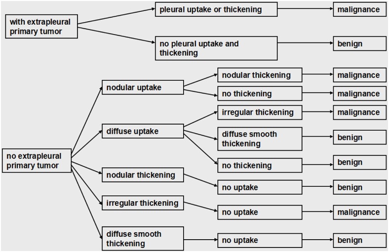
Methods: A total of 176 patients with pleural effusion who underwent 18F-FDG PET/CT examination to differentiate malignancy from benignancy were retrospectively researched. The images of CT imaging, 18F-FDG PET imaging and 18F-FDG PET/CT integrated imaging were visually analyzed. The suspected malignant effusion was characterized by the presence of nodular or irregular pleural thickening on CT imaging. Whereas on PET imaging, pleural 18F-FDG uptake higher than mediastinal activity was interpreted as malignant effusion. Images of 18F-FDG PET/CT integrated imaging were interpreted by combining the morphologic feature of pleura on CT imaging with the degree and form of pleural 18F-FDG uptake on PET imaging.
Results: One hundred and eight patients had malignant effusion, including 86 with pleural metastasis and 22 with pleural mesothelioma, whereas 68 patients had benign effusion. The sensitivities of CT imaging, 18F-FDG PET imaging and 18F-FDG PET/CT integrated imaging in detecting malignant effusion were 75.0%, 91.7% and 93.5%, respectively, which were 69.8%, 91.9% and 93.0% in distinguishing metastatic effusion. The sensitivity of 18F-FDG PET/CT integrated imaging in detecting malignant effusion was higher than that of CT imaging (p = 0.000). For metastatic effusion, 18F-FDG PET imaging had higher sensitivity (p = 0.000) and better diagnostic consistency with 18F-FDG PET/CT integrated imaging compared with CT imaging (Kappa = 0.917 and Kappa = 0.295, respectively). The specificities of CT imaging, 18F-FDG PET imaging and 18F-FDG PET/CT integrated imaging were 94.1%, 63.2% and 92.6% in detecting benign effusion. The specificities of CT imaging and 18F-FDG PET/CT integrated imaging were higher than that of 18F-FDG PET imaging (p = 0.000 and p = 0.000, respectively), and CT imaging had better diagnostic consistency with 18F-FDG PET/CT integrated imaging compared with 18F-FDG PET imaging (Kappa = 0.881 and Kappa = 0.240, respectively).
Conclusion: 18F-FDG PET/CT integrated imaging is a more reliable modality in distinguishing malignant from benign pleural effusion than 18F-FDG PET imaging and CT imaging alone. For image interpretation of 18F-FDG PET/CT integrated imaging, the PET and CT portions play a major diagnostic role in identifying metastatic effusion and benign effusion, respectively.
Conflict of interest statement
The authors have declared that no competing interests exist.
Figures
Similar articles
-
Clinical value of fluorodeoxyglucose-positron emission tomography/computed tomography in differentiation of malignant mesothelioma from asbestos-related benign pleural disease: an observational pilot study.J Thorac Oncol. 2009 Dec;4(12):1480-4. doi: 10.1097/JTO.0b013e3181c0a7ff. J Thorac Oncol. 2009. PMID: 19875971
-
Clinical role of F-18 fluorodeoxyglucose positron emission tomography imaging in patients with lung cancer and suspected malignant pleural effusion.Chest. 2002 Dec;122(6):1918-24. doi: 10.1378/chest.122.6.1918. Chest. 2002. PMID: 12475827
-
The role of FDG PET-CT in differential diagnosis of pleural pathologies.Rev Esp Med Nucl Imagen Mol. 2012 Jul-Aug;31(4):187-91. doi: 10.1016/j.remn.2011.06.002. Epub 2011 Sep 22. Rev Esp Med Nucl Imagen Mol. 2012. PMID: 23067687
-
Does positron emission tomography offer prognostic information in malignant pleural mesothelioma?Interact Cardiovasc Thorac Surg. 2011 May;12(5):806-11. doi: 10.1510/icvts.2010.255901. Epub 2011 Jan 25. Interact Cardiovasc Thorac Surg. 2011. PMID: 21266493 Review.
-
Present and future roles of FDG-PET/CT imaging in the management of malignant pleural mesothelioma.Jpn J Radiol. 2016 Aug;34(8):537-47. doi: 10.1007/s11604-016-0555-1. Epub 2016 May 24. Jpn J Radiol. 2016. PMID: 27222020 Review.
Cited by
-
Treatment of malignant pleural effusion in non-small cell lung cancer with VEGF-directed therapy.Ann Med. 2022 Dec;54(1):1357-1371. doi: 10.1080/07853890.2022.2071977. Ann Med. 2022. PMID: 35543207 Free PMC article.
-
Comment on: "the FDG PET/CT score" for the diagnosis of malignant pleural effusion.Eur J Nucl Med Mol Imaging. 2020 Jan;47(1):5-6. doi: 10.1007/s00259-019-04512-3. Epub 2019 Sep 4. Eur J Nucl Med Mol Imaging. 2020. PMID: 31485682 No abstract available.
-
Contemporary approach to the patient with malignant pleural effusion complicating lung cancer.Ann Transl Med. 2019 Aug;7(15):352. doi: 10.21037/atm.2019.03.61. Ann Transl Med. 2019. PMID: 31516898 Free PMC article. Review.
-
The prognostic value of 18F-fluorodeoxyglucose positron emission tomography/ computed tomography parameters in patients with malignant pleural mesothelioma.Turk Gogus Kalp Damar Cerrahisi Derg. 2021 Jan 13;29(1):92-100. doi: 10.5606/tgkdc.dergisi.2021.20432. eCollection 2021 Jan. Turk Gogus Kalp Damar Cerrahisi Derg. 2021. PMID: 33768986 Free PMC article.
-
Current Applications for Nuclear Medicine Imaging in Pulmonary Disease.Curr Pulmonol Rep. 2020;9(3):82-95. doi: 10.1007/s13665-020-00251-1. Epub 2020 Jul 22. Curr Pulmonol Rep. 2020. PMID: 32837866 Free PMC article. Review.
References
-
- Traill ZC, Davies RJ, Gleeson FV. Thoracic computed tomography in patients with suspected malignant pleural effusions. Clin Radiol. 2001;56:193–6. - PubMed
-
- Gupta NC, Rogers JS, Graeber GM, Gregory JL, Waheed U, Mullet D, et al. Clinical role of F-18 fluorodeoxyglucose positron emission tomography imaging in patients with lung cancer and suspected malignant pleural effusion. Chest. 2002;122:1918–24. - PubMed
-
- Charron M, Beyer T, Bohnen NN, Kinahan PE, Dachille M, Jerin J, et al. Image analysis in patients with cancer studied with a combined PET and CT scanner. Clin Nucl Med. 2000;25:905–10. - PubMed
-
- Toaff JS, Metser U, Gottfried M, Gur O, Deeb ME, Lievshitz G, et al. Differentiation between malignant and benign pleural effusion in patients with extra- pleural primary malignancies: assessment with positron emission tomography- computed tomography. Invest Radiol. 2005;40:204–9. - PubMed
Publication types
MeSH terms
Substances
LinkOut - more resources
Full Text Sources
Other Literature Sources
Medical
