

Effects of IL-1β, IL-6 and IL-8 on erythrocytes, platelets and clot viscoelasticity
- PMID: 27561337
- PMCID: PMC4999875
- DOI: 10.1038/srep32188
Effects of IL-1β, IL-6 and IL-8 on erythrocytes, platelets and clot viscoelasticity
Abstract
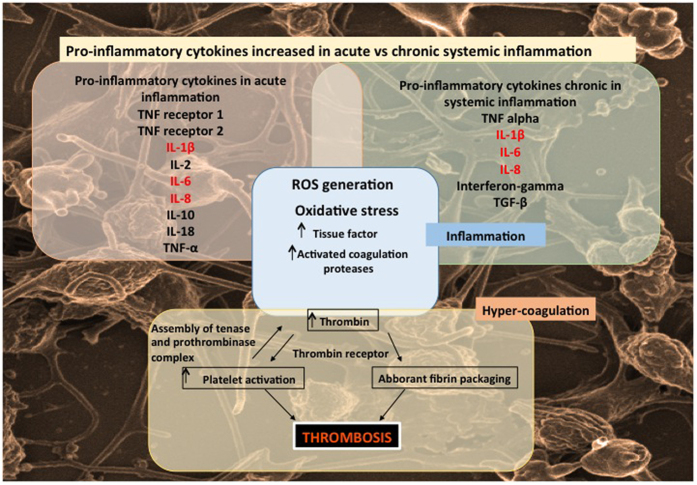
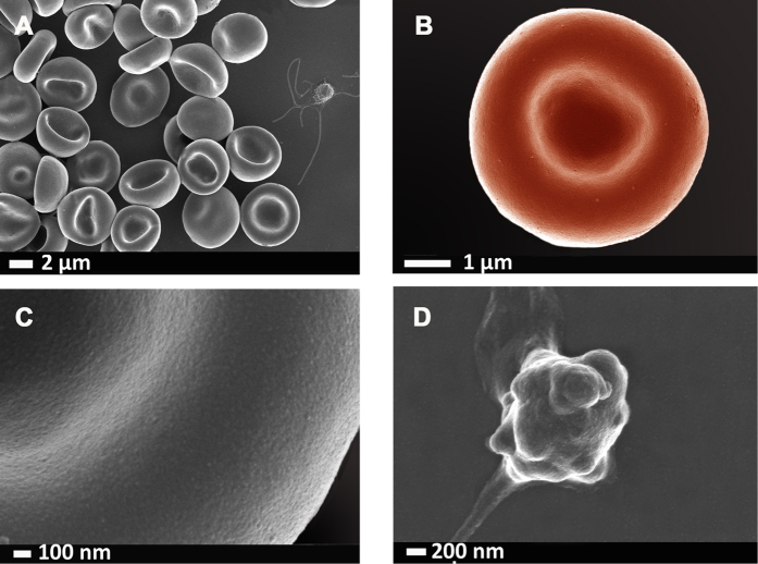
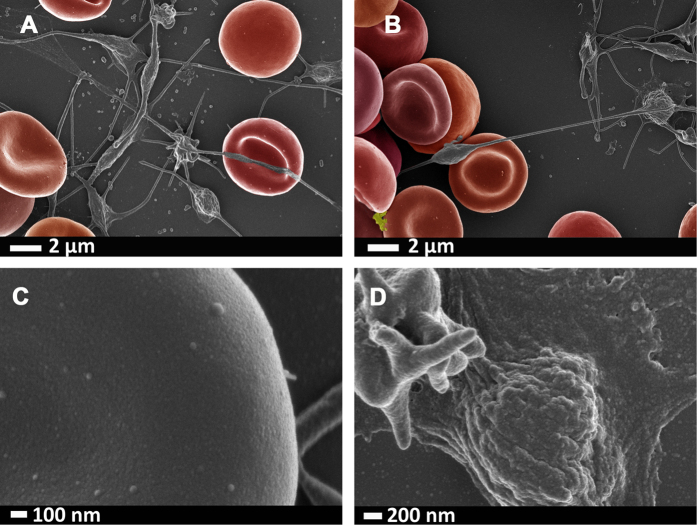
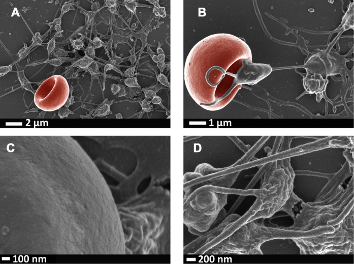
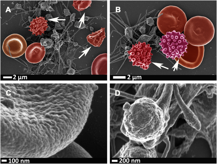
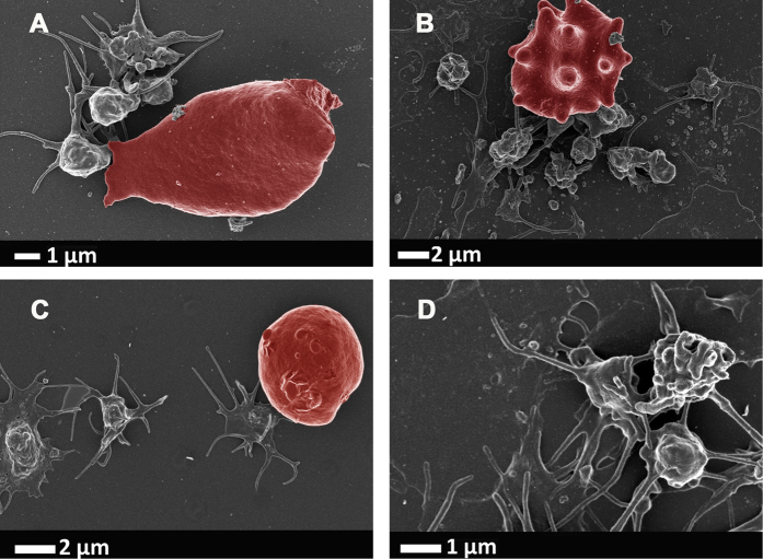
Complex interactions exist between cytokines, and the interleukin family plays a fundamental role in inflammation. Particularly circulating IL-1β, IL-6 and IL-8 are unregulated in systemic and chronic inflammatory conditions. Hypercoagulability is an important hallmark of inflammation, and these cytokines are critically involved in abnormal clot formation, erythrocyte pathology and platelet hyper-activation, and these three cytokines have known receptors on platelets. Although these cytokines are always unregulated in inflammation, we do not know how the individual cytokines act upon the structure of erythrocytes and platelets, and which of the viscoelastic clot parameters are changed. Here we study the effects of IL-1β, IL-6 and IL-8 at low physiological levels, representative of chronic inflammation, by using scanning electron microscopy and thromboelastography. All three interleukins caused the viscoelastic properties to display an increased hypercoagulability of whole blood and pathology of both erythrocytes and platelets. The most pronounced changes were noted where all three cytokines caused platelet hyper-activation and spreading. Erythrocyte structure was notably affected in the presence of IL-8, where the morphological changes resembled that typically seen in eryptosis (programmed cell death). We suggest that erythrocytes and platelets are particularly sensitive to cytokine presence, and that they are excellent health indicators.
Figures
Similar articles
-
Simultaneous presence of hypercoagulation and increased clot lysis time due to IL-1β, IL-6 and IL-8.Cytokine. 2018 Oct;110:237-242. doi: 10.1016/j.cyto.2018.01.007. Epub 2018 Feb 1. Cytokine. 2018. PMID: 29396046
-
The Effect of Endogenous and Synthetic Estrogens on Whole Blood Clot Formation and Erythrocyte Structure.Microsc Microanal. 2017 Jun;23(3):599-606. doi: 10.1017/S1431927617000472. Epub 2017 May 8. Microsc Microanal. 2017. PMID: 28478790
-
Interleukin-12 and its procoagulant effect on erythrocytes, platelets and fibrin(ogen): the lesser known side of inflammation.Br J Haematol. 2018 Jan;180(1):110-117. doi: 10.1111/bjh.15020. Epub 2017 Nov 16. Br J Haematol. 2018. PMID: 29143311
-
Viscoelasticity and Ultrastructure in Coagulation and Inflammation: Two Diverse Techniques, One Conclusion.Inflammation. 2015 Aug;38(4):1707-26. doi: 10.1007/s10753-015-0148-7. Inflammation. 2015. PMID: 25772112 Review.
-
[Effect of proinflammatory factors TNF-α,IL-1β, IL-6 on neuropathic pain].Zhongguo Zhong Yao Za Zhi. 2017 Oct;42(19):3709-3712. doi: 10.19540/j.cnki.cjcmm.20170907.004. Zhongguo Zhong Yao Za Zhi. 2017. PMID: 29235283 Review. Chinese.
Cited by
-
Eryptosis in Peritoneal Dialysis-Related Peritonitis: The Potential Role of Inflammation in Mediating the Increase in Eryptosis in PD.J Clin Med. 2022 Nov 23;11(23):6918. doi: 10.3390/jcm11236918. J Clin Med. 2022. PMID: 36498493 Free PMC article.
-
Correlative Light-Electron Microscopy detects lipopolysaccharide and its association with fibrin fibres in Parkinson's Disease, Alzheimer's Disease and Type 2 Diabetes Mellitus.Sci Rep. 2018 Nov 14;8(1):16798. doi: 10.1038/s41598-018-35009-y. Sci Rep. 2018. PMID: 30429533 Free PMC article.
-
The probable role and therapeutic potential of the PI3K/AKT signaling pathway in SARS-CoV-2 induced coagulopathy.Cell Mol Biol Lett. 2022 Jan 11;27(1):6. doi: 10.1186/s11658-022-00308-w. Cell Mol Biol Lett. 2022. PMID: 35016612 Free PMC article. Review.
-
Pathophysiological Changes in Erythrocytes Contributing to Complications of Inflammation and Coagulation in COVID-19.Front Physiol. 2022 Jun 15;13:899629. doi: 10.3389/fphys.2022.899629. eCollection 2022. Front Physiol. 2022. PMID: 35784888 Free PMC article. Review.
-
The stabilizing effect of an oligomeric proanthocyanidin on red blood cell membrane structure of poorly controlled Type II diabetes.Nutr Diabetes. 2017 May 15;7(5):e275. doi: 10.1038/nutd.2017.25. Nutr Diabetes. 2017. PMID: 28504711 Free PMC article.
References
Publication types
MeSH terms
Substances
LinkOut - more resources
Full Text Sources
Other Literature Sources
Medical
