Review
. 2016 Aug 26;18(1):51.
doi: 10.1186/s12968-016-0269-7.

Principles of cardiovascular magnetic resonance feature tracking and echocardiographic speckle tracking for informed clinical use

Affiliations
Review

Principles of cardiovascular magnetic resonance feature tracking and echocardiographic speckle tracking for informed clinical use

Gianni Pedrizzetti et al. J Cardiovasc Magn Reson. .

Abstract

Tissue tracking technology of routinely acquired cardiovascular magnetic resonance (CMR) cine acquisitions has increased the apparent ease and availability of non-invasive assessments of myocardial deformation in clinical research and practice. Its widespread availability thanks to the fact that this technology can in principle be applied on images that are part of every CMR or echocardiographic protocol. However, the two modalities are based on very different methods of image acquisition and reconstruction, each with their respective strengths and limitations. The image tracking methods applied are not necessarily directly comparable between the modalities, or with those based on dedicated CMR acquisitions for strain measurement such as tagging or displacement encoding. Here we describe the principles underlying the image tracking methods for CMR and echocardiography, and the translation of the resulting tracking estimates into parameters suited to describe myocardial mechanics. Technical limitations are presented with the objective of suggesting potential solutions that may allow informed and appropriate use in clinical applications.

Keywords: Cardiac mechanics; Cardiovascular magnetic resonance; Feature tracking; Myocardial deformation; Strain.


Figures

Fig. 1
Fig. 1
Basic principle of tissue tracking. Tracking the portions of tissue about a series of point (indicated in red on the pictures) is based on defining small square windows centered about such points on a first image (left picture) and searching the as-much-as-possible-similar grayscale pattern on the following image (right picture) in the vicinity of the original window. At the first step, shown here, the search windows have the same position in the pair of images and the feature that was at the center of the window of the first images is sought on the corresponding window on the second image (red dot) to provide an estimate of the local displacement. This procedure is usually repeated moving the second windows at the center of the new position and using a so-called coarse-to-fine approach, reducing progressively the window size. Large windows permit to recognize large gross displacements of the tissue, while the reduction of the window search area about the previous estimated targets allows to improve the accuracy and the locality of the estimation
Fig. 2
Fig. 2
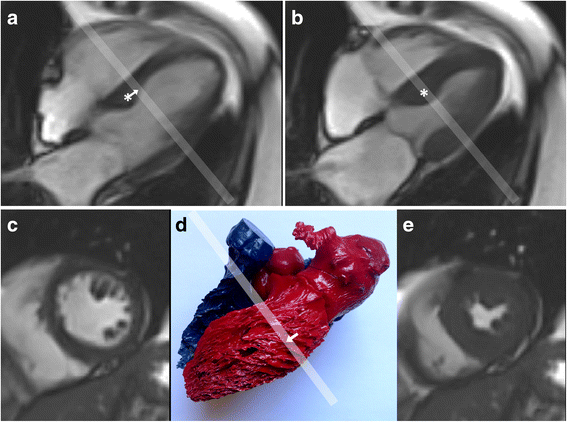
Possible effects on apparent strain of through-plane tissue displacements and trabecular appearances. Panel a This end-diastolic four chamber cine frame shows a basal septal bulge (*) whose long axis displacement causes it to move apically (arrow) to its end systolic position (b). Here it has moved into the short axis plane marked by the pale line. The septum in that short axis plane will therefore appear to have thickened more than it really did so that excess radial strain could be measured by tissue boundary tracking. Conversely, a more basal short axis slice might underestimate local radial strain due to tapering of the septum near the atrio-ventricular junction. Panel c Short axis images typically show trabeculations inside the LV free wall. Panel d this resin cast of human heart cavities shows the typical right handed helical alignments of free wall trabecular indentations. Systolic long axis displacement of these oblique trabecular structures (arrow) could give a false impression of trabecular displacement in the plane indicated by the white bar. A further issue is that trabeculations tend to thicken and move together in systole. This can exclude intervening blood, particularly in a hypertrophied ventricle with good function (panel e, from the same cine as c). If they merge to appear as part of the LV wall, it could result in over-estimation of radial and circumferential strain, if these were based on attempting to track the apparent endocardial boundary

Similar articles

Cited by

References

    1. Singh A. Optic flow computation: a unified perspective. Los Alamitos: IEEE Computer Society Press; 1991.
    1. Barron JL, Fleet DJ, Beauchemin SS. Performance of optical flow techniques. Int J Comput Vis. 1994;12:43–77. doi: 10.1007/BF01420984. - DOI
    1. Adrian RJ. Particle-Imaging Techniques for Experimental Fluid Mechanics. Annu Rev Fluid Mech. 1991;23:261–304. doi: 10.1146/annurev.fl.23.010191.001401. - DOI
    1. Willert CE, Gharib M. Digital particle image velocimetry. Exp Fluids. 1991;10:181–193. doi: 10.1007/BF00190388. - DOI
    1. Hor KN, Gottliebson WM, Carson C, Wash E, Cnota J, Fleck R, et al. Comparison of Magnetic Resonance Feature Tracking for Strain Calculation With Harmonic Phase Imaging Analysis. JACC Cardiovasc Imaging. 2010;3:144–151. doi: 10.1016/j.jcmg.2009.11.006. - DOI - PubMed

Publication types

MeSH terms